Neural Tube Defects Dysraphism Neural Tube Development Normal
")
Neural Tube Defects (Dysraphism )

Neural Tube Development Normal embryological development Neural plate development -18 th day Þ Cranial closure 24 th day (upper spine) Þ Caudal closure 26 th day (lower spine) Þ

Neural Tube defects Definition The most common congenial anomalies of CNS resulting from failure of the neural tube to close between 3 rd-4 th week gestation The exact cause of neural tube defects remains unknown. it appears to result from a combination of genetic and environmental , nutritional factors and family history of neural tube defects, folic acid deficiency and medical conditions such as diabetes and obesity

Neural Tube defects Folic acid deficiency. This vitamin is important to the healthy development of a fetus. Lack of folic acid increases the risk of spina bifida and other neural tube defects. Some medications. Anti-seizure medications, such as valproic acid (Depakene), seem to cause neural tube defects when taken during pregnancy Increased body temperature. Some evidence suggests that increased body temperature (hyperthermia) in the early months of pregnancy may increase the risk of spina bifida.

Types of Neural tube defects 1 - Spina Bifida and it is types 2 - Meningocele 3 - Myelomeningocele 4 - Encephalocele 5 - Anencephaly

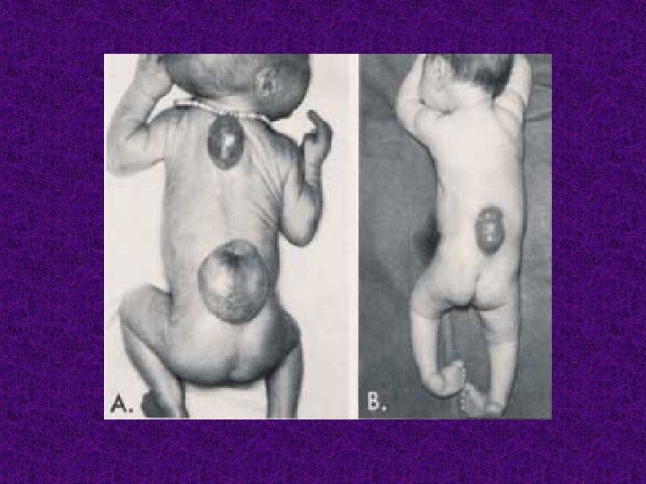
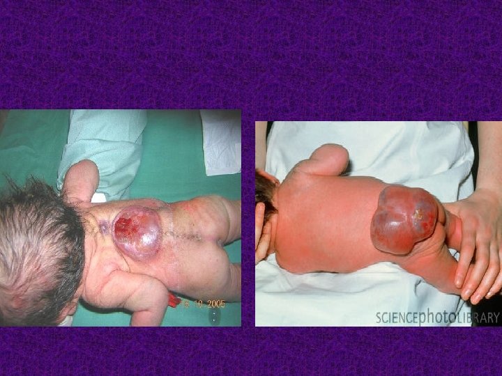
1 - Spina Bifida A- Spina bifida occulta • A condition involving non fusion of the halves of the vertebral arches without disturbance of the underlying neural tissue with tuft of hair and skin dimlpling B- Spina bifida manifesta • Defect of skin , vertebral column and spinal cord and protrusion of meninges and spinal cord through this defect

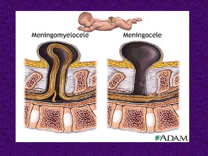
2 - Meningocele : the meninges protrude from the spinal opening, and the malformation may or may not be covered by a layer of skin.

3 - Myelomeningocele • Myelomeningocele; the meninges and spinal tissue protruding through a dorsal defect in the vertebrae • The paralysis may be so severe that the affected individual is unable to walk and may have urinary and bowel dysfunction

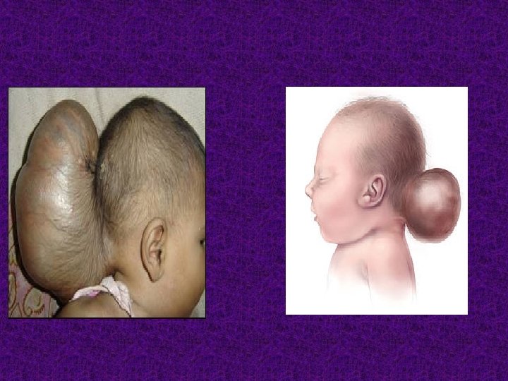
4 - Encephalocele is a hernia of part of the brain, and the membrane covering it (meninges), through a skull defect.
 and absence")
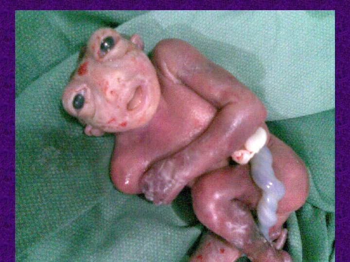
5 - Anencephaly; There is absence of the cranial vault (the skull) and absence of most or all of the cerebral hemispheres of the brain

Clinical picture • Symptoms range from physical and intellectual disability to paralysis, urinary and bowel control problems, blindness, deafness, lack of consciousness, and in many cases, death. • Most children with neural tube defects die or experience serious disability.

Complication Babies born with myelomeningocele also commonly experience accumulation of fluid in the brain, a condition known as hydrocephalus. Most babies with myelomeningocele will need a shunt. some may develop meningitis, an infection in the tissues surrounding the brain Children with myelomeningocele may develop learning disabilities

Diagnosis A- prenatal 1 - Maternal serum ----- second trimester maternal serum alpha fetoprotein (MSAFP)-increased The MSAFP test, however, is not specific for spina bifida. 2 - Ultrasound: An advanced ultrasound can also detect signs of spina bifida. 3 - Amniocentesis An analysis indicates the level of AFP present in the amniotic fluid.

B- postnatal 1 - CT scan for hydrocephalus and others anomalies 2 - MRI

Prevention Folic acid is an important vitamin in the development of a healthy fetus. Recent studies have shown that by adding folic acid to their diets, women of childbearing age significantly reduce the risk of having a child with a neural tube defect, such as spina bifida. Dosage: 400 micrograms of folic acid daily Foods high in folic acid include dark green vegetables, egg yolks, and some fruits.

Treatment There is no cure therapy for spina bifida. The nerve tissue that is damaged or lost cannot be repaired or replaced. Treatment depends on the type and severity of the disorder. children with the mild form need no treatment. Treatment of associated complication as hydrocephalus

Treatment The key priorities for treating myelomeningocele are to prevent infection from developing through the exposed nerves and tissue of the defect , and to protect the exposed nerves and structures from additional trauma. Doctors have recently begun performing fetal surgery for treatment of myelomeningocele.

THANK YOU
- Slides: 23