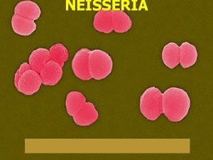
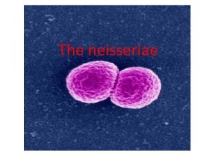
NEISSERIA Introduction The Neisseriae are Gve diplococci Pathogens


- Slides: 34

NEISSERIA

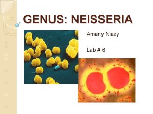
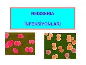
Introduction Ø The Neisseriae are G-ve diplococci Ø Pathogens are: - N. meningitidis Ø N. gonorrhoeae Ø Exacting growth requirements Ø Commensals easy to grow on ordinary media

N. gonorrhoeae Obligate parasite of human urogenital tract. Ø Morphology: Gram -ve diplococci (bean shaped). Ø Culture: enriched media (lysed blood or chocolate), moist aerobic atmosphere +5 -10% CO 2. Temp. 3537 o. C. Ø

Gram stain of N. gonorrhoea

Selective media Ø Thayer-Martin medium contains vancomycin, colistin, nystatin & trimethoprim. Ø Colonies: 48 hrs incubation.

Identification Ø Oxidase +ve. Ø Carbohydrate utilization: N. gonorrhoeae produces acid from glucose only. Ø Slide agglutination with specific antisera (Phadebact test).

Pathogenicity Causes gonorrhoea Ø Arthritis, Ø Septicemia, Ø Ø Ophthalmia neonatorum.

Gonorrhoea Acute pyogenic infection of urethra and (in females) cervix. Ø Acute purulent urethral , vaginal discharge , dysuria Ø Asymptomatic in females Ø Rectum & oropharynx may be involved. Ø

Complications Ø Prostatitis, epididymitis , urethral stricture in males. Ø Salpingitis , infertility in females Ø Septicemia Ø Arthritis Ø Meningitis (rare).

Diagnosis Ø Ø Ø Specimen: urethral, cervical smears &swabs (transport medium). Gram film: intracellular Gram -ve diplococci Culture: selective media Oxidase +ve acid production from glucose Latex agglutination

Treatment of gonorrhoea Ø Ø Ø One curative dose Sens. Testing Blind treatment: ceftriaxone, ciprofloxacin Spectinomycin Penicillin: resistance common.

N. meningitidis Habitat: human nasopharynx (10 -25%) Ø Similar to N. gonorrhoea but less exacting ? Ø Can grow in BA, Chocolate agar without selective media from CSF ? Ø Id. CHO utilization: acid from glucose & maltose. Ø

Gram stain of Neisseria meningitis

Haemorrhagic rash

Death from Waterhouse-Friderichsen syndrome

Neck rigidity

Antigenic structure Ø Polysaccharide antigens Ø Three main groups A, B, C Ø Other groups Y, W 135. Ø Grouping: slide agglutination with specific antisera

Pathogenicity Meningococcal meningitis, as a spread from nasopharynx blood stream meninges in susceptible hosts. Ø Direct spread to meninges Ø Rash Ø Adrenal haemorrhage (Waterhouse-Friderchsen syndrome) Ø

Meningitis Ø Ø Ø Clinically: rapid deterioration of flu like illness Headache, neck stiffness, +ve kerning’s sign, fever, . . … Diagnosis: CSF + blood culture CSF: WBC , RBCs Gram stain: bacteria & cells

Meningitis (Continue) Culture deposit into blood & chocolate agars and glucose broth 7 cooked meat media Ø Incubate in air + 5%CO 2 Ø Id : sugar utilization + latex Ø For partially treated meningitis: detection of bacterial antigen by: latex agglu, CCIE. . for common serogroups of meningitis pathogens. Ø

Treatment Parenteral antimicrobial Ø Start blind treatment after collection of specimens by: Ø § § Ceftriaxone or cefotaxime Change later according to sens. Test. Contacts: rifampicin Prevention: vaccination (polyvalent)

Commensal Neisseriae N. pharyngis, N. flava, N. sicca, . . Ø In mucous mem. Of mouth, nose, pharynx, less common in genital tract. Ø Differ. From pathogenic one: Ø grow in ordinary media( no CO 2) § at room temp. § rough, pigmented § acid from a number of CHOs §


Other causes of meningitis Ø Ø Ø Bacterial causes: Three primary pathogens: N. meningitidis, HI, S. pneumoniae N. menningitidis all ages HI 2 m-5 y S. pneumoniae all ages but more common in adult with underlying illnesses.

Other causative bacteria (Continue) Ø E. coli & other coliforms Ø Listeria Ø Strept. group B Ø Salmonella spp. Ø Favobacteria. . Ø All common in neonates

Other causative bacteria (Continue Ø After surgery or trauma Ø S. aureus Ø S. pneumoniae Ø AFB chronic meningitis Ø Spirochaetes

Other Causes Ø Viral : enterivirus, Paramyxovirus, Herpes viruses, adenoviruses, arboviruses. Ø Fungi: yeasts (Candida, cryptococcus spp. ) Ø Aspergillus spp. Ø Mucor

Findings in CSF Normal CSF: Ø Clear , colorless Ø 0 -5 lymphocytes Ø Sterile Ø 150 -450 mg /l protein Ø 2. 8 -3. 9 mmol/l glucose

CSF in bacterial meningitis Ø Ø Ø Turbid 500 -20, 000 cells mainly polys, few lymphocytes Bacteria in Gram stain Markedly raised protein Reduced or absent glucose

CSF in TB meningitis Ø Ø Ø Clear or slightly turbid 10 -500 cells, mainly lymphocytes( polys early) AFB in Z-N stain Grow in LJ medium Moderately raised protein Sugar reduced

CSF in viral meningitis Ø Clear or slightly turbid Ø 10 -500 cells mainly lymphocytes Ø Stool culture, or serology +ve Ø Normal or slightly raised protein Ø Normal glucose

Cerebral abscess Ø Clear or slightly turbid Ø Bacteria: S. milleri, Bacteroides, S. aureus. Proteus(Causative bacteria) Ø 0 -500 mainly polymorphs Ø Often no organisms in CSF Ø Normal or raised protein Ø Normal glucose

Complication of meningitis Ø Ø Ø Death ( 30% with pneumococci, 10% Hi & N. meningitidis. Ventriculitis hydrocephalus Paralysis Cerebral abscess. .

Treatment of meningitis Ø Depends on age , causal bacteria Ø Urgent , parenteral Ø Ceftriaxone Ø Neonates: amp+ gm (or ceftriaxone) Ø Sens. testing Ø Anti TB
 Neisseria diplococci
 Antigentest åre
 Sve nerve
 Gram negative diplococci
 Gram negative diplococci
 Cocciare
 G+ diplococci
 Neisseria meningitidis prophylaxis
 Meio de cultura para neisseria gonorrhoeae
 Neisseria gonorrhoeae özellikleri
 Neisseria gonorrhoeae aerobic
 Factores de virulencia de neisseria gonorrhoeae
 Youtube.com
 Neisseria gonorrhoeae
 Caracteristicas de neisseria meningitidis
 Neisseria
 Neisseria gonorrhoeae kingdom
 Neisseria gonorrhoeae nitrate test
 Neisseria meningitidis tratamiento
 Neisseria meningitidis
 Gonorrhea
 Neisseri
 Bloodborne pathogens quiz answers
 Unit 3 bloodborne pathogens standard
 Bloodborne pathogens quiz answers
 Blood borne pathogens
 Major human pathogens
 Inhibition of nucleic acid synthesis
 Bloodborne flora
 Bloodborne pathogens know the risk
 Six kingdoms worksheet
 Blood bourne pathogens
 Public employee safety and health
 Food borne pathogens
 Food borne pathogens
