MYOCARDITIS Lakshman Dr S G Shyam Senior Resident


/International Society and")


,")
. • Haemodynamic compromise.")


 EMB samples are positive for myocarditis")

 viral genome expression.")


- Slides: 62
MYOCARDITIS Lakshman Dr. S G Shyam Senior Resident Dept of Cardiology
1. History and introduction 2. Epidemiology and aetiology 3. Pathogenesis 4. Clinical features 5. Diagnosis 6. Management
Old and New…. • 1749 -Jean Baptiste Senac in Versailles, France-Traité des Maladies du Coeur (Treatise on Disease of the Heart). • 1837 -Myocarditis- Joseph Freidrich Sobernheim. • 1980 s - World Health Organization and the International Society and Federation of Cardiology attempted to differentiate both
Definition • 1995, myocarditis was defined by the World Health Organization (WHO)/International Society and Federation of Cardiology (ISFC) as • Inflammatory disease of the heart muscle, diagnosed by established histological, immunological, and immuno histochemical criteria
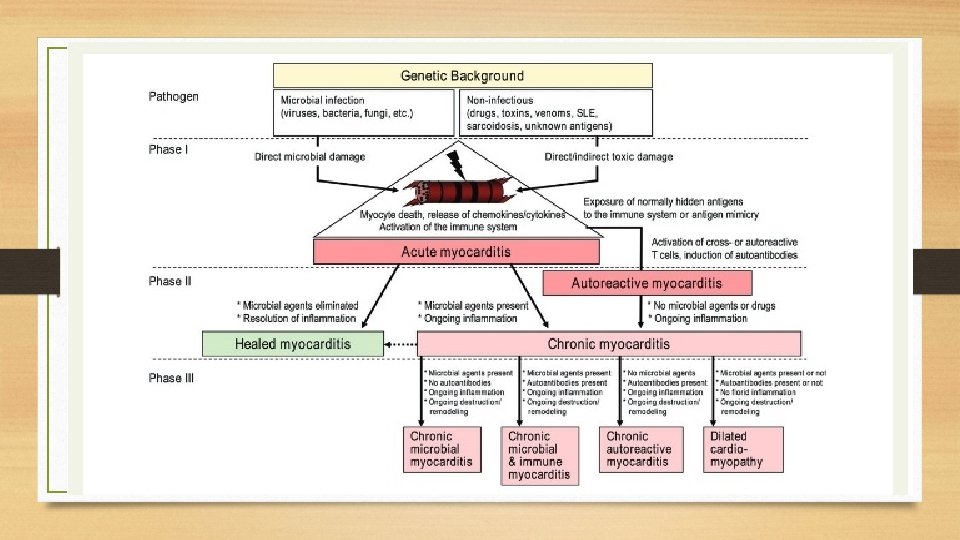
• Inflammatory disease of heart muscle cells • Inflammation – damage- ischemic damage, mechanical trauma, genetic cardiomyopathies. • Classic paradigm of cardiac injury----immunologic response from the host. • Overwhelming or inappropriate immune response may destroy the heart tissue. • Chronically cardiac remodelling leads to DCM, heart failure, or death. • Self limiting.
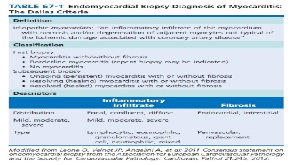
• Dallas criteria. • Criteria criticized because of inter reader variability in interpretation, lack of prognostic value, and low sensitivity due in part to sampling error Caforio et al (2013) in defining myocarditis, using immuno histochemical data • ≥ 14 lymphocytes/mm 2 including ≤ 4 monocytes/mm 2 • CD 3 -positive T lymphocytes ≥ 7 cells/mm 2. • EMB and Post mortem patients. • Useful in sub acute or chronic myocarditis.
Magnitude of the problem • Global prevalence of myocarditis ≈22 of 100 000 patients annually-I C D--International Classification of Diseases (ninth revision). • I C D 9 ---0. 5% and 4% of cases of prevalent heart failure are due to myocarditis. q AHA-myocarditis as the third leading cause of sudden cardiac death in competitive athletes. (Maron et al- Circulation 2015) • Autopsy studies- myocarditis is responsible for 4 -12% of SCD.
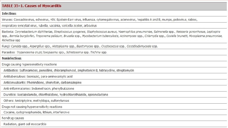
Aetiology • Commonly viruses. • 1950 s to the 1990 s- Entero viruses. • Last 2 decades- PCR and in situ hybridization led to other viruses. • Selected bacteria- diphtheria, Borrelia. • T. cruzi in Endemic areas
• Hypersensitivity myocarditis 1. Vaccines 2. Drugs 3. Systemic autoimmune diseases.
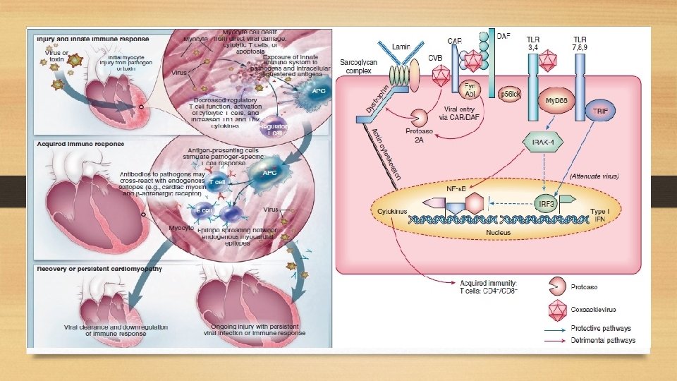
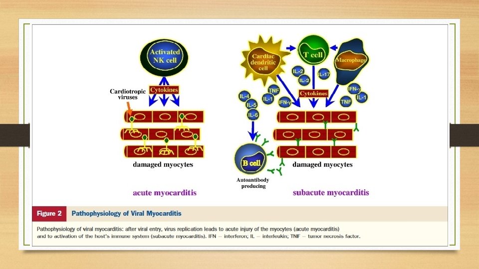
Pathogenesis • Experiments on rodent strains. • Cardio virulent virus strain , parasites, or bacteria. • Delivery of cardiac antigens with a strong adjuvant or carried within dendritic cells in non infectious models. • Viral entry into cardio myocytes → type 1 interferons. • Leads to myocyte death by apoptosis and autophagy.
• Receptors like CAR and CD 55. • Chaga’s Disease-parasites releasing bioactive compounds and promoting oxidative stress. 2 nd Phase. NO, Altered regulatory T-cell, NK cells Type I interferon • Clearance – Normal cardiac function. • 3 rd Phase-------Chronic auto antigen-driven inflammation→ DCM and end-stage heart failure
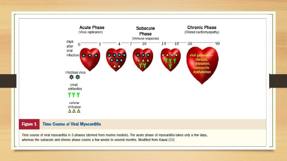
Time Course in Myocarditis Entry of virus------- Receptor mediated Acute-----Virus replication - myocyte necrosis, exposure of intracellular antigens Subacute-----virus-specific T lymphocytes, which may target the host’s organs.
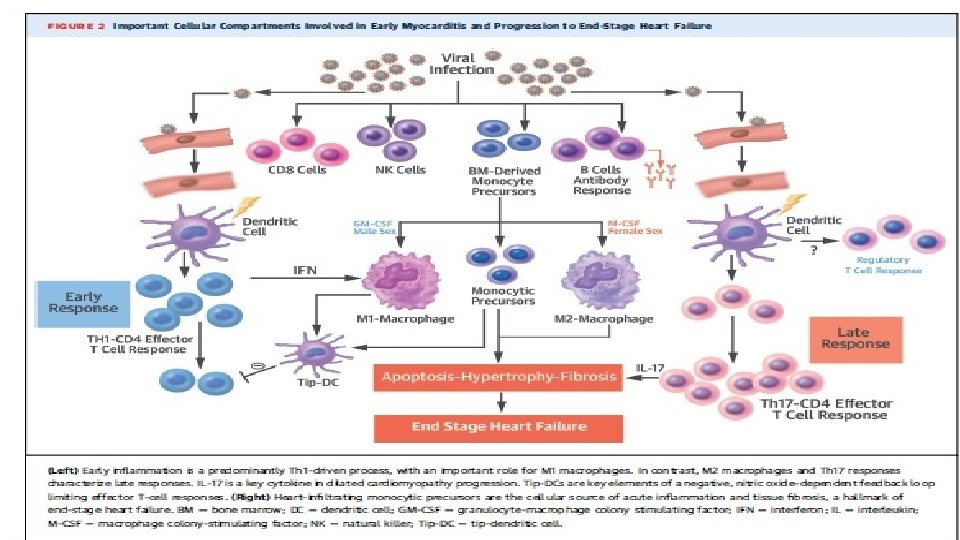
CARDIAC INFLAMMATION CELLULAR EXTRACELLULAR • • • Fibroblasts • Myofibroblasts • Stromal cells T and B Cells Macrophages Dendritic Cells Eosinophils Mast Cells
• Activation, deactivation or trans differentiation of specific cell subsets, are 1. chemokines, 2. cytokines 3. hormones, and degradation or danger • Activate or inhibit gene expression through more or less specific receptors and their intracellular downstream pathways
Mechanisms Activated APC’s CD 4+T Cells Activated TLRs TNF-α, IL-1β, IL-18, or high-mobility group box 1 protein IFN-γ, IL-4, -5, and -13, IL-17 Cardiac fibrosis Heart failure progression Progression to DCM Promote T cell expansion Chronic myocarditis and heart failure
Epigenetics • mi RNA’s • Modulate expression of active proteins • mi RNA’s dysregulation and myocarditis. • mi. RNA-155, -146 b, and -21 in CSB • Modulate virulence. • mi. RNA-221/222
Clinical Features • Chest pain and dyspnoea. • Preceded by viral prodrome URI or GI. • Chest pain resembles MI or pericarditis. q Myo-pericarditis in 11%( Imazio et al-circulation 2013) • Angina Coronary vasospasm Micro vascular dysfunction • Palpitations and syncope. • Heart failure and sudden cardiac death.
• Prevalence of myocarditis as a cause of cardiomyopathy----Bimodal peaking---- first year of life, declines during late childhood, and peaks in the early 20 s. • Men ------severe course------less complete recovery • EMB---------Mobitz type 2 second-degree or higher heart block Sustained or symptomatic ventricular tachycardia not responding to treatment. • GCM, CS, and necrotizing eosinophilic myocarditis have higher incidence of arrhythmias. • Eosinophilic and myocarditis associated with systemic inflammatory disorders
• EMB is infeasible or is not clearly indicated-----non invasive diagnostic ------- probable acute myocarditis. q European Study of Epidemiology and Treatment of Inflammatory Heart Disease, 3055 patients with suspected acute or chronic myocarditis were screened, whereupon 72% had dyspnea, 32% had chest pains, and 18% had arrhythmias (Hufnagel G et al Herz. 2000). • Studies limited- protean manifestations and lack of non invasive diagnostics.
Specific Clinical Presentations Acute Myocarditis • Nonspecific symptoms q Kuhl et al- fatigue (82%), DOE (81%), arrhythmias (55%, both supraventricular and ventricular), palpitations (49%), and chest pain at rest (26%). • Viral pro drome---20 -80%. • Diagnosis of exclusion.
Fulminant Myocarditis • 10% biopsy proven. • Abrupt onset(≤ 2 weeks). • Haemodynamic compromise. • Echo-Global LV dysfunction and myocardial oedema. • EMB- diffuse myocarditis. • Supportive treatment.
Giant Cell Myocarditis • Subtle onset • Present with heart failure, arrhythmia and heart block. • Survival <6 months • Improved with immuno suppressants. • Discontinuation ----- recurrence. • EMB---- Giant cells and scar tissue. • Cardiac transplantation+ mechanical circulatory support.
Chronic Active Myocarditis • Older • Insidious • Features of LV dysfunction. • EMB---fibrosis and myocyte dropout. • 60 -70% of patients-----DCM with unknown aetiology. • MRI, PET-CT, Immunohistochemistry
Differentials • • Ischaemic heart disease. Hypertrophic cardiomyopathy. Cardiac amyloidosis. Arrythmogenic right ventricular cardiomyopathy. q Pieroni et al -patients with right ventricular myocarditis ---indistinguishable from ARVC ---clinical features, presence, and severity of structural and functional right ventricle abnormalities ( J Am Coll Cardiol. 2009).
Diagnostics ECG • Low sensitivity – used for screening. • Nonspecific T waves and ST-segment changes including ST-segment elevation. • Arrhythmias. • Risk stratification and initial screening q Ukena et al - prolonged QRS duration is a significant independent predictor for cardiac death or heart transplantation ( Eur J Heart Fail. 2011)
Echocardiography • Diagnostic assessment- R/O other causes of HF. • Cardiac chamber sizes, wall thicknesses, and systolic and diastolic functions. • Fulminant myocarditis lack cardiac dilation and exhibit increased septal thickness. • Spherical-shaped ventricle ------elliptical -----normal wall thickness. q Speckle tracking-Hsiao et al - LV strain and strain rate may be useful measurements for diagnosis of suspected myocarditis patients, even those with preserved LVEF( Int J Cardio vasc Imaging 2013)
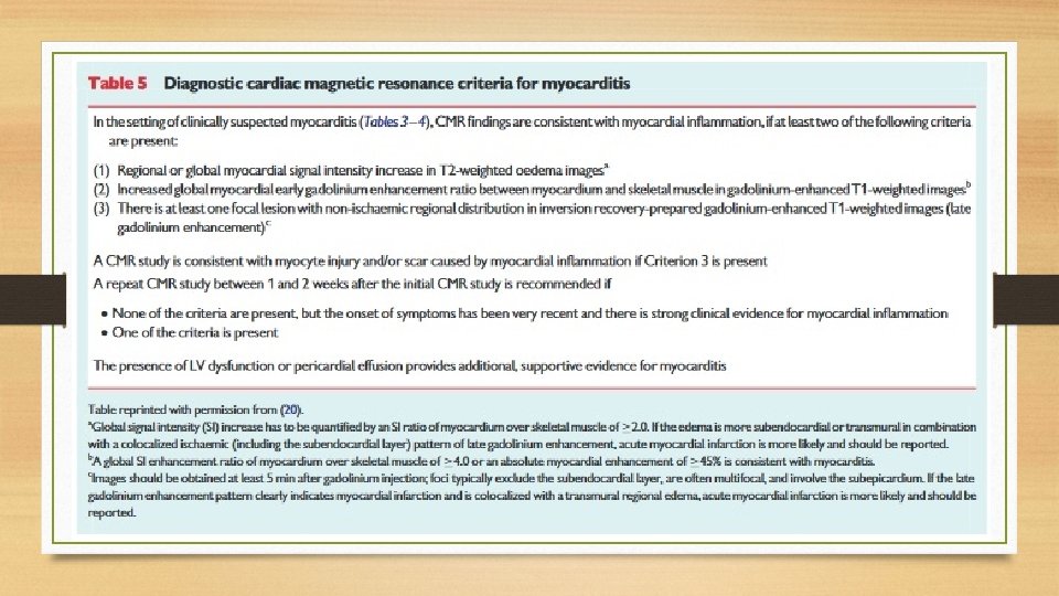
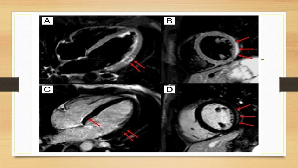
Blood • Cardiac Troponins-Acute or fulminant DCM with heart failure. • Sub acute or chronic myocarditis biomarkers are usually normal. Cardiac Magnetic Resonance • CE-CMR----detect areas of myocardial damage in patients with acute myocarditis. • c. MRI sensitivity and specificity are as high as 100% and 90%.
• T 2 -weighted CMR -oedema or water. • T 1 -weighted sequences -inflammation or fibrosis. • LGE------CS ----multiple, patchy mid myocardial lesions in a non coronary ------septal and RV involvement. • Acute DCM, CMR -myocarditis predict a greater likelihood of recovery, defined as a left ventricular ejection fraction (LVEF) >55% after 24 months
q Regional or global T 2 -weighted “oedema-sensitive” sequences also had a significant improvement in LVEF at 12 -month follow-up(Vermes et al- Eur Heart J Cardio vasc Imaging 2014). • LGE -3. 7%/year risk of a composite of cardiovascular adverse events. q LGE predicted a composite endpoint of cardiac death, heart failure hospitalization, ventricular tachycardia, and sudden death( Schumm J et al-J Cardiovas Magn Reson 2014. )
• Rim-like pattern --- septal wall or a sub epicardial distribution ----free LV lateral wall - PVB 19. • PVB 19 ---- CE -lateral wall • HHV 6 -----CE -mid wall ----inter ventricular septum. • CE- CMR : Guidance for EMB sampling when necessary. • Lacks the ability to determine the magnitude of inflammation. • Acute not in chronic myocarditis. • c MRI is a reliable method of detecting myocardial fibrosis in DCM
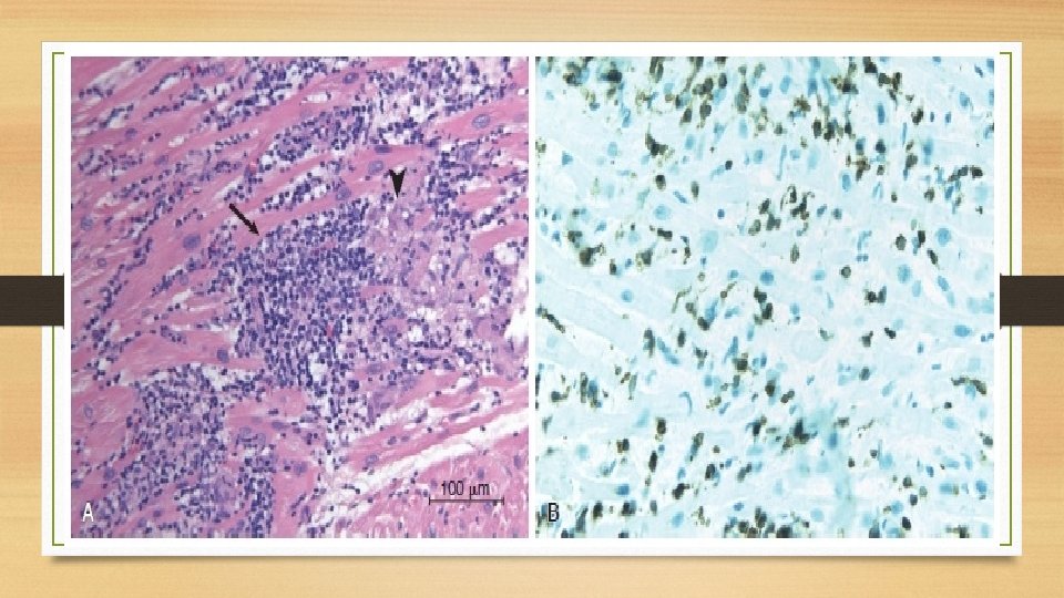
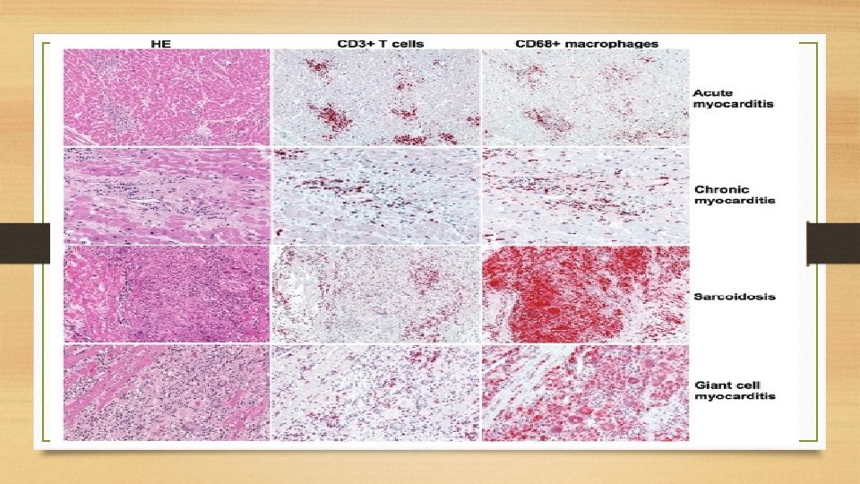
Endomyocardial Biopsy • Lymphocytic infiltration in association with myocyte necrosis and death. • Best method to differentiate between giant cell myocarditis and sarcoidosis. • Eosinophilic myocarditis -myocyte necrosis---intra cavitary thrombi containing eosinophils ----lumen of intra myocardial coronary vessels.
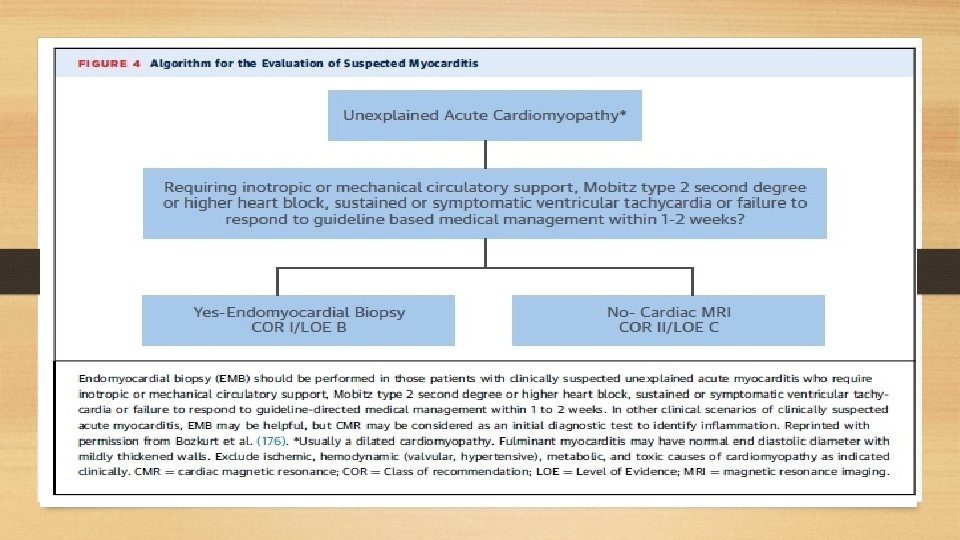
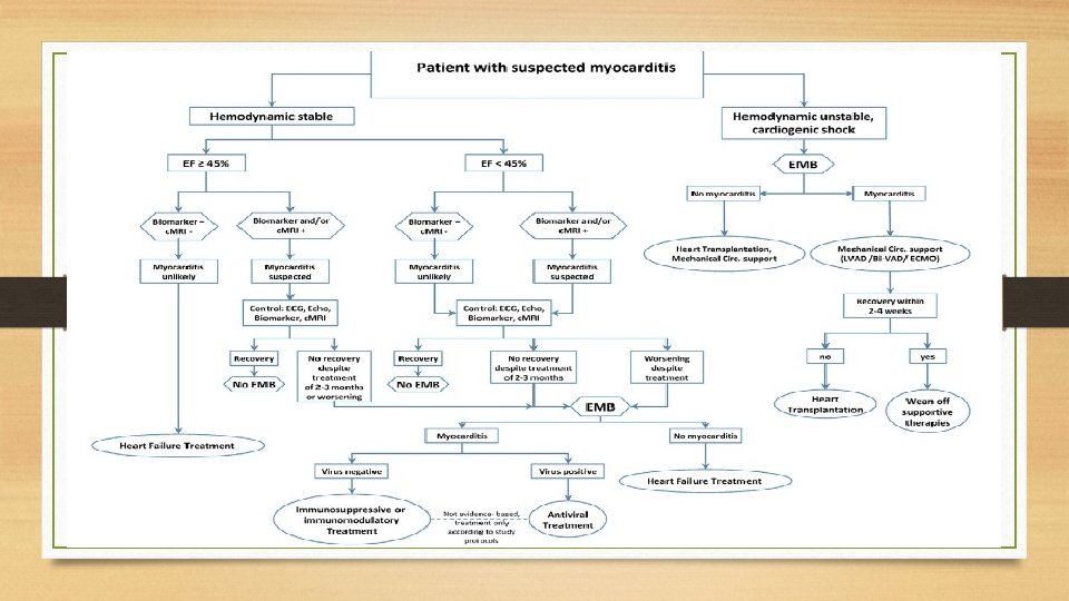
q ESC recommends EMB------ as early as possible. q ACC/AHA recommends EMB based on 3 classes of comprehensive clinical presentations, risks, and measurements using non invasive techniques. • Immunohistochemistry useful.
q Kindermann et al ----69 of 181 (38%) EMB samples are positive for myocarditis using the Dallas criteria, whereas 91 of the 181 (50%) -----CD 3, CD 68 (macrophages), and HLA class II antibodies (Circulation. 2008) • Sensitivity →EMB ------→GCM ---------80% to 93% Lymphocytic & Sarcoid ------20% to 30%
Micro-RNA Profiling • Characterisation of mi RNA profiles among acute, chronic, and fulminant myocarditis including a relationship to the severity of myocardial damage. • mi. R-208 b and mi. R-499 are up regulated upon myocardial damage. • mi. R-155, mi. R-21, mi. R-146 b, mi. R-511, and mi. R-212 are upregulated in myocarditis.
• Cp. G dinucleotide methylation regulates parvovirus 19 (PVB 19) viral genome expression. • Minimum of 4 samples is needed to achieve an acceptable sensitivity for molecular diagnosis of erythro virus PVB 19. • PVB 19 (>250 copies/mg DNA) and for human herpes virus 6 (>500 copies/mg DNA). • Antiviral therapies beneficial in active inflammation.
• FDG-PET----active myocardial inflammation----suspected CS. • FDG-PET +Myocardial perfusion---- active inflammation and scarring. • 18 F-FDG-PET ---extra cardiac inflammation • lymph nodes that are amenable to biopsy q Blankstein et al -------Mismatch of FDG and perfusion measurements predicts adverse cardiac events, and RV involvement is associated with high risk for arrhythmias
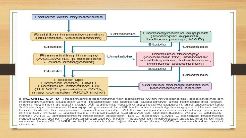
Treatment • Myocarditis presenting as heart failure with reduced systolic function • Current guidelines -gradual titration of ACE inhibitors, ARB’s and beta blockers, to doses used in clinical trials (Yancy CW et al J Am Coll Cardiol 2013). • Virus-negative inflammation-positive patients- Immunosuppression with azathioprine and prednisone for up to 12 months.
Pathogen Inhibition • Antivirals-------Interferons-------INF β and IFN-α 2 a. • Natural products like astragaloside IV -----↓ CVB 3 titres by up regulating IFN-γ. q Chen et al -astragaloside IV ↓ fibrosis of the heart muscle tissue and ↑ the murine survival rate. • Antisense oligo deoxy nucleotides- target viral gene sequences ---inhibit viral gene expression and inhibit viral replication
• Preventing viral entry-------dimeric antibody-like molecule----modified CAR or decay accelerating factor crystallization proteins. • Competitively ----viral particles -----viral-specific activation of the innate and adaptive immunity. • Limitations Nonspecific targeting, Unclear mechanisms Side effect
Doses • Interferon-β---2 × 10⁶ IU alternate day subcutaneous. • Increased at weekly intervals, first to 4 × 10⁶ IU and then to 6– 8 × 10⁶ IU; this is continued for 24 weeks. • Slowly increasing dosage reduces flu like symptoms.
Immune modulation • • Sub acute phase of myocarditis----exaggerated autoimmune responses. Azathioprine Prednisalone Non steroidal anti-inflammatory drugs have shown increased myocardial inflammation and mortality and hence avoided. • Tissue inhibitor of metalloproteinase 1–specific small interfering RNA or polyclonal antisera -------- used as modulators.
Immunoglobulin therapy • Ineffective in acute DCM. • Chronic CMP with viral replication may benefit—Dennert et al (Antivir Ther 2010).
Steroids • Mainstay in CS. • Halt Progression of LV dysfunction q Survival doubtful---- ( Blauwet -Heart Fail Rev 2012 ) • Early initiation • AV block recovery----20 -30% • GCM- cyclosporine based combined immunosuppression beneficial.
Otherapies • Trans venous pacing– Acute lymphocytic myocarditis. • Temporary • LV dysfunction--- wearable external defibrillator. • Class IIa recommendation for an ICD in patients with suspected CS, regardless of LV function or reversibility of heart block
Specific Regimens
Key Messages • Viral infections are the most common triggers of inflammatory myocardiopathies. • Non-specific symptoms are frequently identified and cardiologically evaluated only at an advanced stage. • Pathophysiological processes in myocarditis take place at the cellular and subcellular levels, myocardial biopsy is the only method for confirmation of diagnosis. • Clinical course of myocarditis is unpredictable, all patients with etiologically unexplained heart failure have to undergo myocardial biopsy. • Numerous chronic viral infections and postinfectious or autoimmune inflammations of the myocardium are treatable.