Musculoskeletal infection osteomyelitis 2822019 osteomyelitis Osteomyelitis denotes inflammation


 •")


 Laboratory markers (WBC , fever )")
")

- Slides: 23
Musculoskeletal infection osteomyelitis 28/2/2019
osteomyelitis • • Osteomyelitis denotes inflammation of bone and • marrow, virtually always secondary to infection. • • Osteomyelitis may be a complication of any • systemic infection but frequently manifests as a • primary solitary focus of disease. All types of • organisms, including viruses, parasites, fungi, and • bacteria, can produce osteomyelitis, but • infections caused by certain pyogenic bacteria • and mycobacteria are the most common
Pyogenic osteomyelitis • Pyogenic osteomyelitis is almost always caused by bacterial • infections. • • Organisms may reach the bone by: • (1) hematogenous spread (bacteremia) • (2) extension from a contiguous site • (3) direct implantation
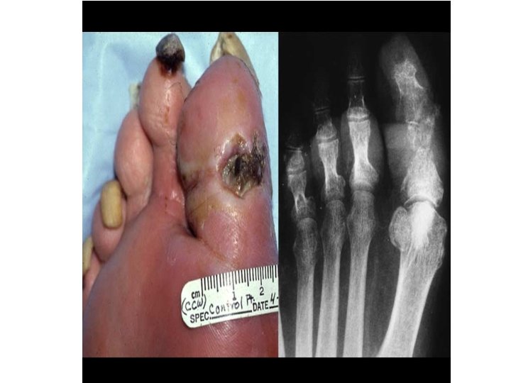
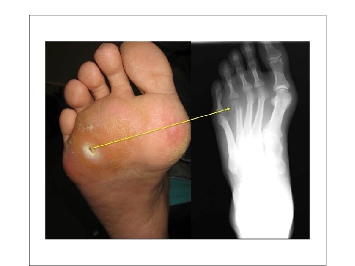
• In otherwise healthy children, most osteomyelitis is • hematogenous in origin and develops in the long bones. • - In adults, however, osteomyelitis more often occurs as a • complication of open fractures, surgical procedures, and • diabetic infections of the feet.
Risk factors • • • 1 - diabetes 2 -HIV 3 -seplenectomy 4 -sickle cell anaemia 4 -TB 5 -surgery
Organisms M. c. pathogen ? ? ? 80_90% In infant, , H. influenzea , group b strep… Iv drug abuser & g. u. t. i ? ? ? Sickle cell ? ? Mixed bacteria in surgery induced osteomyelitis In almost 50% of suspected cases, no organisms can be isolated • • •
• • • • The location of the bone infections is influenced by the osseous vascular circulation, which varies with age: - In the neonate the metaphyseal vessels penetrate the growth plate, resulting in frequent infection of the metaphysis, epiphysis, or both. - In children, localization of microorganisms in the metaphysis is typical. - After growth plate closure, the metaphyseal vessels reunite with their epiphyseal counterparts and provide a route for the bacteria to seed the epiphyses and subchondral regions, which are common sites of infection in the adult
note in infants, but uncommonly in adults, epiphyseal infection spreads through the articular surface or along capsular and tendoligamentous insertions into a joint, producing septic or suppurative arthritis, which can cause destruction of the articular cartilage and permanent disability.
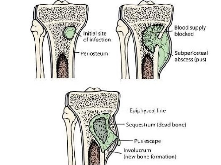
Pathophysiology of acute O. M • • • the acute phase, bacteria proliferate and induce a neutrophilic inflammatory reaction. Necrosis of bone cells and marrow ensues within the first 48 hours. The dead bone is known as a sequestrum. • Rupture of the periosteum leads to a soft tissue abscess which can channel to the skin as a draining sinus. • After the first week, chronic inflammatory cells release cytokines that stimulates osteoclastic bone resorption, ingrowth of fibrous tissue, and the deposition of reactive bone at the periphery. The newly deposited bone can form a shell of living tissue, known as an involucrum.
Presentation and symptoms • The clinical manifestations depend on the cause of osteomyelitis • In haematogenous spread esp. in (infants and children). manifests as an acute systemic illness with • malaise, fever, chills, leukocytosis, and markedto-intense throbbing pain over the affected region
• In other instances, the presentation is subtle, • with only unexplained fever (most often in • infants) or localized pain (most often in adults).
• Acute osteomyelitis develops rapidly over a period of seven to 10 days. The symptoms for acute and chronic osteomyelitis are very similar and include: • • • Fever, irritability, fatigue Nausea Tenderness, redness, and warmth in the area of the infection Swelling around the affected bone Lost range of motion Osteomyelitis in the vertebrae makes itself known through severe back pain, especially at night.
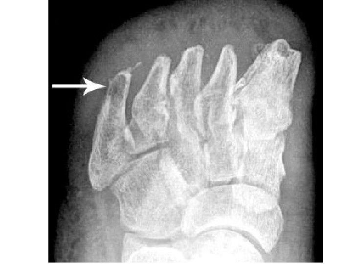
Affected bones • In children and infants (long bones of hands and legs) • In adult ( vertebrae , foot , maxilla and the body of mandible.
Chronic osteomyelitis • • • • In 5% to 25% of cases, acute osteomyelitis fails to resolve and persists as chronic infection. • Chronic infections may develop when there is delay in diagnosis, extensive bone necrosis, inadequate antibiotic therapy or surgical debridement, or weakened host defenses. • The course of chronic infections may be punctuated by acute flare-ups; these are usually spontaneous and may occur after years of dormancy. • Other complications of chronic osteomyelitis include pathologic fracture, secondary amyloidosis, endocarditis, sepsis, and development of squamous cell carcinoma in the draining sinus tracts and sarcoma in the infected bone.
Chronic osteomyelitis • Is due to presence of intracellular bacteria , once intracellular can escape and invade other bone cells. • Bacteria become resistant to some antibiotics. . • Bone destruction and remodelling • Marrow space fibrosis • (very difficult to treat )
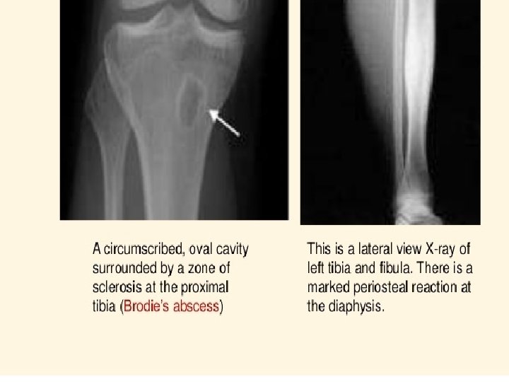
Diagnosis • • Clinical suspicion (signs & symptoms) Laboratory markers (WBC , fever ) Imaging ( xray , MRI ) Biobsy
Management • Antibiotics for at least one month • Surgery (drainage of bus ) esp. . ( failure of conservative treatment ) • Amputation esp. in ischemia &risk of sepsis