OSTEOMYELITIS MYCETOMA Dr Shashidhar M R Osteomyelitis Pyogenic


 : coined osteomyelitis • The root words osteon (bone) and")


 • No abcess, cortical thickening")


- Slides: 35
OSTEOMYELITIS & MYCETOMA Dr Shashidhar M R
ØOsteomyelitis- Pyogenic, Tubercular ØMycetoma
OSTEOMYELITIS
Osteomyelitis • Nelaton (1834) : coined osteomyelitis • The root words osteon (bone) and myelo (marrow) are combined with itis (inflammation) to define the clinical state in which bone is infected with microorganisms.
TYPES §Pyogenic §Tubercular §Chronic Non Specific
PYOGENIC OSTEOMYELITIS Causative agents§Staphylococcus aureus - MC §Neonates : H influenzae §Streptococci in children §Sickle cell anemia: salmonella §E. Coli : GU infections
Why staphylococcus most common? • S. aureus ----- normal skin flora • S. aureus -----increased affinity for host proteins (traumatised bone) • Enzymes (coagulase, surface factor A) ----- hosts immune response. • “Biofilm” (polysaccharide “slime” layer) ---- increases bacterial adherence to any substrate. • Large variety of adhesive proteins and glycoproteins ----mediate binding with bone components.
Pathogenesis: Direct inoculation of microorganisms into bone penetrating injuries and surgical contamination are most common causes Hematogenous spread usually involves the metaphysis of long bones in children or the vertebral bodies in adults Microorganisms in bone Contiguous focus of infection seen in patients with severe vascular disease. Osteomyelitis
Morphology Three stages. Acute Subacute Chronic Acute- acute inflammation and necrosis of the bone. Dead bone - Sequestrum
Metaphysis – infection spreads to subperiosteumsegmental bone necrosis- subperiosteal abscessessinus tracts- discharge dead bone. If crosses the epiphysis, may involve the joint. Chronic. Healing – osteoclasts resorb the bone, chronic inflammatory infiltrate, fibroblasts proliferate in the periphery. New bone formation from the periphery Central dead bone with surrounding new boneinvolucrum
Sites of infection • Metaphysis and epiphysis in infants and adults • Metaphysis in children
Pathogenesis Whatever may be the inciting cause the bacteria reaches the metaphysis of rapidly growing bone & provokes an inflammatory response. why metaphysis is involved 1. 2. 3. Infected embolus is trapped in U-shaped small end arteries located predominantly in metaphyseal region Relative lack of phagocytosis activity in metaphyseal region Highly vascularised region ---minor trauma—hemorrhage ---locus minoris resistantae---excellent culture medium
– sharp hairpin turns – flow becomes considerably slower and more turbulent
PATHOLOGENESIS These are end-artery branches of the nutrient artery acute inflammatory response due to infection tissue necrosis, breakdown of bone Obstruction Avascular necrosis of bone Squestra formation Chronic osteomyelitis
Pathology: � Pathologic features of chronic osteomyelitis are : � The presence of sclerotic, necrotic piece of bone usually cortical surrounded by radiolucent inflammatory exudate and granulation tissue known as sequestrum. � Features: �Dead piece of bone �Pale �Inner smooth , outer rough �Surrounded by infected granulation tissue trying to eat it
Involucrum sequestrum
Pathology: � The involucrum is the sheath of reactive, new, immature, subperiosteal bone that forms around the sequestrum, effectively sealing it off the blood stream just like a wall of abscess. �The involucrum is irregular and is often perforated by openings. �The involucrum may gradually increase in density and thickness. � There is exudation of polymorphonuclear leukocytes joined by large numbers of lymphocytes, histiocytes, and occasionally plasma cells.
local signs • calor, rubor, dolor, tumor • Heat, red, pain or tenderness, swelling • Initially, the lesion is within the medually cavity, there is no swelling, soft tissue is also normal. • The merely sign is deep tenderness.
Clinic picture • It is important to remember that the metaphysis lies within the joint capsule of the hip, shoulder, ankle. Therefore these joints can develop septic arthritis by extension of osteomyelitis. • If the infection and septicemia proceeded unabated, the patient may have toxic shock syndrome. • Sinuses
Approach to diagnosis � The white blood cell count will show a marked leucocytosis as high as 20, 000 or more � The blood culture demonstrates the presence of bacteremia, the blood must be taken when the patient has a chill, especially when there is a spiking temperature. � Radiology: Lytic focus of bone destruction surrounded by zone of sclerotic bone.
Varients
Brodie’s abcess � Bone abscess containing pus or jelly like granulation tissue surrounded by a zone of sclerosis � Age 11 -20 yrs, metaphyseal area, usually upper tibia or lower femur � Deep boring pain, worse at night, relieved by rest � Circular or oval luscency surrounded by zone of sclerosis � Treatment: – Conservative if no doubt - rest + antibiotic for 6 wks. – if no response – surgical evacuation & curettage, if large cavity - packed with cancellous bone graft
Garre’ s osteomyelitis • Sclerosing, nonsuppurative • Jaw (mandible) • No abcess, cortical thickening • Acute local pain, pyrexia subsidefusiform swelling • Acute stage-rest, antibiotics • Sx: Gutter holing, excision+curettage
• Complications of chronic osteomyelitis ØPathologic fracture ØSecondary amyloidosis ØEndocarditis ØSepsis ØSquampous cell carcinoma of the tract ØSarcoma
TUBERCULOUS OSTEOMYELITIS ØBlood borne ØExtension from adjacent sites-ribs ØAdolescents Common sites. Spine- thoracic and lumbar Knees Hips Pott spine- erosion of the intervertrebral discs and involvement of multiple vertebrae, cold abscess-psoas abscess
• Microscopy : tuberculous granuloma • Clinical features : • Complications : 1. Spine : scoliosis, kyphosis, cold & psoas abcsess 2. 3. 4. Tuberculous arthritis Sinus tract formation Amyloidosis
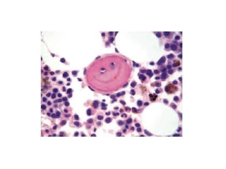
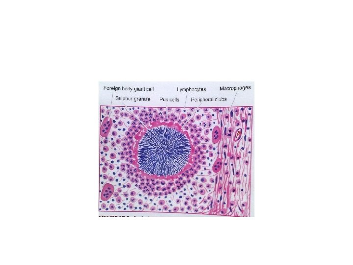
MYCETOMA • Chronic suppurative infection Actinomycosis – bacterial form Eumycetoma – fungal form
Actinomycosis • Actinomycetes israelii • Filamentous & anaerobic • Forms Ø Cervicofacial- lumpy jaw, sinuses Ø Thoracic Ø Abdominal Ø Pelvic
Microscopy
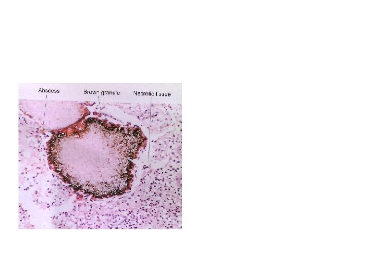
Eumycetoma – Madura foot • Madurella mycetomatis or Madurella griesa • Gross- swelling, black granules from discharging sinuses