Immunological investigations Dr Cismaru Cosmin Andrei Cosmin Cismaruumfcluj


 Biochemistry:")


 interact with antibodies (Ac),")


 c. Western blot d. Imunodifuzia radiala")

 c. Western blot d. Imunodifuzia radiala")


 c. Western blot d. Radial immunodiffusion")
 • Quantitative dosing of the quantity of an antigen in a")

 in")
 • Mixing")


 Negativa R •")

 Ag + Ag • Indirect")
 LES, Sdr. Sj, Sclerodermie Miozita LES, BMTC,")

 Anti-Ig. Eradiolabelled • Direct method Antigen Ig. E Patient serum wash")
 - detection of Ig. E specific to a suspected / known")


 • Main surface markers")


 - Full HLG →")


 • recombinant •")
 este izolată dintr-o bacterie o enzimă taie ADN la")

 • \"A\" - a rat")


- Slides: 58
Immunological investigations Dr. Cismaru Cosmin Andrei Cosmin. Cismaru@umfcluj. ro
Medical investigations • Laboratory tests – important in establishing a diagnosis and conducting treatment strategy • Applicability: - diagnosing or excluding a disease among symptomatic persons - diagnosing a disease among asymptomatic persons (screening) Evaluating a therapeutic response
Characteristics of medical tests • Accuracy • Cost • Interfering factors • Precision • Normal values • Sensitivity • Specificity Sensitivity: The probability of having a positive test when you are sick is the power of a disease test Specificity: The probability of having the negative test when you are healthy, or the proportion of those with a negative test among thehealthy
1. Laboratory investigations § § Hematology: HG with leucocitary formula (complete blood count) Biochemistry: liver tests, kidney, thyroid, metabolic. Imunology-serology: Ab, auto-Ab, tumor markers Microbiology: hemoculture, coproculture, urine culture. 2. Imagery § § § US X-ray CT-scan MRI Scintigraphy 3. Paraclinical investigations § Spirometry § EKG
1. Laboratory investigations § § Hematologice: HLG cu formula leucocitara Biochimie: teste hepatice, renale Imunology-serology: anticorpi, auto-anticorpi, markeri tumorali Microbiologie 2. Imagistica § § Ecografie Radiografie Tomografie Scintigrafie 3. Investigatii paraclinice § Spirometrie § EKG
SEROLOGY AND IMMUNOHISTOCHEMISTRY
Ab detection methods 1. Immunoprecipitation • • • Detects Ab in solutions Quantitative evaluation of the presence of Ab End-point is the visualization of Antigen-antibody precipitates 2. Complement fixation • It is based on fixation / activation of the complement that follows the binding of the complement system components to the immune complexes
3. Neutralization • • • Capacity of specific antibodies to neutralize various toxins / pathogens Rarely used for diagnostic purposes For the detection of post-vaccine antibody synthesis 4. Agglutination • • • Simple and fast test Capable of detecting small antibody concentrations Detection of anti-viral antibodies It uses latex-attached antigenic particles Direct or indirect method
5. Immunofluorescece • • Requires microscope with uv lamp Direct or indirect method 6. Immunoenzymatic Technique (ELISA) • • • The most sensitive Usually the indirect method is the most used and it depends on the use of an anti-human conjugate (Ig. G or Ig. M) Conjugated antibody - an anti-human Ab that has an attached enzyme (HRP) at the Fc portion, the enzyme that is capable of catalysing the conversion of a substrate to a colored product, which will then be read with a spectrophotometer 7. Radio-immune technique • • High sensitivity Increased costs, risk of irradiation
1. Precipitation reactions • When certain molecules of antigen (Ag) interact with antibodies (Ac), they will precipitate out of solution • Indications: • Detection of serum antigens / antibodies
a. Reactions in liquid medium b. Reactions in gel: • Immunoelectrophoresis • Immunofixation • Western blotting • Radial immune diffusion
a. Reactions in liquid medium • Laser nephelometry - based on the property of immune complexes to deviate laser light • Indications: • - quantification of serum proteins: Ig, C 3, CRP, ceruloplasmin.
b. Reactions in gel a. Immunlectrophoresis b. Imunofixarea c. Western blot d. Imunodifuzia radiala
• Immunlectrophoresis allows the evaluation of the gammaglogulins, the immunoglobulins. • M (Ig. M), • G (Ig. G), • A (Ig. A). three major classes of
a. Imunoelectroforeza b. Immunofixation (SDS-PAGE) c. Western blot d. Imunodifuzia radiala
• The serum proteins will be separated on SDS based on molecular weight (Sodium dodecyl sulfate polyacrylamide gel).
a. Imunoelectroforeza b. Imunofixarea (SDS-PAGE) c. Western blot d. Imunodifuzia radiala
• Gel-bound proteins will be transferred to a nitrocellulose membrane by applying an electrical charge. SDS Gel Transfer Membrane Electroblotting Gel after blotting Transfer Membrane After Blotting
1. Blocking 2. 1 st Antibody 3. 2 nd Antibody 4. Detectection Protein X Nitrocellulose membrane
a. Imunoelectroforeza b. Imunofixarea (SDS-PAGE) c. Western blot d. Radial immunodiffusion
Radial immunodiffusion (Mancini) • Quantitative dosing of the quantity of an antigen in a sample. The diameter of the circle increases with time as the antigen diffuses into the medium, reacts with the antibody, and forms insoluble precipitin complexes. [he antigen is quantitated by measuring the diameter of the precipitin circle and comparing it with the diameters of precipitin circles formed by known quantities or concentrations of the antigen 1 1 2 3
Applications • • Quantitative determination of certain proteins: - immunoglobulins, - complement, - alpha-1 antitrypsin
2. Agglutination reactions - Highlighting the presence of particles (bacteria, blood cells, cells) in suspension (serum) Ig. M + Eritrocite aglutinare
• Antibody measurement in patients serum • Serial Dilutions (1: 2) • Mixing with known Ag. • The occurrence of precipitate if Ab is present. • Ab titer Blood typing: yes/no example of agglutination.
ABO blood groups
Blood group AB 0 and Rh testing § Reactives Ø Anti-A, Anti-B and Anti-AB solution of monoclonal antibodies (hybridome technqiue)
Indications • Establishment of blood groups ABO, Rh • Demonstration of antibodies to certain bacteria • Detection of rheumatoid factor
3. Complement Fixation Human serum Complement Positive R Antigen (viral, bacterian) Negativa R • indications • - Diagnosis of viral and bacterial infections
4. Reactions with marked reagents • The immunofluorescence reaction • Radio-immunological analysis • Immunoenzymatic analysis
a. The immunofluorescence reaction • Direct- Antigen detection (biopsy) Ag + Ag • Indirect - detection of antibodies (serum) Ag + Ag
Indications • Detection of antinuclear antibodies (ANA) LES, Sdr. Sj, Sclerodermie Miozita LES, BMTC, Sdr. Raynaud, Psoriazis Sclerodermie, Miozita, Sclerodermie CBP, Sdr. Sj Sdr. CREST Hepatita cronica, Vasculita, Trombocitopenie
b. Radioimmunological tests Detection of Ig. E specific to a suspected / known antigen - total / specific Ig. E count 1 k. UI / l = 2. 4 μg / l Antigen Patient serum Anti-Ig. Eradiolabel Ig. E wash
b. Radioimmunoassay (RIA) Anti-Ig. Eradiolabelled • Direct method Antigen Ig. E Patient serum wash • Indirect method Antigen Ig. E Patient serum wash
RAST (radio-alergo-sorbent test) - detection of Ig. E specific to a suspected / known Ag - quantification of total / specific Ig. E Indications: Quantitative determination of different antigens: bacterial, viral, tumor, hormone Medicines (digital, theophylline, cortisone) Determination of allergens in allergic diseases
c. Immunoenzymatic analysis • ELISA, RIST
ELISA - classical- quantification of Ab Ser uman HRP Ac anti Ig. G-HRP - sandwich- quantification of antigen Ac anti Ag-HRP HRP
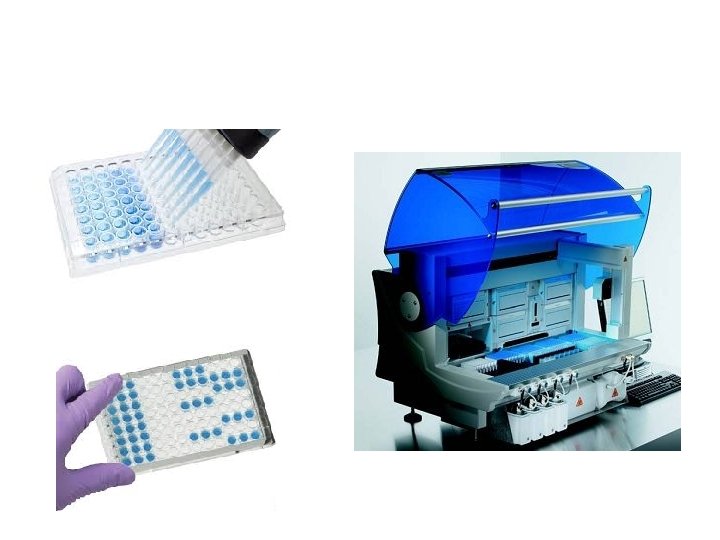
Indications: • Quantitative determination of some important diagnostic proteins: alpha-fetoprotein, carcinoembryonic antigen, HBs antigen, etc. • Quantitative determination of bacterial, viral, fungal, parasitic or appropriate antigens in serum of patients for diagnostic purposes • Evidence and titration of autoantibodies as markers of autoimmune diseases
5. Flowcitometry Procedure: A cell suspension is passed through a special detector, which allows multiparametric (physico-chemical) detection of thousands of cells in a relatively short time Fluid cells susp Light detectors FL 1 FITC Computer Laser Light 488 nm
Flow-citometry (FCM) • Main surface markers
6. Investigations in hypersensitivity • in vivo - skin tests - Challenge test: oral, nasal, bronchial - Hot, cold test • In vitro - total Ig. E - specific Ig. E (RAST, ELISA) - mediators determinations: triptase
Indications: • • Diagnosis of Allergic Diseases: - drug allergy / food allergy contact dermatitis / atopic - allergic asthma • Triptase - diagnostic and forensic value • - VN: 1 -10 ng / ml (> 20 ng / ml- mastocytosis, anaphylaxis)
7. Detection of immune complexes • Physical methods: - gel filtration, - precipitation, - ultracentrifugation Detecting cryoglobulins radiolabelled C 1 q
Indications • • Estimating the severity of chronic infections: Viral Hepatitis B, C Bacterial endocarditis parasitosis • Estimating the severity of autoimmune disorders: • Systemic lupus erythematosus • Rheumatoid arthritis
8. Investigations in autoimmune diseases • Specific organ autoantibodies • ELISA, IFI → Ab anti-Thyroglobulin (Hashimoto's thyroiditis) • ELISA, RIA → Ab anti-Acetyl Choline Receptor (MG - miastenia • gravis) • IFI → Ab Anti Mitochondria (Primary Biliary Cirrhosis) • Non-specific organ autoantibodies • IF → ANA (LES, Sdr Sj, Polymositis, Dermatomyositis)
9. Investigation of immunodeficiencies • Humoral immunity (Lymphocytes B, Antibodies) - Full HLG → leukocyte formula - Radial gel immuno-diffusion → Ig dosing - Flow cytometry with Ac anti-CD 19 → LB separation • Cellular Immunity (Lymphocyte T) - Full HLG → leukocyte formula - Flow cytometry → Population tracking of T lymphocytes (LTh, LTc, )
Evaluation of lymphocytes • Lymphocyte populations: LB, LT, NK Subpopulations: LTHelper, LT cytotoxic • Quantitative assessment - Flow cytometry • Qualitative / functional evaluation - Activation tests - Proliferation tests - Differentiation tests - The leukocyte adhesion inhibition test
Lymphocyte activation test • NBT Reduction Test • Determination of the percentage of granulocytes producing intracellular superoxide anion due to the activation of cellular oxidases • The optical microscope determines the percentage of cells that contain blue intracellular formazan deposits • Blast transformation test of lymphocytes • Determine the number of blast cells formed in response to in vitro stimulation with a specific allergen
Indications • Diagnosis of immunodeficiencies primary secondary • Diagnosis of late-onset hypersensitivity • Evaluation of post-vaccination immune response
Mo. Ab • humanized • chimeric (have fragments from the mouse) • recombinant • generically called by suffix: • "Mab" - Mo Ab
Ingineria Genetica plasmida (inel ADN) este izolată dintr-o bacterie o enzimă taie ADN la locul specific o genă pt proteină, este luată dintr-o altă celulă, este tăiată cu aceeaşi enzimă gena este inserată în plasmidă, unde se potriveşte perfect ADN-ul recombinant bacteria se divide şi se replică se copiază pe sine şi ADN-ul recombinant plasmida recombinantă este inserată din nou în bacterie noua genă determină bacteria să sintetizeze o proteină nouă (de ex. IFN)
Tehnologia Hibridomului Ag fuziunea cel pt hibridoma cel canceroase din plasmă plasmocite cu sinteza Ac clone testate pt Ac dorit cel hibridom cresc în cultură cel hibridom individual sunt clonate clone dorite sunt cultivate şi îngheţate hibridomul este păstrat viabil în şoarece Ac monoclonali sunt purificaţi
MAb • 1 -2 letters previously (for Ab origin) • "A" - a rat • "Axo" - rat-murine hybrid • "E" - hamster • "I" - primate • "O" - mouse • "U" - human • "Xi" - chimeric • "Zu" - humanized • a distinct, compatible syllable: ne-bac-u-mab; omal-zu-mab; Ritu-xi-mab • an internal syllable shows the mab target: • "Bac" - bacterium • "Ci (r)" - cardiovascular • "Col" - cc. colon • "Got" - glandular tumors (testis) • "Gov" - glandular tumors (ovary) • "Forest" - infections • "Lim" - immunomodulation • "Mar" - breast tumors • "Mel" - melanoma • "Pr (o)" - you prostate • "Toxa" - a toxin • "Tum" - tumor • "Vir" - virus
Usage • Diagnostic • Treatment
Diagnostic Ø Ø Infectious diseases produced by bacteria, viruses or protozoa: rapid typing of microorganisms from tissues, biological fluids detecting circulating molecules (eg HBs in hepatitis B) isolation and characterization of bacterial and viral Ag Ø Hematologie: the typing of the A, B, O system; Rh etc typing populations and lymphocyte subpopulations T (CD 4, CD 8, CD 3 etc. ) B (CD 19, CD 20, m. Ig etc. ) NK (CD 56, CD 57) etc diagnosis of leukemias Ø Ø Ø
Ø Internal Medicine: myocardial infarction - Anti-myosin; thrombosis - anti-trypsin, etc. Ø Pathological anatomy: d 44 (carcinoma, lymphoma, sarcoma, melanoma) and / or metastases, their origin. Ø Surgery: HLA typing of donor and recipient for organ transplants; Ø Oncology: in vivo tumor diagnosis by radioscintigraphy. Ø Laboratory: Various immunological dosing methods (RIA, ELISA) for: hormones, enzymes, proteins, coagulation factors, drugs (digoxin)
Treatment: Ø inactivation or neutralization of toxins; Ø passive immunization in some bacterial or viral infections (tetanus, rabies); Ø in medullary transplants and inhibition of graft rejection; Ø some autoimmune diseases; Ø in cancer and leukemias, in the form of immunoconjugates (by coupling Ac. Mo with cytostatics, toxins or radioactive isotopes); Purging: Ø purification of some molecules used in therapy (IFN, hormones, etc. ); Ø purifying some populations and cell subpopulations (stem cells, lymphocytes, etc. )
Dicussions, questions