How does the cell manufacture these magnificent machines

 bonds are called")


 • Polar uncharged (hydrophilic)")


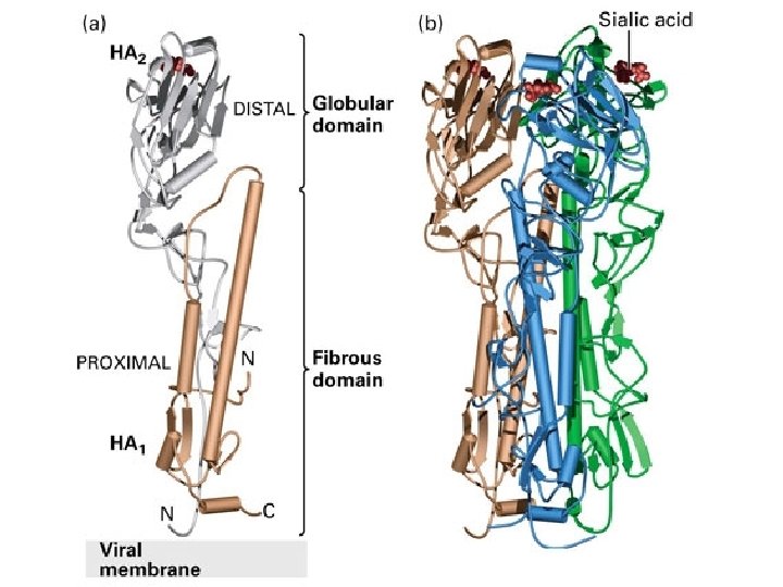
 binding with the globular HA 2 domain of Hemagglutinin (space-filled model)")


- Slides: 39
How does the cell manufacture these magnificent machines? Proteins, that is…
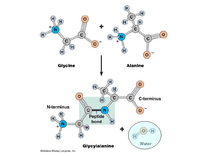
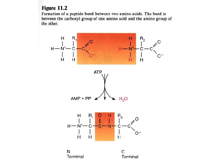
Proteins • Long polymers of amino acids, joined by peptide (amide) bonds are called polypeptides • Polypeptides fold into stable threedimensional shapes and are called proteins • Shape determines the function of proteins (active sites are on the surface)
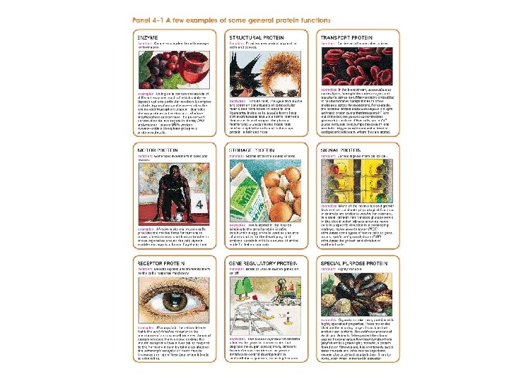
Proteins - classified by functions Enzymes - catalytic activity and function Transport Proteins - bind & carry ligands Storage Proteins - ovalbumin, gluten, casein, ferretin Contractile (Motor): can contract, change shape, elements of cytoskeleton (actin, myosin, tubulin) Structural (Support): collagen of tendons & cartilage, elastin of ligaments (tropoelastin), keratin of hair, feathers, & nails, fibroin of silk & webs Defensive (Protect): antibodies (Ig. G), fibrinogen & thrombin, snake venoms, bacterial toxins Regulatory (Signal): regulate metabolic processes, hormones, transcription factors & enhancers, growth factor proteins Receptors (detect stimuli): light & rhodopsin, membrane receptor proteins and acetylcholine or insulin.
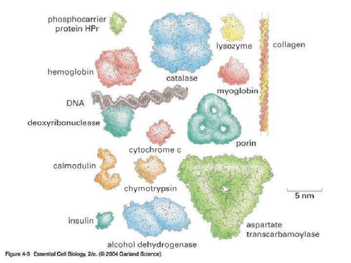
Structure of Proteins the Variety of Protein Structures may be INFINITE. . . average protein has 300 -400 amino acid's & has a MW of 30 k. D to 45 k. D a PROTEIN of 300 amino acids made with 20 different kinds of amino acids can have 20300 different linear arrays of aa's [10390 different proteins] 1 st protein sequenced was Beef Insulin by Fred Sanger - 1958 Nobel Prize winner to date about 100, 000 protein have been sequenced only about 10, 000 structures known [2 K/yr] E. coli make about 3, 000 proteins, humans make about 100, 000 proteins.
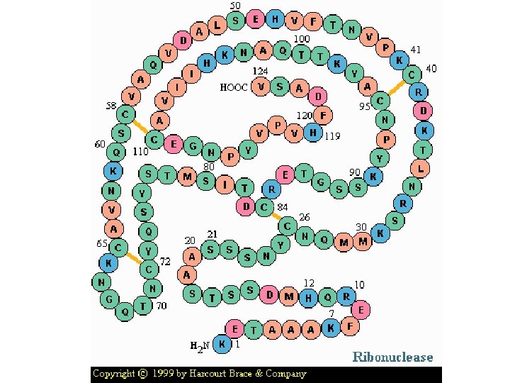
4 levels of protein structure are recognized primary - linear sequence of aa's secondary - regular, recurring orientation of aa in a peptide chain due to H-bond tertiary - complete 3 -D shape of a peptide quaternary - spatial relationships between different polypeptides or subunits Start with the building blocks: amino acids (aa’s)
Two views of an amino acid
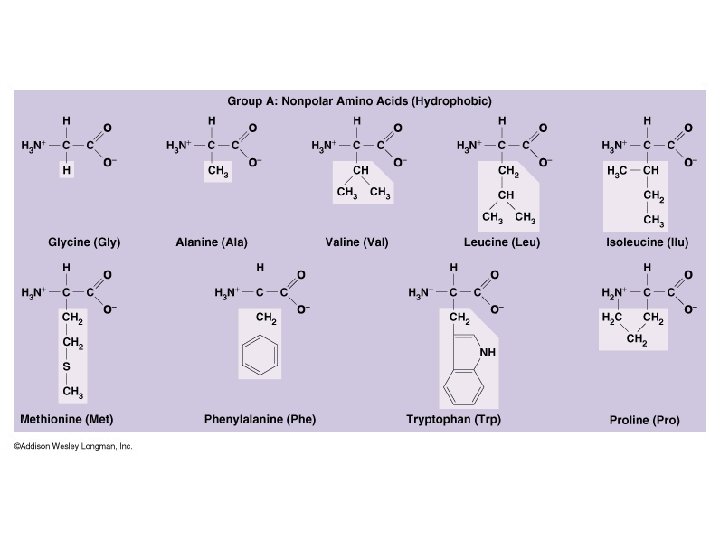
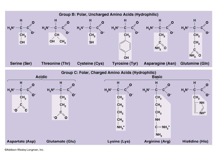
There are three types of side chains…. • Nonpolar (hydrophobic) • Polar uncharged (hydrophilic) • Polar charged (hydrophilic)
Single-letter code: M D L Y
Primary sequence… Linear sequence of amino acids in a polypeptide repeated peptide bonds form the back bone of the polypeptide chain R side groups project outward on alternate side Chain. . . one end of polypeptide chain has a free (unlinked) amine group: N-terminus other end has a free (unlinked) carboxyl group: C-terminus N-C-C-N-C-C-N-C-C-N-C-C Size… protein size is specified by mass (MW in daltons = 1 amu) average MW of a single amino acid ≈ 113 Da thus if a protein is determined to have a mass of 5, 763 Da ≈ 51 amino acids average yeast protein = 52, 728 Da [52. 7 k. Da] with about 466 amino acids Protein Primary Sequence today is determined by reading the GENOME Sequence Function is derived from the 3 D structure (conformation) specified by the primary amino acid sequence and the local environs interactions.
Four levels of protein structure
-helix = Pitch 3. 6 aa per turn
In a Beta sheet, R-groups of alternating amino acids protrude above and below the sheet
Proteins are 3 -dimensional molecules Primary structure = Amino acid sequence Secondary structure = 1. Alpha helix 2. Beta sheet -sheet Tertiary structure = 3 -D shape Quaternary structure = ? ? -helix
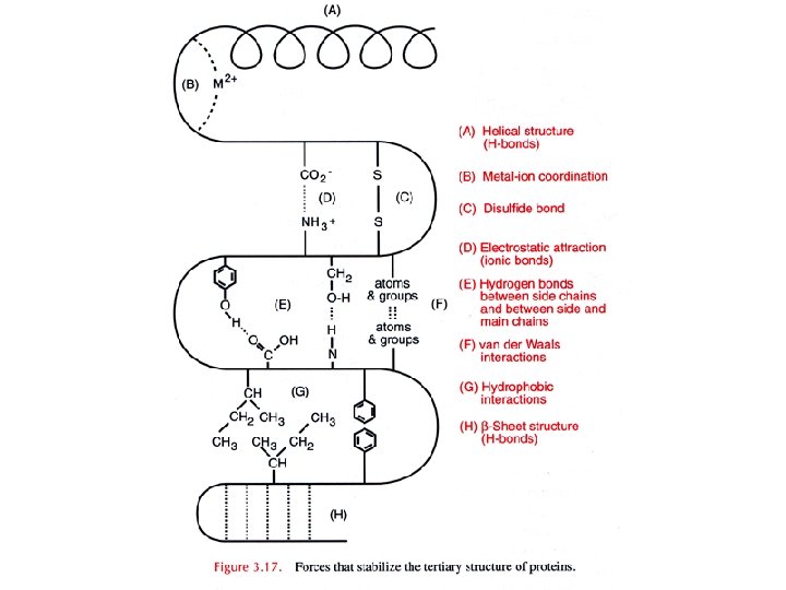
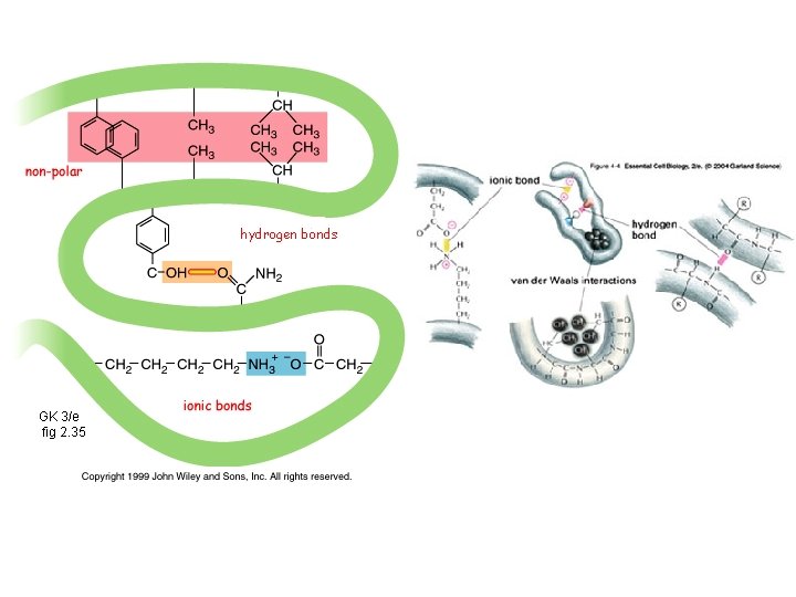
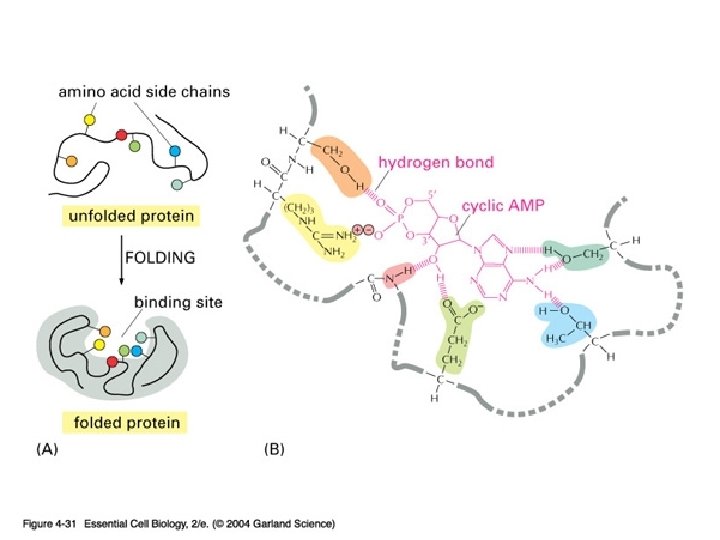
Tertiary level most responsible for 3 -D orientation of proteins in space is thermodynamically most stable conformation of a protein. . . and is due to – weak non-covalent interactions - hydrophobic interior & hydrophilic exterior - via H-bonds - & S-S bridges results in Protein Folding into specific 3 D shapes & unique binding sites
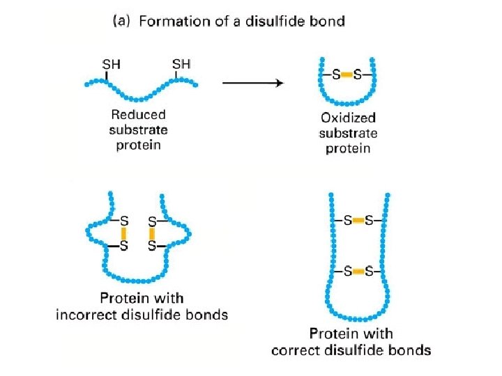
Disulfide bridge formation stabilizes protein structure Cys - S - H + H - S - Cys
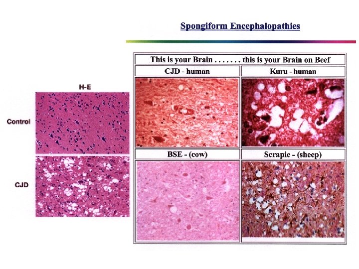
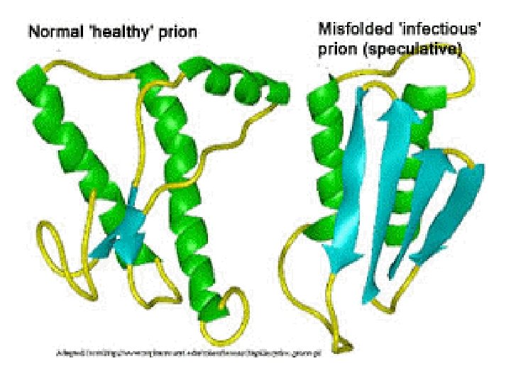
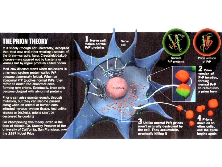
denaturation
An antibody (right) binding with the globular HA 2 domain of Hemagglutinin (space-filled model)