HODJKINS LYMPHOMA Dr M Sofi MD FRCP London








- Slides: 21
HODJKIN’S LYMPHOMA Dr. M. Sofi MD; FRCP (London); FRCPEdin; FRCSEdin
Hodgkin’s Lymphoma: Hodgkin’s lymphoma is a malignant disease that affects the lymphatic system. In the majority of cases the neoplastic cells can be derived from B lymphocytes. • Most common primary • Characteristic feature is localizations are cervical a low number of (60 -80%), mediastinal, and malignant Hodgkininguinal lymph nodes. Reed-Sternberg (H-RS) • Hodgkin’s lymphomas cells surrounded by disseminate both via numerous reactive cells lymphatic vessels or per (bystander cells). continuitatem into lymphatic organs as well as by blood vessels or per continuitatem into extralymphatic organs.
Hodgkin’s Lymphoma Epidemiology • The incidence rate is at 2 -3/100, 000 per year. The age peak lies at approx. 32 years. • Hodgkin lymphoma is one of the most treatable forms of cancer. • About 75 percent of people diagnosed with Hodgkin lymphoma can be cured with treatment. • Over 90 percent of people live at least 10 years after treatment.
Classification of Hodgkin’s lymphomas is according to the WHO classification: I. Nodular Lymphocyte-predominant Hodgkin’s lymphoma (NLPHL) • The NLPHL encompasses almost 5% of all Hodgkin’s lymphomas. • As opposed to the classical Hodgkin’s lymphomas the malignant cells are referred to as L&H (lymphocytic and histiocytic) • For therapeutic purposes, nodular lymphocytepredominant HL is managed in the same way as indolent non-Hodgkin lymphoma
Classification II. Classical Hodgkin’s lymphoma (c. HL) • • Nodular sclerosing (NSHL) Mixed cellularity (MCHL) Lymphocyte-rich (LRHL) Lymphocyte-depleted (LDHL) Tumor cells of the classical Hodgkin’s lymphomas are referred to as Hodgkin and Reed-Sternberg cells (H-RS) Stage-adapted therapy allows long term cure in more than 80 percent of all patients. Hodgkin’s lymphoma is one of the oncological diseases with the highest cure rates in adults
Features of Hodgkin lymphoma include: • Asymptomatic lymphadenopathy may be present (above the diaphragm in 80% of patients) • Constitutional symptoms • unexplained weight loss [>10% of total body weight], • unexplained fever, night sweats) are present in 40% of patients; collectively, these are known as "B symptoms" • Intermittent fever is observed in approximately 35% of cases • Pel-Ebstein fever is observed (high fever for 1 -2 wk, followed by an afebrile period of 1 -2 wk)
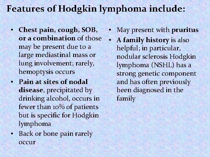
Features of Hodgkin lymphoma include: • Chest pain, cough, SOB, • May present with pruritus or a combination of those • A family history is also may be present due to a helpful; in particular, large mediastinal mass or nodular sclerosis Hodgkin lung involvement; rarely, lymphoma (NSHL) has a hemoptysis occurs strong genetic component • Pain at sites of nodal and has often previously disease, precipitated by been diagnosed in the drinking alcohol, occurs in family fewer than 10% of patients but is specific for Hodgkin lymphoma • Back or bone pain rarely occur
Physical Examination Physical examination include: • Superior vena cava syndrome may develop in • Painless lymphadenopathy patients with mediastinal can be seen in the cervical lymphadenopathy area (neck, 60 -80%), axilla • CNS symptoms or signs may (armpit, 6 -20%), and, less be due to paraneoplastic commonly, in the inguinal syndromes, including: area (groin, 6 -20%) • Cerebellar degeneration, • Waldeyer ring (back of the throat, including the tonsils) neuropathy or occipital (lower rear of the • Guillain-Barre syndrome head) or epitrochlear areas is • Multifocal infrequently observed leukoencephalopathy • Splenomegaly and/or hepatomegaly may be present
Differential Diagnoses • Cytomegalovirus • Infectious Mononucleosis • Non-Hodgkin Lymphoma • Systemic Lupus Erythematosus • • • Sarcoidosis Serum Sickness Small Cell Lung Cancer Syphilis Toxoplasmosis Tuberculosis
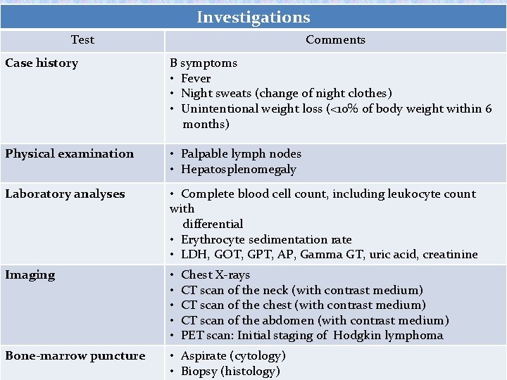
Investigations Test Comments Case history B symptoms • Fever • Night sweats (change of night clothes) • Unintentional weight loss (<10% of body weight within 6 months) Physical examination • Palpable lymph nodes • Hepatosplenomegaly Laboratory analyses • Complete blood cell count, including leukocyte count with differential • Erythrocyte sedimentation rate • LDH, GOT, GPT, AP, Gamma GT, uric acid, creatinine Imaging • Chest X-rays • CT scan of the neck (with contrast medium) • CT scan of the chest (with contrast medium) • CT scan of the abdomen (with contrast medium) • PET scan: Initial staging of Hodgkin lymphoma Bone-marrow puncture • Aspirate (cytology) • Biopsy (histology)
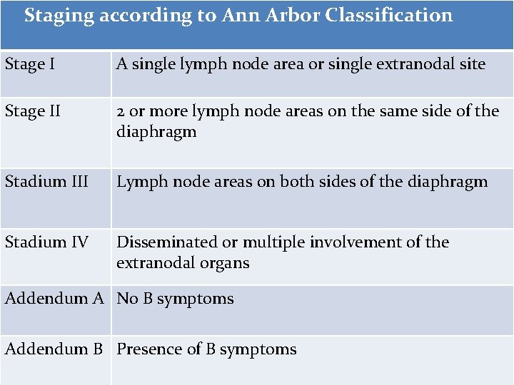
Staging according to Ann Arbor Classification Stage I A single lymph node area or single extranodal site Stage II 2 or more lymph node areas on the same side of the diaphragm Stadium III Lymph node areas on both sides of the diaphragm Stadium IV Disseminated or multiple involvement of the extranodal organs Addendum A No B symptoms Addendum B Presence of B symptoms
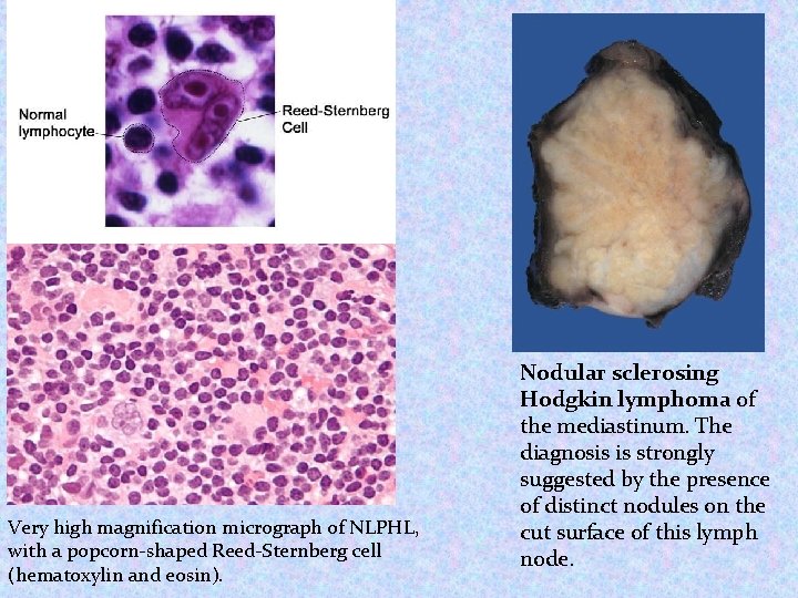
Very high magnification micrograph of NLPHL, with a popcorn-shaped Reed-Sternberg cell (hematoxylin and eosin). Nodular sclerosing Hodgkin lymphoma of the mediastinum. The diagnosis is strongly suggested by the presence of distinct nodules on the cut surface of this lymph node.
Non-Hodgkin lymphomas (NHLs) are tumors originating from lymphoid tissues, mainly of lymph nodes. These tumors may result from chromosomal translocations, infections, environmental factors, immunodeficiency states, and chronic inflammation Signs and symptoms • Peripheral adenopathy: Painless and slowly progressive; can spontaneously regress • Primary extranodal involvement and B symptoms: Uncommon at presentation; however, common with advanced, malignant transformation or end-stage disease • Bone marrow: Frequent involvement; may be associated with: • Cytopenias(s) • Fatigue weakness more common in advancedstage disease
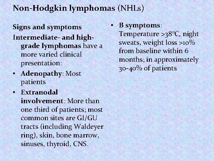
Non-Hodgkin lymphomas (NHLs) • B symptoms: Signs and symptoms Temperature >38°C, night Intermediate- and highsweats, weight loss >10% grade lymphomas have a from baseline within 6 more varied clinical months; in approximately presentation: 30 -40% of patients • Adenopathy: Most patients • Extranodal involvement: More than one third of patients; most common sites are GI/GU tracts (including Waldeyer ring), skin, bone marrow, sinuses, thyroid, CNS.
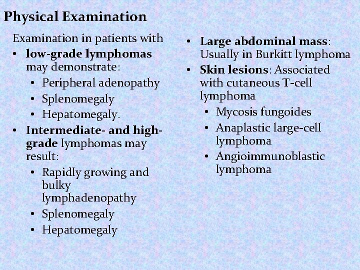
Physical Examination in patients with • low-grade lymphomas may demonstrate: • Peripheral adenopathy • Splenomegaly • Hepatomegaly. • Intermediate- and highgrade lymphomas may result: • Rapidly growing and bulky lymphadenopathy • Splenomegaly • Hepatomegaly • Large abdominal mass: Usually in Burkitt lymphoma • Skin lesions: Associated with cutaneous T-cell lymphoma • Mycosis fungoides • Anaplastic large-cell lymphoma • Angioimmunoblastic lymphoma
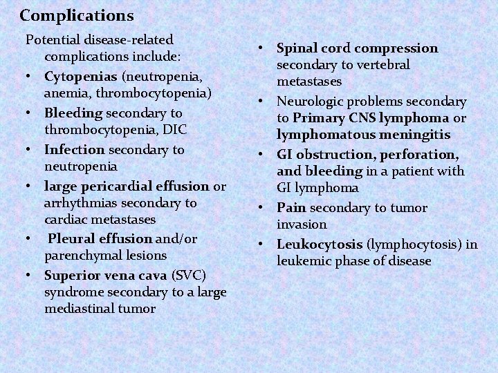
Complications Potential disease-related complications include: • Cytopenias (neutropenia, anemia, thrombocytopenia) • Bleeding secondary to thrombocytopenia, DIC • Infection secondary to neutropenia • large pericardial effusion or arrhythmias secondary to cardiac metastases • Pleural effusion and/or parenchymal lesions • Superior vena cava (SVC) syndrome secondary to a large mediastinal tumor • Spinal cord compression secondary to vertebral metastases • Neurologic problems secondary to Primary CNS lymphoma or lymphomatous meningitis • GI obstruction, perforation, and bleeding in a patient with GI lymphoma • Pain secondary to tumor invasion • Leukocytosis (lymphocytosis) in leukemic phase of disease
Testing: Laboratory studies with suspected NHL include: • CBC count: May show anemia, thrombocytopenia leukopenia/pancytopenia, thrombocytosis • Serum chemistry: May show elevated LDH, LFT and calcium levels • HIV serology: Especially in patients with diffuse large cell immunoblastic or small non cleaved histologies • Human T-cell lymphotropic virus-1 serology: Patients with adult T-cell leukemia/lymphoma • Hepatitis B testing: In patients in whom rituximab therapy is planned • Immunophenotypic analysis of lymph node, bone marrow, peripheral blood • Cytogenetic studies: NHL associated with monoclonal gammopathy
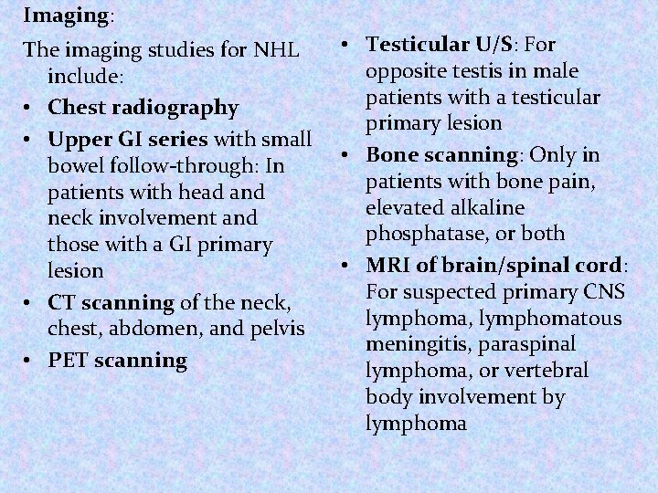
Imaging: The imaging studies for NHL include: • Chest radiography • Upper GI series with small bowel follow-through: In patients with head and neck involvement and those with a GI primary lesion • CT scanning of the neck, chest, abdomen, and pelvis • PET scanning • Testicular U/S: For opposite testis in male patients with a testicular primary lesion • Bone scanning: Only in patients with bone pain, elevated alkaline phosphatase, or both • MRI of brain/spinal cord: For suspected primary CNS lymphoma, lymphomatous meningitis, paraspinal lymphoma, or vertebral body involvement by lymphoma
Procedures: • Bone marrow aspiration and biopsy: For staging rather than diagnostic purposes • Excisional lymph node biopsy (extranodal biopsy): For lymphoma protocol studies • Lumbar puncture for CSF analysis in patients with: • Diffuse aggressive NHL with bone marrow, epidural, testicular, paranasal sinus, or nasopharyngeal involvement. • High-grade lymphoblastic lymphoma • High-grade small noncleaved cell lymphomas • HIV-related lymphoma • Primary CNS lymphoma • Neurologic signs and symptoms
Management: The treatment of NHL varies greatly and include: • Chemotherapy: Most common; usually combination regimens • Radiation therapy • Rituximab administration • Bone marrow transplantation: Possible role in relapsed high-risk disease • Radioimmunotherapy • Transfusions of blood products • Antibiotic Pharmacotherapy • Medications include: • Cytotoxic agents • Histone deacetylase inhibitors (eg, vorinostat) • Colony-stimulating factor (eg, epoetin filgrastim) • Monoclonal antibodies (eg, rituximab, ibritumomab) • Proteasome inhibitors (eg, bortezomib) • Immunomodulators (eg, interferon alfa-2 a or alfa-2 b) • Corticosteroids (prednisone, dexamethasone,
THANK YOU FOR YOUR ATTENTION