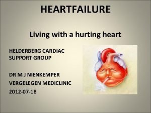
Heart Failure Tom Heaps Consultant Acute Physician Heart


- Slides: 22

Heart Failure Tom Heaps Consultant Acute Physician

Heart Failure in the UK prevalence ~900, 000 people average of diagnosis 76 most common cause is CAD 30 -40% mortality at 1 y after diagnosis (worse than most cancers) 5% of all acute admissions (projected to rise by 50% over next 25 years) 1 million inpatient bed days per annum 2% of annual NHS budget (70% of cost due to hospitalizations) 1 in 4 patients are readmitted within 3 m of discharge

NYHA Classification Class Symptoms I No limitation of physical activity: ordinary physical activity does not cause fatigue, palpitations, dyspnoea or angina II Slight limitation of physical activity: comfortable at rest but ordinary activity causes fatigue, palpitations, dyspnoea or angina III Marked limitation of physical activity: comfortable at rest but less than ordinary activity causes fatigue, palpitations, dyspnoea or angina IV Unable to carry out any physical activity without discomfort: symptoms of cardiac insufficiency at rest and discomfort increases with any physical activity

Case Study 67 -year-old male, diabetes, COPD, MI 2005, CKD 2 ECHO 2008 mild AS and LVEF 33% NYHA II at last heart failure clinic appointment aspirin 75 mg OD, simvastatin 40 mg ON, metformin 500 mg BD, furosemide 40 mg OD, ramipril 2. 5 mg OD, bisoprolol 1. 25 mg OD admitted to AMU with 1/52 Hx of increasing SOBOE (ET reduced from 200 to 50 yds), orthopnoea and oedema RR 20, Sp. O 2 94% on air, BP 156/82 mm. Hg, HR 94/min 1. WHAT IS THE DIAGNOSIS? 2. WHAT ARE THE POTENTIAL TRIGGERS? 3. WHAT INVESTIGATIONS ARE REQUIRED?

Acute Heart Failure Syndrome of rapid onset of symptoms and signs secondary to cardiac dysfunction due to: 1. Acute myocardial injury – ischaemia, acute valvular dysfunction, pericardial effusion/tamponade, myocarditis, aortic dissection, acute VSD/ventricular wall rupture, cardiac contusion 2. Afterload/chronotropy/inotropy/lusitropy mismatch – hypertensive crisis, arrhythmia, thyrotoxicosis 3. Decompensation of chronic heart failure

Decompensation of CHF Poor compliance with HF treatment Excessive salt intake Addition of new drug e. g. NSAID, steroid, thiazolidinedione, diltiazem Alcohol or drug abuse Uncontrolled hypertension Infection/sepsis esp. pneumonia AKI AECOPD Arrhythmia e. g. AF Ischaemia/ACS or valvular dysfunction Hyper- or hypothyroidism Anaemia Electrolyte disturbances e. g. hypocalcaemia, hypophosphataemia Iatrogenic fluid overload

Investigation FBC, U&E, LFT, CRP, Ca 2+, Mg 2+, PO 43 -, lipids, glucose, TFTs Infection screen CXR ECG ECHO c. MRI CT or conventional coronary angiography Spirometry NT-pro. BNP or BNP and hs. Tn. I in selected cases

Case Study cont. CXR – venous congestion, upper lobe diversion, enlarged heart ECG – AF rate 98/min urea 14, creatinine 155 (138), other bloods unremarkable suddenly deteriorates at 02: 00 with MEWS = 6 RR 34, Sp. O 2 88% on 2 l O 2, HR 144, BP 169/102, no urine since admission cyanosed, clammy, agitated, JVP +5 cm, bibasal crackles ++ ABG: p. H 7. 29, p. O 2 7. 6, p. CO 2 7. 2, BE -8. 3, lactate 3. 2 1. WHAT IS THE DIAGNOSIS? 2. WHAT IS THE TREATMENT NOW?

ACUTE HEART FAILURE IV diuretic bolus e. g. furosemide 50 mg +/- IV morphine 2. 5 -5 mg s. BP >110 mm. Hg: IV vasodilator e. g. GTN 50 mg in 50 ml @ 0. 6 -6 ml/h (10 -100μg/min) Inadequate ventilation or oxygenation Oxygen NIV ETT and IV Lifethreatening tachy- or bradyarrhythmia Systolic BP <85 mm. Hg or shock DCCV Pacing Stop vasodilators and βblockers Inotrope / Vasopressor Consider IABP Inadequate diuresis Acute mechanical cause or valvular dysfunction ACS Catheterize Increase diuretics Consider low-dose dopamine or UF Urgent ECHO Surgical or percutaneous intervention Antithrombotic therapy Coronary reperfusion

IV Diuretics vs. Nitrates in AHF limited evidence for beneficial ‘venodilatory’ effects of IV diuretics high dose diuretics increase fluid loss but reduction in circulating volume may cause organ hypoperfusion (e. g. AKI) and increased myocardial stress (activation of RAAS and SNS) increased risk of other SE e. g. ototoxicity with high-dose furosemide continuous IV infusion of diuretics increases diuresis with lower cummulative doses but no effect on symptoms or safety at 72 h IV GTN causes venodilatation and reduced LV filling pressures (i. e. preload); arterial dilatation (reduced afterload) at higher doses may decrease myocardial O 2 demand improve CO better outcome (decreased rates of intubation and MI) with high-dose nitrates plus low-dose diuretics vs. low-dose nitrates plus high-dose diuretics in one study risk of sudden hypotension and increased myocardial ischaemia with nitrates

NIV in AHF Consider as adjunct to pharmacological Rx if severe respiratory distress, refractory hypoxaemia or T 2 RF Contraindicated if significant hypotension CPAP and Bi. PAP are probably equivalent Improves symptoms, respiratory parameters and ABGs Earlier meta-analyses showed improved outcomes with NIV More recent meta-analyses (including results of large 3 CPO trial) failed to show any effect on intubation rates, LOS or mortality

2 Minute Haemodynamic Profiling in AHF Congestion at rest? orthopnoea, elevated JVP, oedema, rales, ascites Yes Poor perfusion at rest? cold extremities, narrow pulse pressure, drowsiness A. B. C. D. No Yes No A: Warm & Wet C: Warm & Dry B: Cold & Wet D: Cold & Dry diuretics > nitrates > diuretics, withold β-blockers and ACE-i, consider inotropes target profile, optimize and titrate chronic therapy exclude hypovolaemia, consider invasive monitoring, cautious filling, inotropes, IABP

Case Study 2 76 -year-old Afro-Caribbean female HTN, CKD 3 (renovascular disease) and OA diagnosis of HF with LVEF 26% on ECHO GFR fell from 44 to 31 m. L/min on initiation of ACE-i - stopped admitted with worsening oedema and SOBOE (NYHA III) bisoprolol 5 mg, doxazosin 4 mg, furosemide 80 mg, simvastatin 40 mg, naproxen 500 mg BP 143/88 mm. Hg, HR 84/min ECG: sinus rhythm, non-specific BBB (QRS 0. 16 s) 1. WHAT ARE THE TREATMENT OPTIONS?

Chronic Management of HF Diuretics ACE-inhibitor ARB if ACE-i not tolerated β-blocker H-ISDN if ACE-i AND ARB not tolerated MR antagonist Ivabradine if in SR and HR >70/min CRT-P or CRT-D LVAD +/- heart transplant Digoxin if in AF or HR still >70

Cardiac Resynchronization Therapy (CRT) CRT-P reduces hospital admissions and mortality and improves symptoms and exercise capacity if symptomatic despite maximal medical therapy if: life expectancy >1 y, good functional status, sinus rhythm AND NYHA III-IV with EF ≤ 35% AND QRS ≥ 120 ms (LBBB) OR QRS ≥ 150 ms (non-LBBB) OR NYHA II with EF ≤ 30% AND QRS ≥ 130 ms (LBBB) OR QRS≥ 150 ms (non-LBBB) Possible benefit from CRT-P if: permanent AF AND NYHA III-IV with EF ≤ 35% AND QRS ≥ 120 ms AND AV nodal ablation OR pacing required for slow AF OR HR ≤ 60 resting and ≤ 90 on exertion indication for conventional pacing AND NYHA II-IV AND EF ≤ 35% irrespective of QRS duration CRT-D if: previous ventricular arrhythmia OR symptomatic (NYHA II-III) with EF ≤ 35% after ≥ 3 m of maximal medical therapy

Case Study 2 cont. started on hydralazine 37. 5 mg TDS and ISDN 20 mg TDS α-blocker stopped ivabradine 2. 5 mg BD added for rate control dose of furosemide increased to 120 mg OD fluid restricted to ≤ 2 l/d referred to HFNS for consideration of CRT-P no significant decline in renal function but no significant diuresis no improvement in oedema or reduction in body weight over next 4 d 1. WHY IS SHE NOT RESPONDING TO INCREASED DIURETICS? 2. WHAT ARE THE OPTIONS TO TREAT HER FLUID OVERLOAD?

Diuretic Resistance: Mechanisms Poor compliance, excess salt intake, concomitant NSAID use Renal impairment (reduced active secretion of diuretics and decreased peak urinary concentrations) Compensatory hyperplasia/hypertrophy of epithelial cells in DCT and increased Na+ reabsorption with chronic loop diuretic Reduced biovailability or delayed absorption of diuretics due to intestinal mucosal oedema Compensatory post-diuretic salt retention (when urinary drug concentrations < diuretic threshold)

Diuretic Resistance: Management Reduce dietary salt intake (<2 g/d) and stop NSAIDs Increase diuretic dose in renal failure (e. g. furosemide 240 mg daily) Add in thiazide diuretic e. g. metolazone, bendroflumethiazide or indapamide (monitor U&E closely) Switch to bumetanide or torasemide (bioavailability 80% compared with 40% for furosemide) or IV diuretics Split dosing i. e. BD/TDS or switch to torasemide (duration of action 18 -24 h compared with 4 -6 h for furosemide) or give continuous IV infusion e. g. furosemide 10 mg/h consider ultrafiltration with CVVH: increased weight loss and reduced readmissions c/w diuretics (UNLOAD)

Case Study 3 82 -year-old female T 2 DM, HTN, AF 2 x admissions this year with pulmonary oedema review in AEC with results of outpatient ECHO breathless after walking 50 yds, mild ankle oedema, raised JVP ramipril 2. 5 mg, gliclazide 40 mg BD, furosemide 40 mg OD BP 159/91, HR 88/min irregularly irregular, CBG 15. 1 mmol/L BNP 252 pg/m. L (<35 pg/m. L), Hb. A 1 c 9. 5%, other bloods normal ECHO: good LV systolic function (LVEF 63%), moderate LVH, E/A reversal of doppler waveform across mitral valve 1. WHAT IS THE DIAGNOSIS 2. HOW SHOULD SHE BE TREATED

Diastolic HF (HF-PEF) symptoms and signs of CHF with EF >40% normal LVEF on ECHO in up to 50% with ACPOE (rule out reversible cause of LVSD) older, female, hypertensive, diabetic, AF (CAD less common) similar prognosis to HF-REF impaired LV relaxation during diastole and E/A reversal on ECHO no treatment proven to reduce mortality perindopril (PEP-CHF), irbesartan (i-PRESERVE) and candesartan (CHARMPRESERVED) may improve symptoms and reduce admissions β-blockers (e. g. nebivolol), rate-limiting CCBs (e. g. verapamil) and digoxin may increase diastolic filling times by reducing ventricular rate diuretics for congestive symptoms optimize Rx of comorbidities e. g. HTN, diabetes, AF (and CAD)

Case Study 4 78 -year-old male, previous CABG EF 15% NYHA IV (breathless at rest) LBBB on ECG admitted with worsening oedema unable to tolerate ACE-i/ARB and spironolactone due to CKD 4 s. BP 90 mm. Hg, Sp. O 2 90% on air asked to see urgently by nurse ? dying (DNACPR in place) asleep, Cheyne-Stokes respiration, apnoeic periods with Sp. O 2 84% 1. WHAT IS THE DIAGNOSIS? 2. WHAT ARE THE TREATMENT OPTIONS?

Cheyne-Stokes Respiration (CSR) in HF Periodic breathing or central sleep apnoea (CSA) Occurs in up to 50% with severe HF (NYHA III-IV) during sleep Heightened chemoreceptor sensitivity to CO 2, increased circulating catecholamine levels and hypoxaemia Hyperventilation Hypocapnia – CO 2 levels fall below apnoeic threshold Apnoea Sympathetic activity ++ Pa. CO 2 levels rise above apnoea threshold Daytime somnolence, fatigue, PND, hypoxaemia/diastolic dysfunction, increase in fatal arrhythmias and mortality Nocturnal O 2 < CPAP < Bi. PAP < adaptive servoventilation (ASV)
 Acute vs chronic heart failure
 Tom heaps
 Cushings triad
 Alain sauvage
 Treatments for acute renal failure
 Acute brain failure
 West haven criteria hepatic encephalopathy
 Non conducted pac ecg
 Failure to pace
 Ductile fracture surface
 What does tom symbolize in the devil and tom walker
 Tomtom go 910 update
 Skew heaps
 Heap's law
 Geetika tewari
 Love heaps
 Heaps cs
 Binomial heap delete min
 Soft heaps of kaplan and zwick uses
 Stratus cumulus nimbus
 Vetsulin dosage chart for dogs
 Heart failure complications
 Dr nienkemper

