GRAM POSITIVE COCCI STREPTOCOCCUS PNEUMONIAE Dr R S
 Professor")


 Bacterial growth in normally sterile fluids Blood (pneumonia, meningitis, endocarditis)")


- Slides: 20
GRAM POSITIVE COCCI STREPTOCOCCUS PNEUMONIAE Dr. R. S. Gopika M. D. (Hom. ) Professor & Ho. D Dept. of Pathology SKHMC
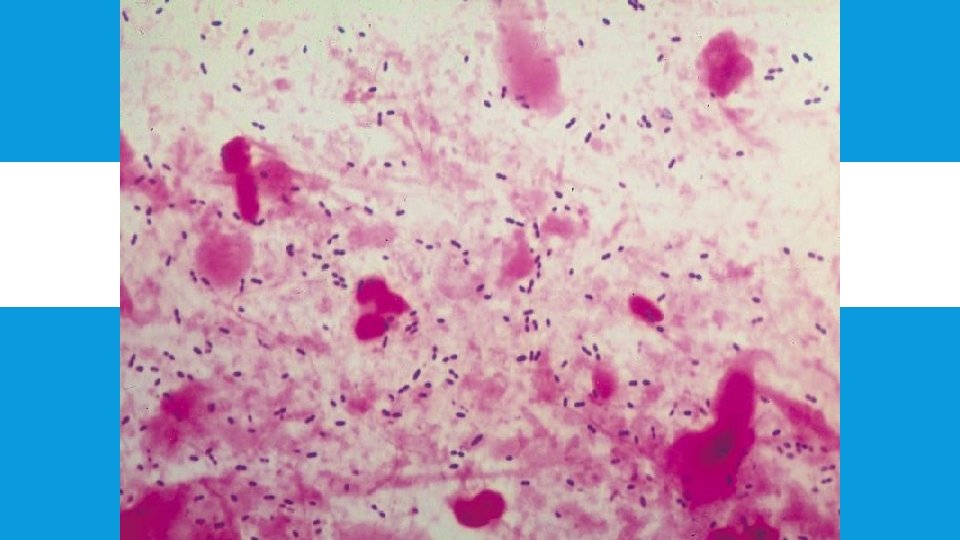
STREPTOCOCCUS PNEUMONIAE pneumococcus Gram-positive encapsulated Diplococcus flame shaped 1µm normally found in the nasopharynx
QUELLUNG REACTION Neufeld/ capsular swelling reaction To the pneumococcal colonies type specific antiserum along with methyelene blue is added. In presence of homologus antiserum capsule swells, sharply delineated. Bed side sputum test.
QUELLUNG REACTION
CULTURE- CHARACTERS 37ºC, p. H- 7. 8 , CO 2 -5 - 10% Glucose broth - uniform turbidity Blood Agar 18 hrs – small dome shaped, glistening, alpha haemolysis. further incubation – colonies flat , raised edges, central umbonation – carrom coin/draught man’s colonies.
BLOOD AGAR 5% CO 2, overnight. Colonies are surrounded by zones of alpha-hemolysis.
RESISTANCE 52º C -15 min
VIRULENCE FACTORS Polysaccharide capsule—prevents phagocytosis Pneumolysin -pore-forming protein that can cause lysis of host cells and activate complement Autolysin -activation of this protein lyses the bacteria releasing its internal contents (pneumolysin) Hydrogen peroxide—causes damage to host cells (can cause apoptosis in neuronal cells during meningitis) Ig. A protease – cleaves resp mm Ig. A
Pili—colonization of upper respiratory tract and increase the formation of large amounts of TNF by the immune system during sepsis, raising the possibility of septic shock Choline binding protein A / Pneumococcal surface protein A -an adhesin that can interact with carbohydrates on the cell surface of pulmonary epithelial cells and can inhibit complement-mediated opsonization of pneumococci
PATHOGENESIS normal colonization can become infectious if the organisms are carried into areas such as the Eustachian tube or nasal sinuses where it can cause otitis media and sinusitis,
PNEUMONIA occurs if the organisms are inhaled into the lungs and not cleared (viral infection, or smoking-induced ciliary paralysis might be contributing factors). capsule makes it resistant to phagocytosis, alveolar macrophages cannot adequately kill the pneumococci. The organism spreads to the blood stream/ bacteremia and is carried to the meninges, joint spaces, bones, and peritoneal cavity, and may result in meningitis, brain abscess, septic arthritis or osteomyelitis.
Age groups at highest risk for disease: Infants and children < 2 years of age Adults > 65 years of age
ACUTE OTITIS MEDIA From colonisation to invasion of middle ear through the eustachian tube Facilitated by previous viral infection Mostly in young children with immature immune defence
PNEUMOCOCCAL PNEUMONIA The incubation period -1 to 3 days. Symptoms abrupt onset of fever and chills or rigors. Other common symptoms include pleuritic chest pain, cough productive of mucopurulent, rusty sputum, dyspnea , tachypnea , hypoxia , tachycardia and weakness. Nausea, vomiting, and headaches occur less frequently.
Complications Pleural effusions Septicemia Endocarditis Meningitis
INVASIVE PNEUMOCOCCAL DISEASE (IPD) Bacterial growth in normally sterile fluids Blood (pneumonia, meningitis, endocarditis) CSF (meningitis) Joint fluids (artritis) Pleural fluid (pleuritis) Peritoneal fluid (peritonitis)
LAB
ENTEROCOCCUS Normal flora of human intestine Diplococci Spectacle shapedappearnce E. Faecalis UTI, intra abdominal, pelvic and soft tissue infections Infection on burn surfaces.
commons. wikimedia. org en. wikipedia. org Text Book of Microbiology-Anandanarayanan