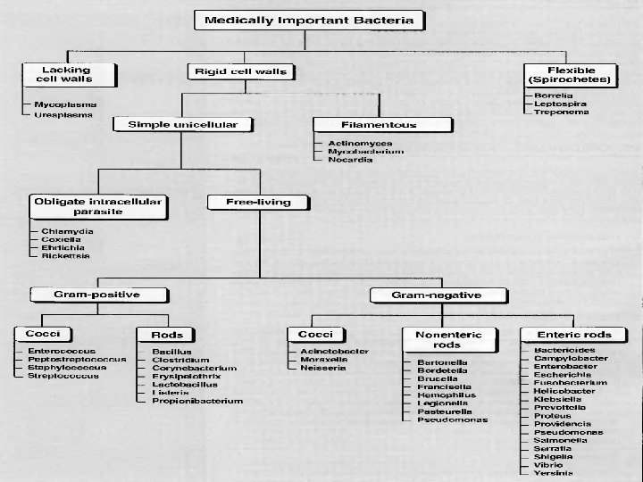
Genus Streptococcus Gram positive Catalase negative cocci objectives

Genus Streptococcus Gram positive, Catalase negative cocci

objectives * Study systemic bacteriology * Describe Genus Streptococcus, its types, pathogenesis, and lab diagnosis.

Gram positive cocci Staphylococci Streptococcus Catalase -ve Catalase +ve
 Hemolysis β α hemolysis S. Pyogenes S. Pneumoniae S. agalactiae")
Genus Streptococcus Lancefield (serology) Hemolysis β α hemolysis S. Pyogenes S. Pneumoniae S. agalactiae Viridans Streptococci γ hemolysis S. bovis Serogroups (Capsular Ag) Group A S. Pyogenes 80 Serotypes, (M-protein) Group B S. agalactiae Group D S. bovis

When grown on sheep blood agar, streptococci display one of three types of hemolysis of the red blood cells in the agar. ü Alpha hemolysis--The red blood cells in the media are partially digested producing a greening of the agar. ü Beta hemolysis--The red blood cells in the media are completely digested producing a clearing of the agar. ü Gamma hemolysis--No change is noted in the agar. The red blood cells are not lysed. Expected Hemolysis: Ø Streptococcus pyogenes always beta hemolylic Ø Streptococcus agalactiae usually beta hemolytic Ø Streptococcus pneumoniae and Viridans streptococci are always alpha hemolylic Ø Enterococcus faecalis gamma hemolytic

Hemolysis patterns on blood agar 7
 clear zone around colonies S. pyogenes on blood agar (beta hemolysis)")
(Beta hemolysis) clear zone around colonies S. pyogenes on blood agar (beta hemolysis)
")
Alpha hemolysis (green discoloration)

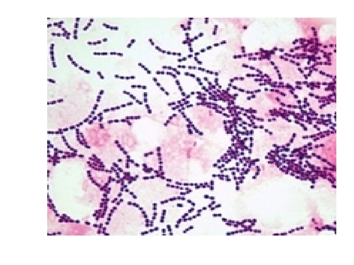
GENERAL CHARACTERISTIC: * G+ve cocci , arrange in chains or pairs. * Some strains are capsulated * Majority are facultative anaerobic, few are obligatory anaerobic. * Catalase –ve * Non motile. * Non spore forming * Fastidious microorganism
: M-protein 80 serotypes Reservoir Human throat and")
S. pyogenes (Group A β- hemolytic, GABH): M-protein 80 serotypes Reservoir Human throat and skin(N. F. ) Transmission Spread by respiratory droplets or direct contact
. •")
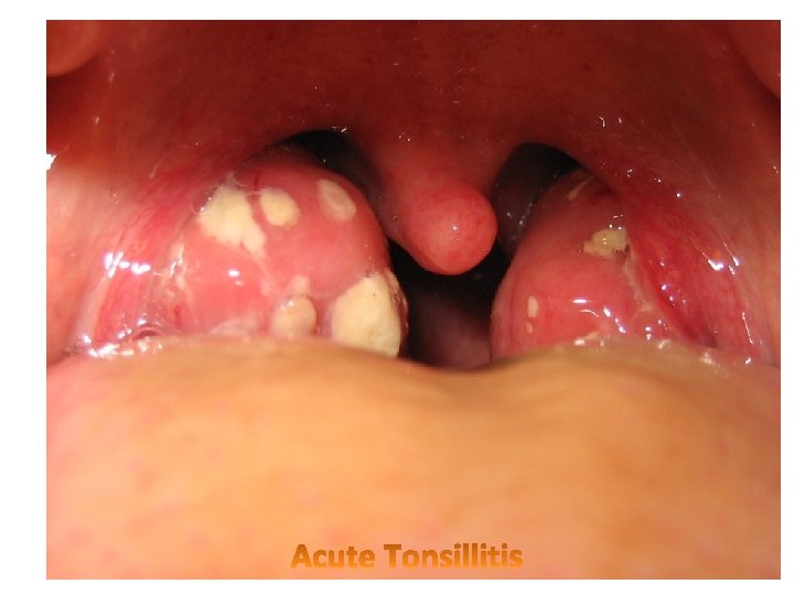
DISEASES S. pyogenes SYSTEMIC INFECTION LOCAL INFECTIONS • Sore throat (acute pharyngitis, pharyngotonsillitis). • Wound infection, cellulitis, fasciitis and myonecrosis. • Impetigo. • Erysipelas. • • Sepsis. Meningitis. Puerperal Sepsis. Streptococcus toxic shock syndrome • Scarlet fever POST STREPTOCOCCAL • Rheumatic fever • Acute Glomerulonephritis

Laboratory Diagnostic steps Specimens Direct Gram’s Slide Biochemical Culture Serology Antibiotics sensitivity

Lab dx. Specimens: sputum, throat swab, nasopharyngeal swab, blood, CSF…etc. Gram stain: G+ve cocci, arrange in chains. Culture: on blood agar pinpointed, Grayish white, translucent, matte or glossy colonies with large zone of βhemolysis. Bacitracin disc (0. 04 U) sensitive causes zone of growth inhibition. Serology: Lancefield grouping, M-protein serotyping and ASO test (Antistreptolysin-O test).

ASO test: * Measure Ab against Streptolysin O *ASO test uses in post streptococcal infection complication. This test used to determine significance streptococcal infection by measuring the ASOT: * ASOT (Ab Titer): Normal < 200 < significance result
")
S. pyogenes on blood agar (beta hemolysis)
: Normal flora of female genital tract (15 -20%")
S. agalactia (Group B β- hemolytic): Normal flora of female genital tract (15 -20% of woman), male urethra, GIT. Leading Cause fo f r neonatal sepsis & meningitis. Bacitracin resistant
 test – Detects the production of enhanced hemolysis")
Biochemical Identification • Christie-Atkins, Munch-Petersen (CAMP) test – Detects the production of enhanced hemolysis that occurs when b-lysin and the hemolysins of Group B streptococci come in contact Group B streptococci showing the classical “arrow-shaped hemolysis near the staphylococcus streak

Identification of group D Streptococci: Lancefield Group D streptococci are divided into two groups: (1) Enterococci, and (2) nonenterococci. Enterococcus faecalis and Enterococcus faecium are the species of Enterococcus. Strep. bovis and Strep. equinus are species of Enterococci". "Group-D streptococci, not
 Previously regarded as part of streptococci, today regarded as")
Enterococcus (E. faecalis, E. faecium) Previously regarded as part of streptococci, today regarded as separated genus. Normal flora of GIT, oral mucosa Causes UTI, wound infection, bed sore, endocarditis. Varies Hemolysis

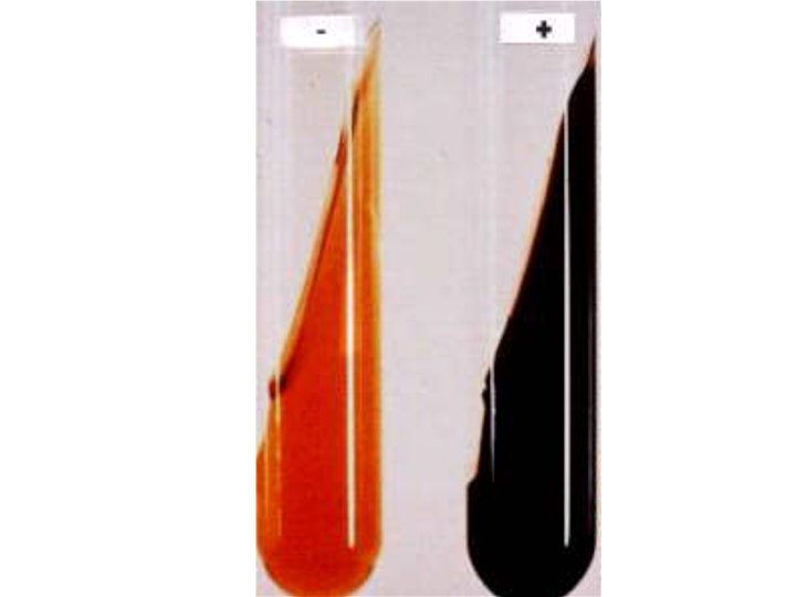
Bile-Esculin Hydrolysis Test: The purpose of this test is to determine the ability of an organism to hydrolyze the glycoside esculin to esculatin and glucose in the presence of bile (10 - 40%). This test aids in the differentiation of group D streptococci from other "not group D streptococci". Procedure: 1. Inoculate the organism to be tested into the bile esculin medium. Incubate at 37 o. C for 24 hours (stab into medium, then streak on slant). Positive Test: Presence of a black to dark brown color on the slant -(Enterococcus faecalis) Negative Test: No blackening of the medium - (Streptococcus agalactiae or Streptococcus pyogenes)

Viridans Streptococcus They contain many species, they are untypable i. e. no group specific Ag, they are α- hemolytic Streptococcus It is present as a Commensal on mucosa of mouth, nasopharynx, and saliva. Tooth extraction enters human body subacute bacterial endocarditis (SBE) in patients with abnormal heart valves and no antibiotic prophylaxis. S. mutans causes tooth carries.
")
Alpha hemolysis (green discoloration)
 Pneumococci are typed to 83 serotypes according to the")
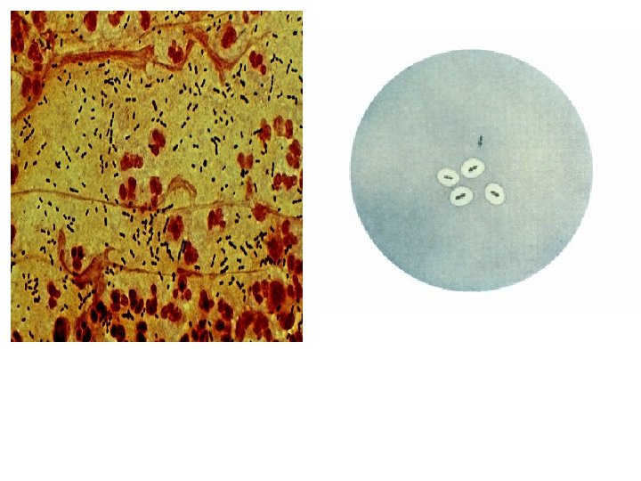
S. pneumoniae (Pneumococci, Diplococcus pneumoniae) Pneumococci are typed to 83 serotypes according to the nature of capsular polysaccharide antigen. Its N. F of URT(nosopharyngeal &oropharyngeal flora). It can be provoked by predisposing factors (influenza, common cold or other bac. Infec. In adults, types 1 -8 are responsible for about 70% of pneumococcal pneumonia and for 5% of fatalities due to pneumococcal bacteremia. In CHILDREN, type 6, 14, 19 and 23 are frequent causes. Capsular polysaccharide Gram +VE diplococcus

Diseases Pneumonia Meningitis. Sinusitis. Otitis media. Sepsis.

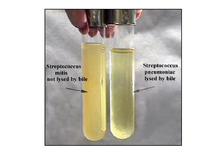
CHARACTER PNEUMOCOCCI VIRIDANS STREPTOCOCCI Ovoid or lanceolate diplococci Rounded cocci in short or long chains. Present Absent Optochin sensitivity +ve -ve Bile solubility +ve -ve Capsular swelling test (Quelling reaction) +ve -ve Virulence in mice +ve -ve Morphology Capsule
 Gram stain 3) Biochemical tests: • Optochin sensitivity disc • Bile")
Lab dx. 1) Gram stain 3) Biochemical tests: • Optochin sensitivity disc • Bile Solubility test 5) Virulence in mice: mice will die, while when inject Viridans the mice will survive. 2) Culture: on Chocolate or blood Agar αhemolysis small (pinpointed), gray, colonies may be mucoid. 4) Serology Quelling reaction test Latex particle agglutination test 6) Antimicrobial susceptibility testing.

Optochin disc for S. pneumococci

Question? Name this test

THANKS
- Slides: 35