FOUNDATION BLOCK 2019 Dr Malak M ElHazmi MICROBIOLOGY
 Dr. Malak M. El-Hazmi")
FOUNDATION BLOCK (2019) Dr. Malak M. El-Hazmi

MICROBIOLOGY
.")
Laboratory diagnosis of infections. ID Ø Microscopic examination. Ø culture. Ø Serological tests (Ab). Ø Detection of Ag. Ø Molecular method.

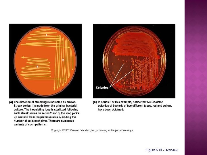
Types of specimens

BACTERIOLOG Y

GRAM STAIN G- bacilli

BACTERIAL CELL WALL

BACTERIAL SHAPES AND ARRANGEMENTS

GRAM STAIN Gram-positive cocci Gram-positive bacilli Gram-negative cocci Gram-negative bacilli

GRAM POSITIVE BACTERIA

GRAM POSITIVE COCCI

GRAM POSITIVE COCCI

GRAM NEGATIVE BACTERIA
 e. g Neisseria Gram negative bacilli e. g E.")
Gram negative cocci (Diplococci ) e. g Neisseria Gram negative bacilli e. g E. coli Salmonella

Gram positive cocci in chain Gram positive cocci in clusters Streptococci Staphylococci Penicillin Cephalosporin Rx cloxacillin Cephalosporin if MRSA vancomycin

A gram-stained smear of a CSF sample from a 3 year old child seen in the emergency department presenting with fever and neck stiffness. Gram-positive diplococci & pus cells Streptococcus pneumoniae

This is a bacterium isolated from a child with sore throat and tonsillitis. A: Describe the Gram stain Gram positive B: Describe the shape and arrangement of the bacteria Cocci in chains

Following is the Gram stained smear of an organism isolated from a wound infection. Describe what you see in the slide above. What is the likely organism ? Gram-positive cocci in clusters Staphylococcus aureus

Following is the Gram-stained smear of from urethra of a 25 –year old male complaining of urethral discharge Describe the Gram stain of the intracellular bacteria Describe the shape of the bacteria Gram negative cocci ( diplococci)

Describe the Gram stain of this organism: Describe its shape Gram negative bacilli ( rods )

BACTERIAL CULTURE MEDIA Type of Media Purpose Selective Suppression of unwanted microbes; encouraging desired microbes. Differential Differentiation of colonies of desired microbes from others. Enrichment Similar to selective media but designed to increase number of desired microbes to detectable levels.
 Enriched medium (Chocolate Agar) Differential medium")
BACTERIAL CULTURE MEDIA General culture medium (Blood Agar) Enriched medium (Chocolate Agar) Differential medium (Mac. Conkey Agar) Selective medium (Thiosulphate citrate bile salt sucrose TCBS)

BACTERIA CULTURING Laboratory Incubator
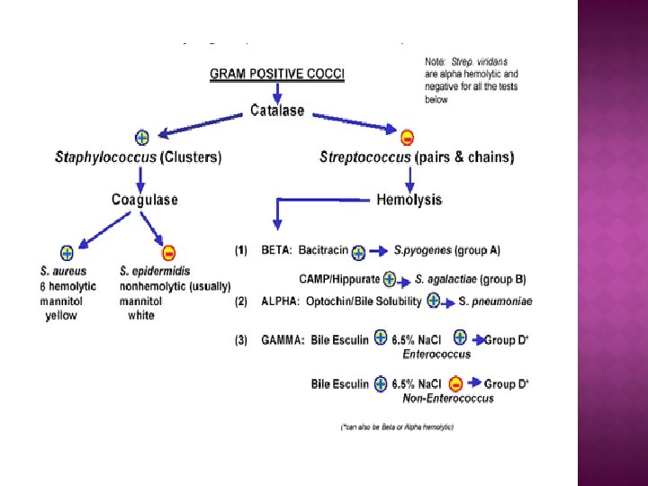

Identification of streptococci by hemolytic reaction Colonies are surrounded by clear zone of hemolysis complete hemolysis Colonies are surrounded by partial hemolysis with greenish color Beta-hemolytic Streptococcus colonies St. pyogenes Alpha-hemolytic Streptococcus colonies St. pneumoniae No haemolysis Gamma-hemolytic Streptococcus colonies Enterococcus faecalis

Identification of streptococci by hemolytic reaction Beta-hemolytic Streptococcus colonies Alpha-hemolytic Streptococcus colonies Gamma-hemolytic Streptococcus colonies

This is a blood agar growing beta hemolytic streptococci.

This culture was grown from a sputum specimen of a 60 year old man complaining of cough, fever and chest pain. α hemolytic streptococci on blood agar

GRAM NEGATIVE BACTERIA
 Lactose fermenting colonies E. coli non-lactose fermenting colonies salmonella")
Mac. Conkey's agar (DEFERENTIAL MEDIUM) Lactose fermenting colonies E. coli non-lactose fermenting colonies salmonella

Biochemical testings To confirm the identity of bacteria. Antibiotic susceptibility testings

Automated instrument for identification and susceptibility testings

VIROLOGY

VIRAL STRUCTURE Helical Virus Icosahedral Virus

VIRAL CLASSIFICATION

VIRAL ELECTRON MICROGRAPHS Herpes virus Adenovirus Rabies virus Influenza Viruses

Herpes simplex virus -1 : Herpesviridae Enveloped virus Icosahedral capsid d. s DNA genome Loose envelope

These are electron micrographs of a virus Q 1: Name this virus Q 2: Describe its structure. Herpes virus Enveloped virus , Icosahedral capsid, d. s DNA genome

Adenovirus : Adenoviridae Nonenveloped virus Icosahedral capsid d. s DNA genome Only V with fiber

This is an electron micrograph of a virus Q 1: Name this virus Q 2: Describe its structure. Adenovirus Nonenveloped virus, with fiber Icosahedral capsid & d. s DNA genome

Rabies virus: Rhabdoviridae Enveloped virus Helical capsid s. s RNA genome Bullet shape

This is an electron micrograph of a virus Q 1: Name this virus Q 2: Describe its structure. Rabies virus Enveloped virus , Helical capsid & s. s RNA genome

Influenza Viruses : Orthomyxoviridae Enveloped V & spikes Helical capsid Segmented s. s RNA Pleomorphic shape

This is an electron micrograph of a virus Q 1: Name this virus Influenza Viruses Q 2: Describe its structure Enveloped Virus with spikes , Helical capsid , Segmented s. s RNA

PARASITOLOG Y

Classification of Parasites Protozoa Unicellular Single cell for all function Amoebae: move by psudobodia. Flagellates: move by flagella. Ciliates : move by cilia Apicomplexa (sporozoa) Tissue parasites Helminths Mulicellular Specialized cells Round worms (Nematodes) cylindrical, unsegmented Flat worms 1 -Trematodes: leaf-like, unsegmented. 2 -Cestodes: tape-like, segmented

Giardia lamblia trophozoite Two nuclei, each with central karyosome Four pairs of flagella

Giardia lamblia cyst • Mature, infective cyst, containing 4 nuclei • Note a straight axoneme running longitudinally

Following is the microphotograph of an organism found in the upper part of the small intestine. Name the Organism Giardia lamblia What is the Stage? Trophozoite stage

Following is the microphotograph of an organism found in stools Name the Organism Giardia lamblia What is the Stage? Cyst stage

Classification of Parasites Protozoa Unicellular Single cell for all function Amoebae: move by psudobodia. Flagellates: move by flagella. Ciliates : move by cilia Apicomplexa (sporozoa) Tissue parasites Helminths Mulicellular Specialized cells Round worms (Nematodes) cylindrical, unsegmented Flat worms 1 -Trematodes: leaf-like, unsegmented. 2 -Cestodes: tape-like, segmented
 Ascaris adult")
Nematodes Ascaris lumbricoides (roundworm) Ascaris adult
")
Cestodes Taenia saginata (tapeworm)
")
Trematodes Fasciola hepatica (Flukes)

 , Lice (pleural) Pediculus humanus")
LICE Louse(singular) , Lice (pleural) Pediculus humanus
")
Phlebotomus ( sand fly)

Mosquitoes :

MYCOLOGY

Fungi can be divided to two types based on morphology A B Based on morphology, name the two fungal structures in A and B? A: Yeast e. g. Candida B: Mould fungi e. g. Aspergillus

Microscopic appearance of yeast and mould fungi A B Name the two fungal structures in A and B? A: Budding yeast cells e. g. Candida B: Branching Fungal hyphae e. g. Aspergillus

B
- Slides: 64