Genetic Disorders Etiolgy of Mental Retardation Syndromes with


- Slides: 52
Genetic Disorders Etiolgy of Mental Retardation Syndromes with Mental Retardation
Mental Retardation Mental retardation is a particular state of functioning that begins in childhood and is characterized by limitation in both intelligence and adaptive skills (daily living, communication, social).
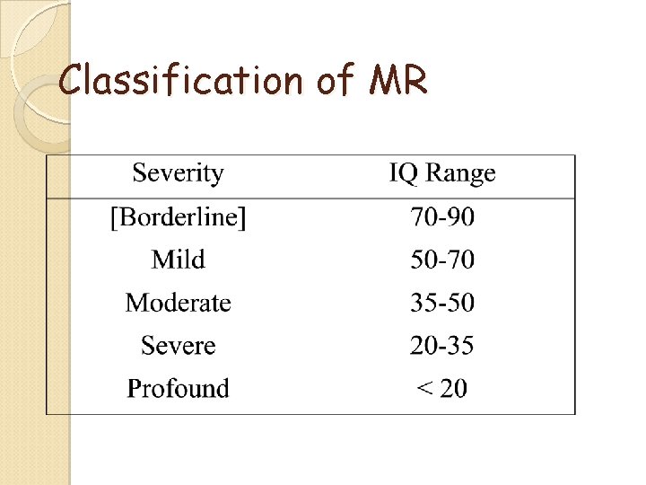
Classification of MR
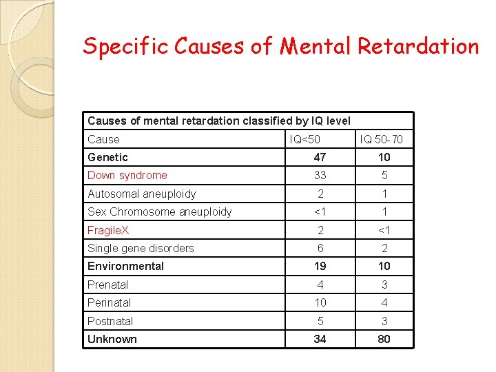
Specific Causes of Mental Retardation Causes of mental retardation classified by IQ level Cause IQ<50 IQ 50 -70 Genetic 47 10 Down syndrome 33 5 Autosomal aneuploidy 2 1 Sex Chromosome aneuploidy <1 1 Fragile. X 2 <1 Single gene disorders 6 2 Environmental 19 10 Prenatal 4 3 Perinatal 10 4 Postnatal 5 3 Unknown 34 80
The Diagnostic Process: Clinical Evaluation Clinical history, prenatal and birth ◦ birth measurements extremely important Family pedigree ◦ 3 -generation, learning problems, psychiatric disorders, autism, mental retardation Physical and neurological examination ◦ assess minor anomalies, growth, development
Major categories of genetic disorders include; ◦ Chromosomal -Aneuploidy, Deletions, Duplications/Insertions ◦ Single Gene -Autosomal Dominant, Recessive, X-linked -Nonmedelian inheritance ( Triplet repeat disorders, Imprinting, Mitochondrial ) ◦ Multifactorial – central nervous system abnormalities
Diagnostic Tests Chromosome analysis Molecular analysis
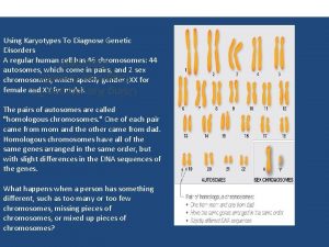
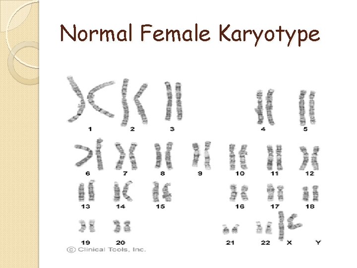
Normal Female Karyotype
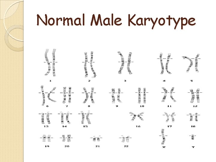
Normal Male Karyotype
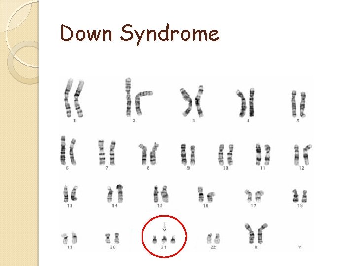
Down Syndrome
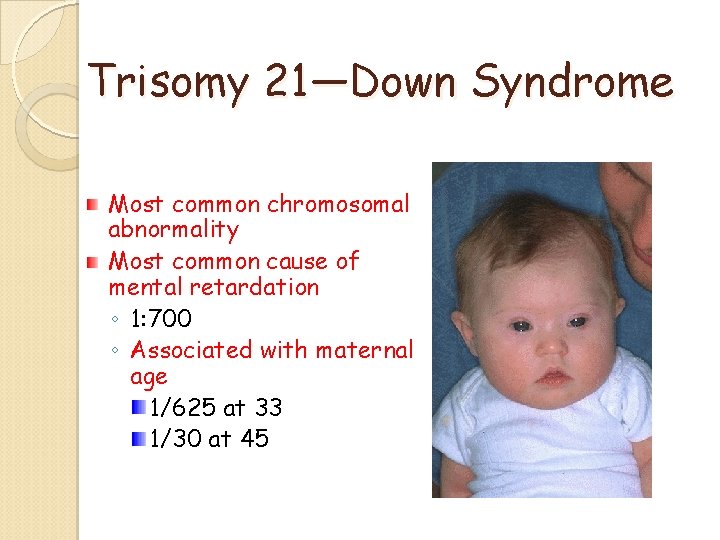
Trisomy 21—Down Syndrome Most common chromosomal abnormality Most common cause of mental retardation ◦ 1: 700 ◦ Associated with maternal age 1/625 at 33 1/30 at 45
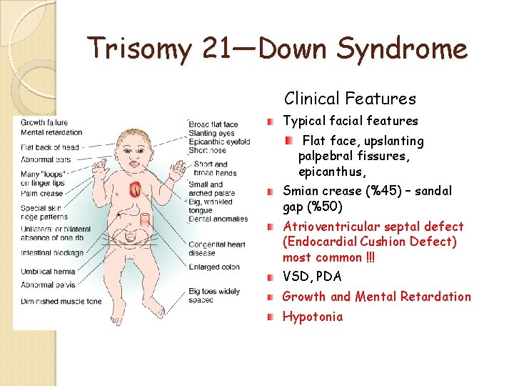
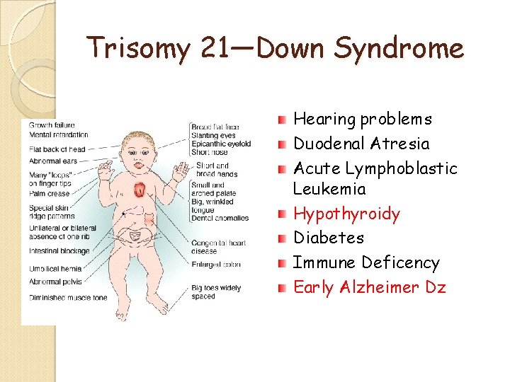
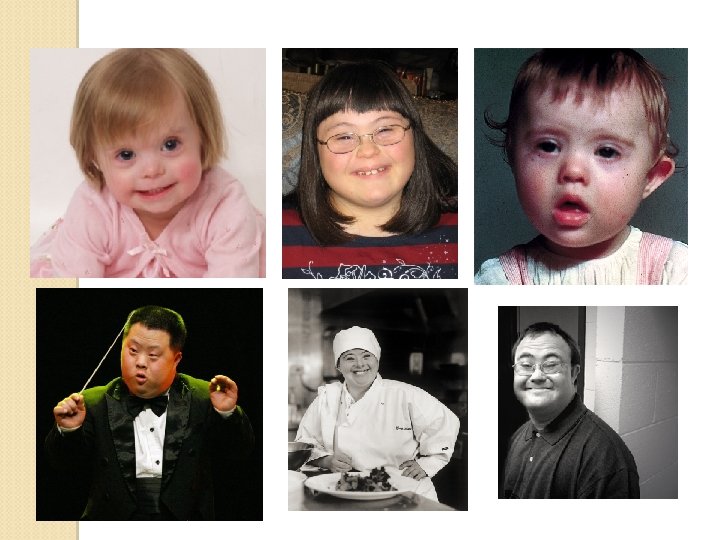
Trisomy 21—Down Syndrome Clinical Features Typical facial features Flat face, upslanting palpebral fissures, epicanthus, Smian crease (%45) – sandal gap (%50) Atrioventricular septal defect (Endocardial Cushion Defect) most common !!! VSD, PDA Growth and Mental Retardation Hypotonia
Trisomy 21—Down Syndrome Hearing problems Duodenal Atresia Acute Lymphoblastic Leukemia Hypothyroidy Diabetes Immune Deficency Early Alzheimer Dz
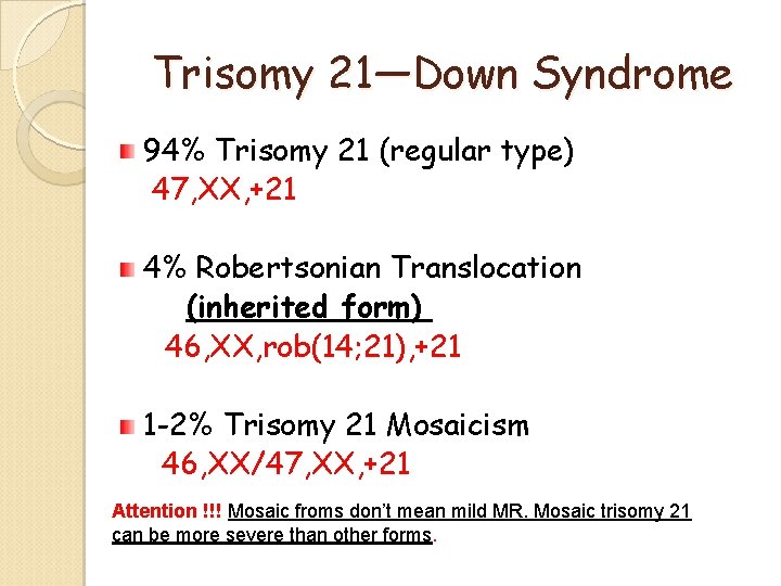
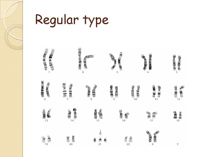
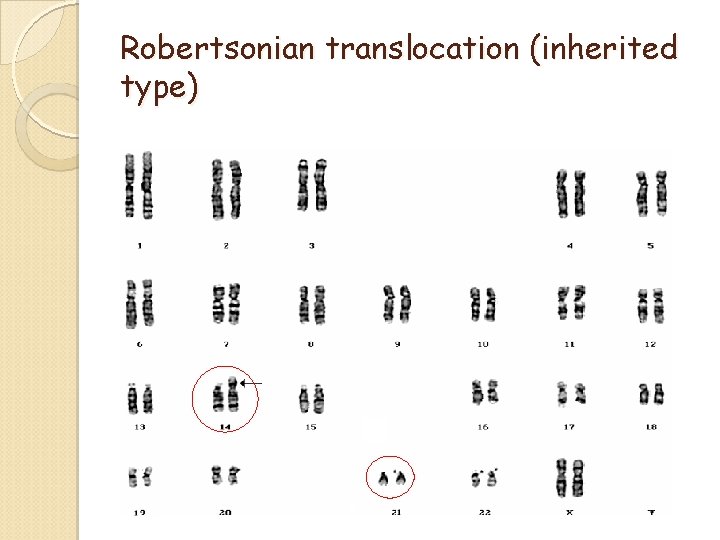
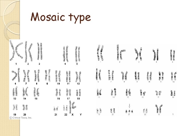
Trisomy 21—Down Syndrome 94% Trisomy 21 (regular type) 47, XX, +21 4% Robertsonian Translocation (inherited form) 46, XX, rob(14; 21), +21 1 -2% Trisomy 21 Mosaicism 46, XX/47, XX, +21 Attention !!! Mosaic froms don’t mean mild MR. Mosaic trisomy 21 can be more severe than other forms.
Regular type
Robertsonian translocation (inherited type)
Mosaic type
Trisomy 21—Down Syndrome can be diagnosed 95% at clinical level. Karyotype analysis is important for genetic counseling of recurrence risk. Recurrence risks for regular type and mosaic forms are lower than 1 %. Recurrence risk for Down syndrome due to robertsonian translocation is much higher. (10 -15 %)
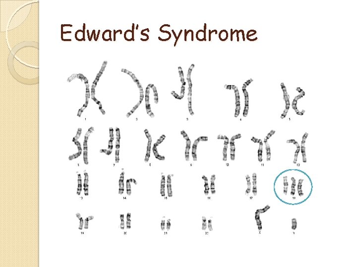
Edward’s Syndrome
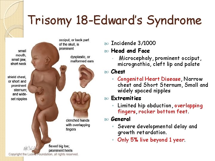
Trisomy 18 -Edward’s Syndrome Incidende 3/1000 Head and Face ◦ Microcephaly, prominent occiput, micrognathia, cleft lip and palate Chest ◦ Congenital Heart Disease, Narrow chest and Short Sternum, Small and widely spaced nipples Extremities ◦ Limited hip abduction, overlapping fingers, rocker bottom feet. General ◦ Severe developmental delay and growth retardation. ◦ Only 5% live beyond 1 year.
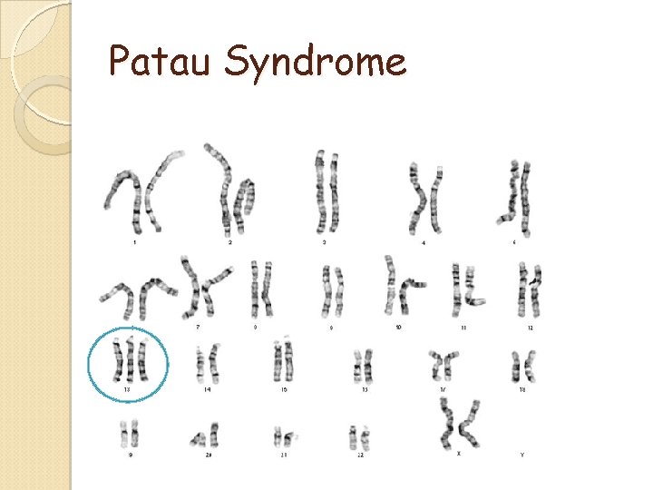
Patau Syndrome
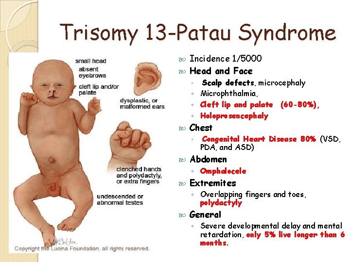
Trisomy 13 -Patau Syndrome Incidence 1/5000 Head and Face Scalp defects, microcephaly ◦ Microphthalmia, ◦ Cleft lip and palate (60 -80%), ◦ Holoprosencephaly ◦ Chest ◦ Congenital Heart Disease 80% (VSD, PDA, and ASD) Abdomen ◦ Omphalocele Extremites ◦ Overlapping fingers and toes, polydactyly General ◦ Severe developmental delay and mental retardation, only 5% live longer than 6 months.
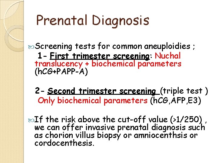
Prenatal Diagnosis Screening tests for common aneuploidies ; 1 - First trimester screening: Nuchal translucency + biochemical parameters (h. CG+PAPP-A) 2 - Second trimester screening (triple test ) Only biochemical parameters (h. CG, AFP, E 3) If the risk above the cut-off value (>1/250) , we can offer invasive prenatal diagnosis such as chorion villus biopsy or amniocenthsis or cordocenthesis.
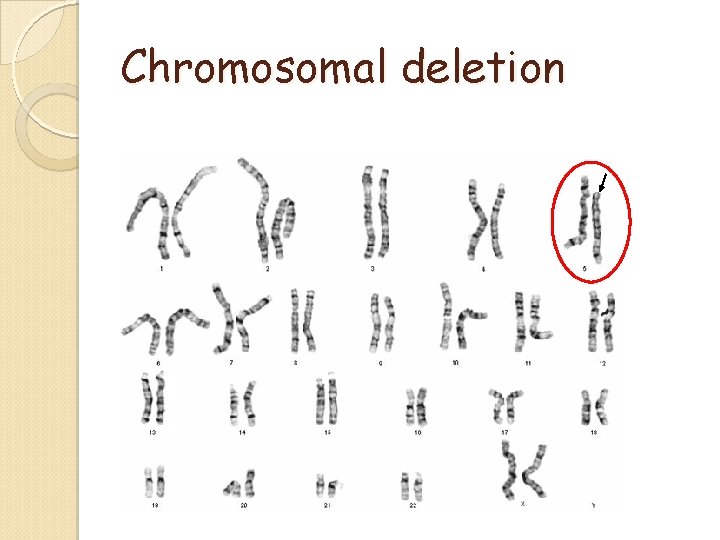
Chromosomal deletion
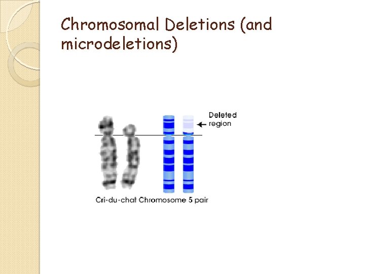
Chromosomal Deletions (and microdeletions)
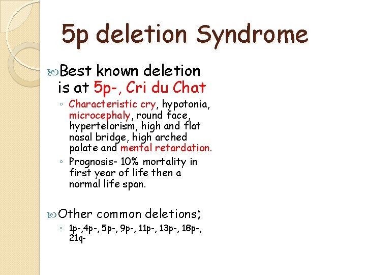
5 p deletion Syndrome Best known deletion is at 5 p-, Cri du Chat ◦ Characteristic cry, hypotonia, microcephaly, round face, hypertelorism, high and flat nasal bridge, high arched palate and mental retardation. ◦ Prognosis- 10% mortality in first year of life then a normal life span. Other common deletions; ◦ 1 p-, 4 p-, 5 p-, 9 p-, 11 p-, 13 p-, 18 p-, 21 q-
Chromosomal Microdeletions are small chromosomal deletions which can not be detected by convetional karyotype analysis. (only high resolution banding procedures or FISH) These deletions produce syndromes that are usually clinically recognizible.
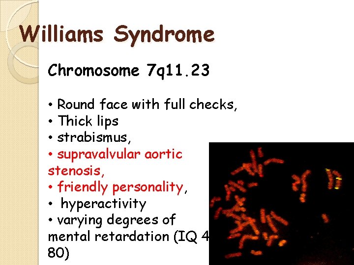
Williams Syndrome Chromosome 7 q 11. 23 • Round face with full checks, • Thick lips • strabismus, • supravalvular aortic stenosis, • friendly personality, • hyperactivity • varying degrees of mental retardation (IQ 4180)
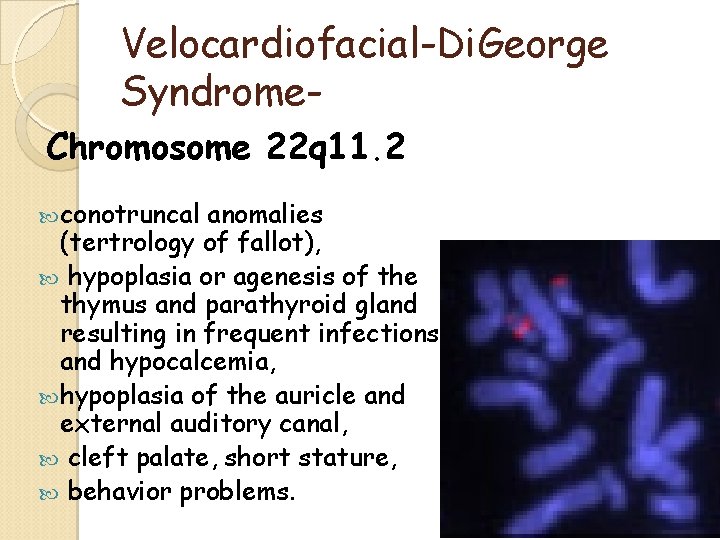
Velocardiofacial-Di. George Syndrome. Chromosome 22 q 11. 2 conotruncal anomalies (tertrology of fallot), hypoplasia or agenesis of the thymus and parathyroid gland resulting in frequent infections and hypocalcemia, hypoplasia of the auricle and external auditory canal, cleft palate, short stature, behavior problems.
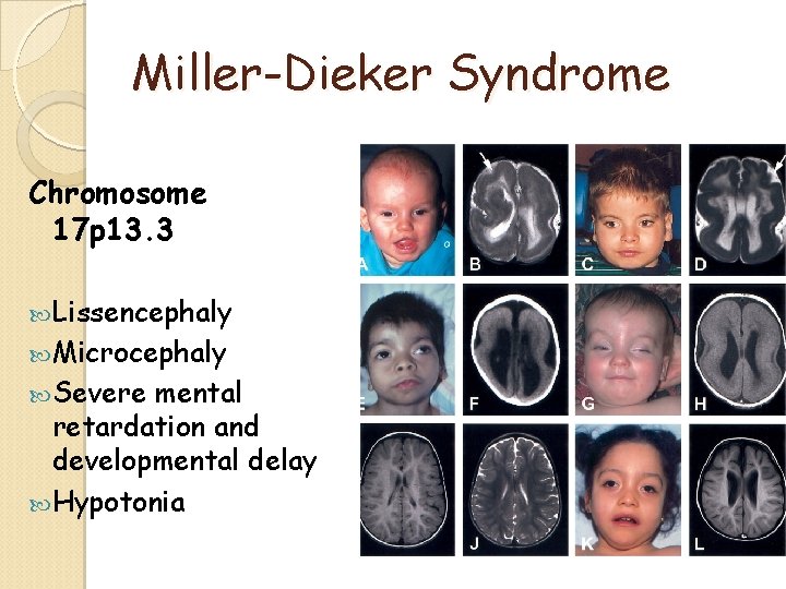
Miller-Dieker Syndrome Chromosome 17 p 13. 3 Lissencephaly Microcephaly Severe mental retardation and developmental delay Hypotonia
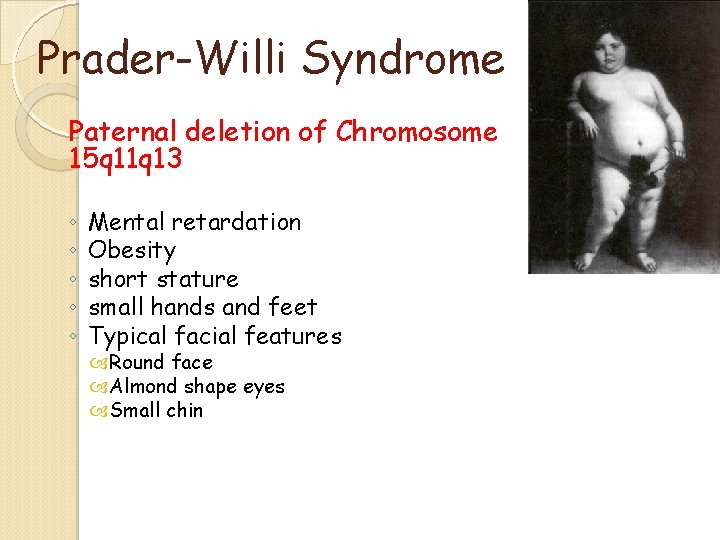
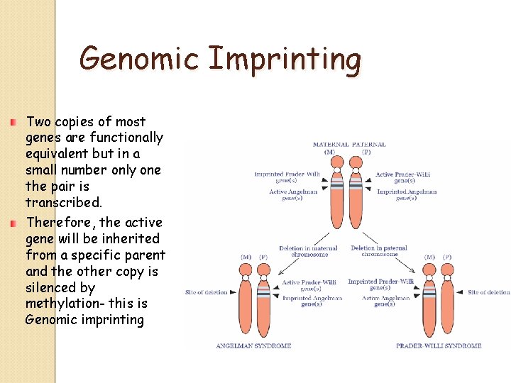
Prader-Willi Syndrome Paternal deletion of Chromosome 15 q 11 q 13 ◦ ◦ ◦ Mental retardation Obesity short stature small hands and feet Typical facial features Round face Almond shape eyes Small chin
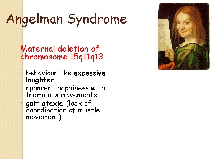
Angelman Syndrome Maternal deletion of chromosome 15 q 11 q 13 ◦ behaviour like excessive laughter, ◦ apparent happiness with tremulous movements ◦ gait ataxia (lack of coordination of muscle movement)
Genomic Imprinting Two copies of most genes are functionally equivalent but in a small number only one the pair is transcribed. Therefore, the active gene will be inherited from a specific parent and the other copy is silenced by methylation- this is Genomic imprinting
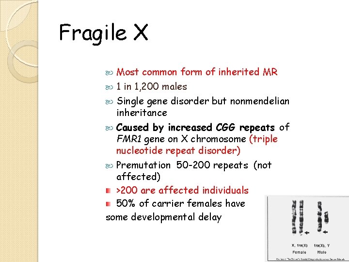
Fragile X Most common form of inherited MR 1 in 1, 200 males Single gene disorder but nonmendelian inheritance Caused by increased CGG repeats of FMR 1 gene on X chromosome (triple nucleotide repeat disorder) Premutation 50 -200 repeats (not affected) >200 are affected individuals 50% of carrier females have some developmental delay
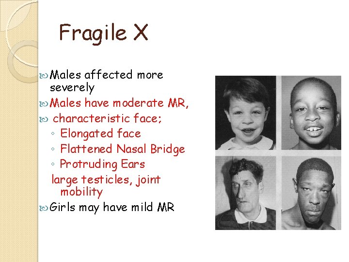
Fragile X Males affected more severely Males have moderate MR, characteristic face; ◦ Elongated face ◦ Flattened Nasal Bridge ◦ Protruding Ears large testicles, joint mobility Girls may have mild MR
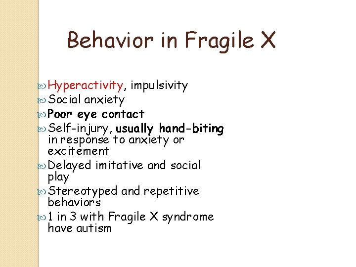
Behavior in Fragile X Hyperactivity, impulsivity Social anxiety Poor eye contact Self-injury, usually hand-biting in response to anxiety or excitement Delayed imitative and social play Stereotyped and repetitive behaviors 1 in 3 with Fragile X syndrome have autism
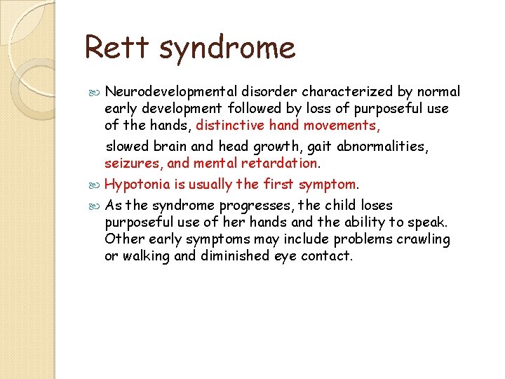
Rett syndrome Neurodevelopmental disorder characterized by normal early development followed by loss of purposeful use of the hands, distinctive hand movements, slowed brain and head growth, gait abnormalities, seizures, and mental retardation. Hypotonia is usually the first symptom. As the syndrome progresses, the child loses purposeful use of her hands and the ability to speak. Other early symptoms may include problems crawling or walking and diminished eye contact.
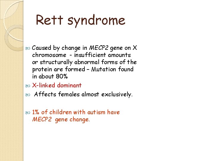
Rett syndrome Caused by change in MECP 2 gene on X chromosome - insufficient amounts or structurally abnormal forms of the protein are formed – Mutation found in about 80% X-linked dominant Affects females almost exclusively. 1% of children with autism have MECP 2 gene change.
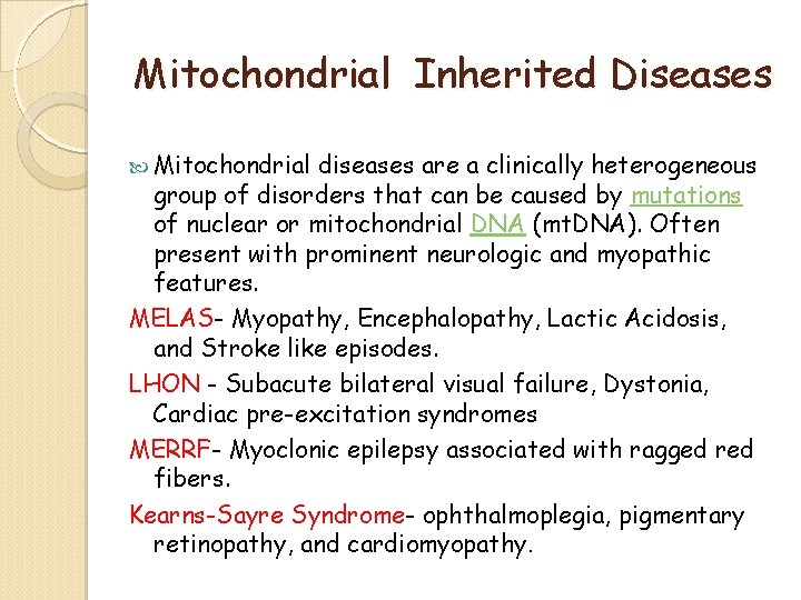
Mitochondrial Inherited Diseases Mitochondrial diseases are a clinically heterogeneous group of disorders that can be caused by mutations of nuclear or mitochondrial DNA (mt. DNA). Often present with prominent neurologic and myopathic features. MELAS- Myopathy, Encephalopathy, Lactic Acidosis, and Stroke like episodes. LHON - Subacute bilateral visual failure, Dystonia, Cardiac pre-excitation syndromes MERRF- Myoclonic epilepsy associated with ragged red fibers. Kearns-Sayre Syndrome- ophthalmoplegia, pigmentary retinopathy, and cardiomyopathy.
Neurocutaneous Syndromes Familial/ primitive ectoderm All AUTOSOMAL DOMINANT ◦ ◦ ◦ ◦ Neurofibromatosis I/II Tuberous Sclerosis Sturge-Weber Von Hippel-Lindau Ataxia Telengiectesia Linear Nevus Syndrome Hypomelanosis of Ito Incontinentia Pigmenti
Environmental factors Infectious Radiation Chemical agents Agents Hormones Maternal Disease Nutritional Deficiencies Hypoxia
Infectious Agents Rubella ◦ Malformations of the eye Cataract (6 th week) Microphthalmia ◦ Malformations of the ear (9 th week) Congenital deafness Due to destruction of cochlea ◦ Malformations of the heart (5 th -10 th week) Patent ductus arteriosis Atrial septal defects Ventricular septal defects ◦ May be responsible for some brain abnormalities Mental retardation ◦ Intrauterine growth retardation ◦ Myocardial damage ◦ Vascular abnormalites ◦ Incidence 47%- during 1 st four weeks 22% - 5 th – 8 th weeks 13% - 9 th – 16 th week
Infectious Agents Cytomegalovirus ◦ Malformations Microcephaly ◦ Cerebral calcifications ◦ Blindness Chorioretinitis ◦ Kernicterus (a form of jaundice) ◦ multiple petechiae of skin ◦ Hepatosplenomegaly ◦ Mother asymptomatic
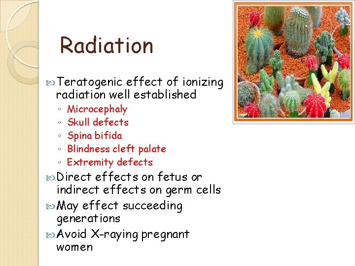
Radiation Teratogenic effect of ionizing radiation well established ◦ ◦ ◦ Microcephaly Skull defects Spina bifida Blindness cleft palate Extremity defects Direct effects on fetus or indirect effects on germ cells May effect succeeding generations Avoid X-raying pregnant women
Drugs Thalidomide ◦ Antinauseant & sleeping pill ◦ Found to cause amelia & meromelia Total or partial absence of the extremities ◦ Intestinal atresia ◦ Cardiac abnormalities ◦ Many women had taken thalidomide early in pregnancy (in Germany in 1961) Anticonvulsants ◦ Diphenylhydantoin (phenytoin) Craniofacial defects Nail & digital hypoplasia Growth abnormalities Mental retardarion The above pattern is know as “fetal hydantoin syndrome” ◦ Valproic acid Neural tube defects Heart defects Craniofacial & limb anomalies
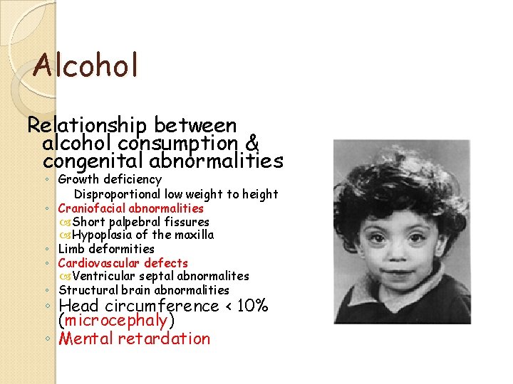
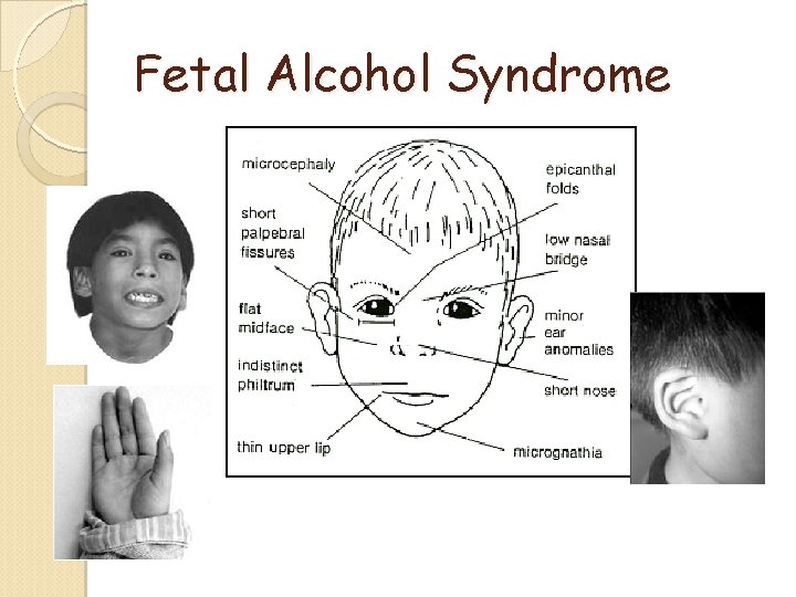
Alcohol Relationship between alcohol consumption & congenital abnormalities ◦ Growth deficiency Disproportional low weight to height ◦ Craniofacial abnormalities Short palpebral fissures Hypoplasia of the maxilla ◦ Limb deformities ◦ Cardiovascular defects Ventricular septal abnormalites ◦ Structural brain abnormalities ◦ Head circumference < 10% (microcephaly) ◦ Mental retardation
Fetal Alcohol Syndrome
Cigarette Smoking Has not been linked to major birth defects ◦ Smoking does contribute to intrauterine growth retardation & premature delivery ◦ Some evidence that is causes behavioral disturbances
Maternal Disease Maternal Phenylketonuria (PKU) ◦ Enzyme phenylalanine hydroxylase is deficient phenylalanine (PA) concentrations Developmental delay and Mental retardation Microcephaly Congenital heart disease Facial dismorphism ◦ Risk can be with low PA diet