Formation of Urine Filtration Each nephron has its


- Slides: 19
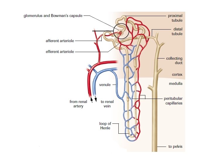
Formation of Urine
Filtration • Each nephron has its own individual blood supply • The afferent arteriole carries blood to the glomerulus; a highpressure filter Capillary bed = 25 mm Hg Glomerulus = 65 mm Hg
Urine Formation The formation of urine occurs as the result of three processes. 1. Filtration- the movement of fluids from the blood into the Bowman’s capsule 2. Reabsorption – the transfer of essential solutes and water from the nephron back into the blood 3. Secretion – the movement of materials from the blood back into the nephron
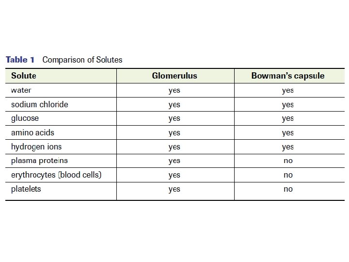
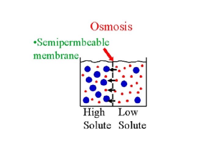
Solutes • Dissolved solutes pass through the walls of the glomerulus into the Bowman’s capsule (from an area of high concentration to an area of low concentration) • Not all materials enter the Bowman’s capsule
Reabsorption • The concentration of the fluids change as they move through the kidneys • 600 m. L of fluid flows through the kidneys every minute • 20% of this volume is filtered into the nephron (120 m. L) • If none of this liquid was reabsorbed by the body, you would form 120 m. L of urine each minute! • Luckily for us, only 1 m. L of urine is formed for every 120 m. L of fluid filtered into the nephron
• Where does the rest of the 119 m. L volume go? • It is (selectively) reabsorbed!
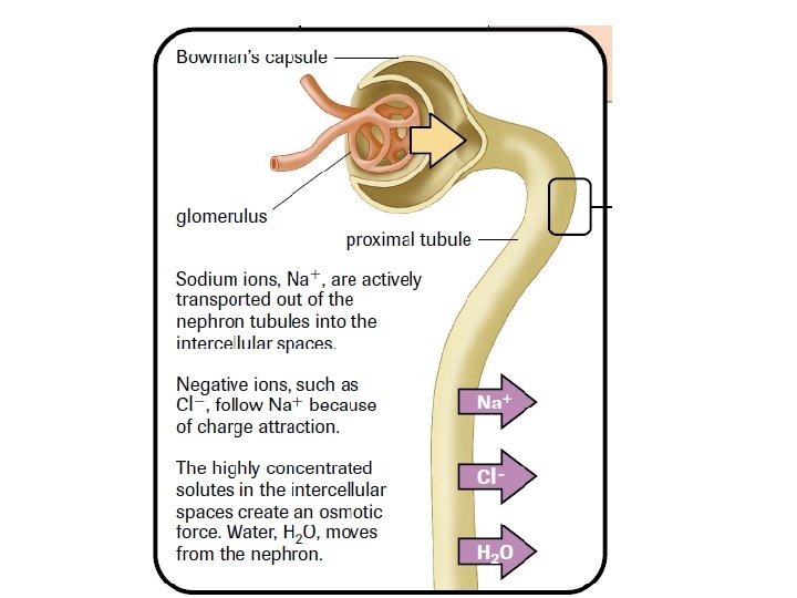
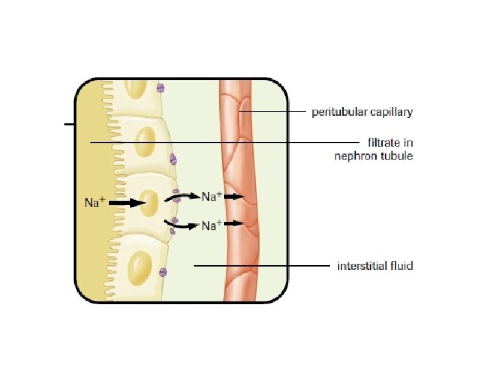
Selective Reabsorption • Occurs by both active and passive transport • Mitochondria provide energy for the active transport • Carrier molecules move Na+ ions across the cell membranes of the cells that line the nephron • Negative ions, such as Cl- and HCO 3 - follow positive Na+ ions because of charge attraction
• Other molecules are actively transported from the proximal tubule into the surrounding capillaries • Glucose and amino acids attach to specific carrier molecules which shuttle them out of the nephron
Threshold Level - The maximum amount of material that can be moved across the nephron • Once threshold level has been reached, the excess solute is excreted in the urine
Osmotic Gradient • As solutes move out of the nephron into the capillaries, an osmotic gradient is created • This means that there is a greater concentration of solutes on the outside of the nephron compared to the inside • Water moves out of the nephron by osmosis (high [ ] to low [ ])
• A Second osmotic force is created by the proteins that still remain in the blood stream • Recall: these proteins are too large to be filtered into the Bowman’s capsule • This helps draw water from the interstitial fluid into the blood Interstitial fluid – fluid that surrounds the body’s cells
• As water is reabsorbed and removed from the nephron, the remaining solutes in the tubules become more concentrated
Secretion – is the movement of wastes from the blood into the nephron • Nitrogen-containing wastes, excess H+ ions, and minerals such as K+ ions are examples of substances secreted • Secretion occurs by active transport