Urine Formation by the Kidneys I Glomerular Filtration

Urine Formation by the Kidneys: I. Glomerular Filtration, Renal Blood Flow and Their Control Yanal A Shafagoj. MD, Ph. D 1

Body fluid regulation. 2

Summary of Kidney Functions • Excretion of metabolic waste products urea, creatinine, bilirubin, hydrogen • Excretion of foreign chemicals: drugs, toxins, pesticides, food additives • Secretion, metabolism, and excretion of hormones - renal erythropoetic factor (erythropoietin) - 1, 25 dihydroxycholecalciferol (Vitamin D) - renin Hormones metabolized and excreted by the kidney - Most peptide hormones (e. g. insulin, angiotensin II, etc • Regulation of acid-base balance • Gluconeogenesis: glucose synthesis from amino acids • Control of arterial pressure • Regulation of water & electrolyte excretion 3
 • Uric acid (from")
Excretion of Metabolic Waste Products • Urea (from protein metabolism) • Uric acid (from nucleic acid metabolism) • Creatinine (from muscle metabolism) • Bilirubin (from hemoglobin metabolism) 4

Excretion of Foreign Chemicals • Pesticides • Food additives • Toxins • Drugs 5

Regulation of Erythrocyte Production O 2 Delivery Kidney Erythropoetin Erythrocyte Production in Bone Marrow 6

Regulation of Vitamin D Activity • Kidney produces active form of vitamin D (1, 25 dihydroxy vitamin D 3 ) • Vitamin D 3 is important in calcium and phosphate metabolism 7

Regulation of Acid-Base Balance • Excrete acids (kidneys are the only means of excreting non-volatile acids) • Regulate body fluid buffers (e. g. Bicarbonate) 8
 during")
Glucose Synthesis Gluconeogenesis: kidneys synthesize glucose from precursors (e. g. , amino acids) during prolonged fasting 9

Regulation of Arterial Pressure Endocrine Organ • renin-angiotensin system • prostaglandins • kallikrein-kinin system Control of Extracellular Fluid Volume 10

Regulation of Water and Electrolyte Balances • Sodium and Water • Potassium • Hydrogen Ions • Calcium, Phosphate, Magnesium 11

Effect of increasing sodium intake 10 -fold on urinary sodium excretion and extracellular fluid volume 12

Organs of the urinary system University of Jordan 13

Internal anatomy of the kidneys University of Jordan 14

Blood supply of the kidneys University of Jordan 15

Functions of Nephron Structures n Afferent Arteriole – Transports arterial blood to the glomerulus for filtration

Functions of Nephron Structures n Efferent Arteriole – Transports filtered blood from the glomerulus , through the peritubular capillaries and the vasa recta, and to the kidney venous system

Functions of Nephron Structures n Glomerulus – The site for blood filtration – operates as a nonspecific filter; in that, it will remove both useful and nonuseful material – the product of the glomerulus is called filtrate

Functions of Nephron Structures n Bowman’s Capsule – A sac that encloses Bowman’s Capsule and transfers filtrate from the glomerulus to the Proximal Convoluted Tubule (PCT)
 – A thick, constantly actively")
Functions of Nephron Structures n Proximal Convoluted Tubule (PCT) – A thick, constantly actively segment of the nephron that reabsorbs most of the useful substances of the filtrate: sodium (65%), water (65%), bicarbonate (90%), chloride (50%), glucose (nearly 100%!), etc. – The primary site for secretion (elimination) of drugs, waste and hydrogen ions

Functions of Nephron Structures n Descending Limb of the Loop of Henle – A part of the counter current multiplier – freely permeable to water and relatively impermeable to solutes (salt particles) – receives filtrate from the PCT, allows water to be absorbed and sends “salty” filtrate on the next segment. “Saves water and passes the salt”

Functions of Nephron Structures n Ascending Limb of the Loop of Henle – part of the counter current multiplier – impermeable to water and actively transports (reabsorbs) salt (Na. Cl) to the interstitial fluid of the pyramids in the medulla. “Saves salt and passes the water. ” – the passing filtrate becomes dilute and the interstitium becomes hyperosmotic
 – receives dilute fluid from")
Functions of Nephron Structures n Distal Convoluted Tubule (DCT) – receives dilute fluid from the ascending limb of the Loop of Henle – Variably active portion of the nephron – When aldosterone hormone is present, sodium is reabsorbed and potassium is secreted. Water and chloride follow the sodium.

Functions of Nephron Structures n Collecting Duct – receives fluid from the DCT – variably active portion of the Nephron – when antidiuretic hormone (ADH) is present, this duct will become porous to water. Water from the collecting duct fluid then moves by osmosis into the “salty” (hyperosmotic) interstitium of the medulla. – The last segment to save water for the body

Functions of Nephron Structures Peritubular Capillaries – transport reabsorbed materials from the PCT and DCT into kidney veins and eventually back into the general circulation – help complete the conservation process (reabsorption) that takes place in the kidney

Path of Blood Through Kidney n Renal artery n interlobar arteries (up to renal columns, between lobes) arcuate arteries (over pyramids) interlobular arteries (up into cortex) afferent arterioles glomerulus (cluster of capillaries) efferent arterioles (near medulla vasa recta) peritubular capillaries interlobular veins arcuate veins interlobar veins Renal vein

n n The kidney weighs 113 -170 gm. Renal artery arises as the fifth branch of the abdominal aorta. The renal artery arises from the aorta at the level of the second lumber vertebra. Because the aorta is to the left of the midline, the right renal artery is longer. The inferior vena cava lies to the right midline making the left renal vein two times longer than the right renal vein. For this reason it is better to take the donor left kidney (short artery, long vein) & place it in the right pelvis of the recipient. Multiple arteries & veins can supply the kidney. 27

Nephron Tubular Segments 28

Cortical and juxtamedullary nephrons 29

Basic Mechanisms of Urine Formation 30

Structures and functions of a nephron Renal tubule and collecting duct. Epithelium to modify the ultrafiltrate Renal corpuscle Ultrafiltration device Afferent arteriole Glomerular capsule Urine (contains excreted substances) Fluid in renal tubule 1 Filtration from blood plasma into nephron 2 Tubular reabsorption from fluid into blood Efferent arteriole Peritubular capillaries University of Jordan 3 Tubular secretion from blood into fluid Blood (contains reabsorbed substances) 31

Functional Anatomy of the Kidney Structure & function of the kidney are closely matched. The kidney is a combination of: 1. Ultrafiltation device (the glomerular apparatus). 2. Epithelium (tubules), which modifies the ultrafiltarte by: - addition (secretion) or - removal (reabsorption). n

n n n Filtration in kidney is also affected, as systemic capillaries, by Starling forces (Hydrostatic & Osmotic pressure in & out… 4 forces). Bowman’s capsule stands for the interstitium To be discussed with regulation of GFR

RBF n n Kidneys are reconditioning organs i. e. Receive too much blood. RBF= 20 -25% of Q →→ This makes the A-V oxygen difference small (1. 4 ml/dl). Nevertheless, kidneys consume twice O 2/per gm tissue as brain. This O 2 consumption is directly related to Na+ reabsorption. If GFR is high → Na+ reabsorption is high→O 2 consumption is high. When GFR is severely depressed (Acute RF) → decrease need for O 2 …. nothing to reabsorb If RBF decreases →→acute renal Failure

Renal Function n Renal Plasma Flow = ~650 cc / min total n Renal Cortex Outer Medulla Inner Medulla n n = ~500 cc / min = ~125 cc / min = ~25 cc / min
 Heart 0. 8 A-V difference Vol% 11 Brain 0. 5")
Tissue Blood flow (ml/g/min) Heart 0. 8 A-V difference Vol% 11 Brain 0. 5 6. 2 Sk muscles 0. 03 6 Liver 0. 6 3. 4 Kidney 4. 2 1. 4 Carotid bodies 20 0. 5
 : n n RPF : how much")
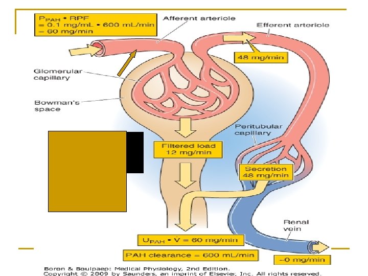
How to measure Renal Plasma Flow (RPF) : n n RPF : how much plasma enter both kidneys per minute. We use a substance X that is completely removed (cleaned) from the blood once it reaches the kidneys: i. e. Renal vein concentration of X = 0 i. e; once comes to the peritubular capillaries is completely secreted. The substance used commonly here is the PAH (para-aminohippuric acid. )

Use of Clearance to Estimate Renal Theoretically, if a substance is completely cleared Plasma Flow from the plasma, its clearance rate would equal renal plasma flow Cx = renal plasma flow
 is freely")
Use of PAH Clearance to Estimate Renal Plasma Flow Paraminohippuric acid (PAH) is freely filtered and completely secreted and thus is almost completely cleared from the renal plasma 1. amount enter kidney = RPF x PPAH 2. amount entered ≈ amount excreted 3. ERPF x Ppah ERPF = = UPAH x V PPAH ERPF = Clearance PAH ~ 10 % PAH remains

To calculate actual RPF , one must correct for incomplete extraction of PAH APAH =1. 0 EPAH = APAH - VPAH APAH = 1. 0 – 0. 1 = 0. 9 1. 0 normally, EPAH = 0. 9 i. e PAH is 90 % extracted RPF = ERPF EPAH VPAH = 0. 1

Calculation of Tubular Reabsorption = Filtration -Excretion Filt s = GFR x Ps Excret s = Us x V

Calculation of Tubular Secretion = Excretion - Filtration Filt s = GFR x Ps VPAH = 0. 1 Excret s = Us x V

Question The maximum possible clearance rate of a substance that is completely cleared from the plasma by the kidneys would be equal to 1. glomerular filtration rate 2. the filtered load of the substance 3. urine excretion rate of the substance 4. renal plasma flow 5. none of the above

Use of Clearance to Estimate Renal Plasma Flow Theoretically, if a substance is completely cleared from the plasma, its clearance rate would equal renal plasma flow Cx = renal plasma flow

Clearances of Different Substances Substance Clearance (ml/min inulin 125 PAH 585 glucose 0 sodium 0. 9 urea 70 Clearance of inulin (Cin) = GFR if Cx < Cin : indicates reabsorption of x if Cx > Cin : indicates secretion of x Clearance creatinine (Ccreat) ~ 140 (used to estimate GFR) Clearance of PAH (Cpah) ~ effective renal plasma flow

Measurement of RPF: CPAH underestimate the RPF. The CPAH is the effective RPF. The true RPF is 10% higher. 10% of RBF goes to the medulla, hilum, pelvis etc. p-aminohippuric acid PAH is an exogenous organic anion and not produced in our body. Freely filtered, not reabsorbed, completely secreted. In the kidney it is not metabolized, accumulated, or produced. Tm = 80 mg/min Extraction ratio of PAH 46
. How")
RPF n n n 650 ml is the RPF. 125 is filtered (GFR). How much is the filtration fraction? . . . ≈ 20% 525 leave through efferent arteriole and go to peritubular capillaries. 1 ml/min is the urine output. Renal vein carries 649 ml/min

Example n n n n NOTE : RPF is not a routine clinical test. So. . if we inject a certain amount of PAH in blood to achieve certain blood concentration. If RPF is 650 ml/min. And urine output is 1 ml/min Then, the concentration of PAH will increase 650 times in the urine. But actually this is not the case. Because 10 % of renal blood goes to nourish the kidney i. e. don’t participate in the renal function…. don't reach the glomeruli So 585 ml/min (90%) is the effective RPF and 650 ml/min (100%) is called true or total RPF. True RBF = effective RBF 0. 9.

PAH CURVE for FILTRATION and SECRETION : n n n n Filtration is passive. Secretion is active (saturation and Tmax). Filtered load of “x” : is how much of “x” is being filtered/minute. Filtered load is proportional to its plasma conc. (linear) Since GFR = 125…………filtration counts for 125/650 = 20 % of excretion of PAH in urine. Secretion counts for 80 %. Secretion exhibit Tmax; (Transport maximum. ) The excretion curve is the sum of the above two curves.

The source of PAH in the urine: 1. 2. 3. filtration 20% secretion 80% without any reabsorption.
 from plasma in the kidney… But only under")
Remember: PAH is completely cleared (cleaned) from plasma in the kidney… But only under one condition…name it……… - When we increase the PAH in the plasma, the filtration will increase proportionally (because filtration is a passive process). (remember: 20% of the PAH is filtered) F+S Filtered Secreted - But in the case of secretion (which is an active process) after reaching Tmax…. No more increase in secretion…plateau phase - So at a certain concentration the kidney will not be able to clear the whole plasma from that substance (Cr). - If PAH delivered to the peritubular capillaries exceeds Tmax (80 mg/min) →→ PAH clearance becomes less than RPF (underestimation of RPF)

Let us look at it from different angle: We will use “Law of Conservation of Mass”: Amount excreted in the urine/min = Amount provided for excretion (by artery)/min - A x: is the amount of X entering the kidneys through the renal artery - Ux: is the amount of X leaving the kidneys through urine ren - “X” leaves the kidney through: 1. renal vein or 2. through urine Thus: Ax = Vx (we consider this portion equals zero) + Ux • Conditions must be met before using “x” as RPF marker: “X” does not accumulate, made, or catabolized by the kidney If we assume that Vx equals zero, then: Ux = Ax
 = Urine output (V) * Ux Amount provided for")
Amount Excreted of X (mg/min) = Urine output (V) * Ux Amount provided for excretion (mg/min) = RPF * Px P=plasma So… RPF = (Ux/Px) * V

PHA CURVE for FILTRATION and SECRETION : 140 120 100 80 60 excreted 40 Secreted 20 Filtrated 140 20 40 60 80 100 120

Excretion = Filtration - Reabsorption + Secretion Filtration : somewhat variable, not selective (except for proteins), averages 20% of renal plasma flow Reabsorption : highly variable and selective most electrolytes (e. g. Na+, K+, Cl-) and nutritional substances (e. g. glucose) are almost completely reabsorbed; most waste products (e. g. creatinine, urea) are not reabsorbed or poorly reabsorbed Secretion : highly variable; important for rapidly excreting some waste products (e. g. H+and K+), foreign substances (including drugs), and toxins 56

Renal Handling of Different Substances 57
 178. 5")
Renal Handling of Water and Solutes Filtration Reabsorption Excretion Water 180 (liters/day) 178. 5 1. 5 Sodium 25, 560 (mmol/day) 25, 410 Glucose (gm/day) 180 0 0 1. 8 180 Creatinine 1. 8 (gm/day) 150 58

Glomerular Filtration GFR = 125 ml/min = 180 liters/day • Plasma volume is filtered 60 times per day • Glomerular filtrate composition is about the same as plasma, except for large proteins • Filtration fraction (GFR / Renal Plasma Flow) = 0. 2 (i. e. 20% of plasma is filtered) 59

Glomerular capillary filtration barrier 60

Effects of size and electrical charge of dextran on filterability by glomerular capillaries. 61

Determinants of Glomerular Filtration Rate 62

Determinants of Glomerular Filtration Rate Normal Values: GFR = 125 ml/min Net Filt. Press = 10 mm. Hg Kf = 12. 5 ml/min per mm. Hg, or 4. 2 ml/min per mm. Hg/ 100 gm (400 x greater than in many tissues) 63
 • Kf = hydraulic conductivity x surface area. Cannot")
Glomerular Capillary Filtration Coefficient (Kf) • Kf = hydraulic conductivity x surface area. Cannot b measured directly • Normally is not highly variable. 400 times as high as Kf of other tissues • Disease that can reduce Kf and GFR - chronic hypertension - obesity / - glomerulonephritis diabetes mellitus increases the thickness of the basement membrane 64

GFR/1. 73 m 2 Age Males Females 20 -29 94 -140 72 -110 30 -39 59 -137 71 -121 40 -49 76 -120 50 -102 50 -59 67 -109 50 -102 60 -69 54 -98 45 -75 70 -79 49 -79 37 -61 80 -89 30 -60 27 -55 90 -99 26 -44 26 -42

n Use of GFR to classify renal impairment n 1. Decreased Renal reserve. When 50% of the nephrons are destroyed (One kidney). GFR drops to 50%. Homeostasis is perfectly maintained. Urea and creatinine can be within the normal range. n 2. Renal Insufficiency: When GFR drops to 20 -50%. The earliest sings is isosthenuria or polyuria with isotonic urine. Azotemia, and hypertension appear too. n 3. Renal Failure: GFR drops to less than 20% N. All signs and symptoms of uremia (urine in the blood) are present. n 4. End-stage Renal Disease ESRD: Occurs when GFR drops to less than 5% N. At this stage, dialysis or transplantation are necessary for survival. Is an administrative term rather than medical term. It means that person should be covered by government insurance, because replacement therapy is mandatory.

Ccreatinine…instead of inulin we use creatinine… we either use t. GFR or e. GFR (“t” stands for true and “e” stands for ) estimated n GFR = [(140 -age in yr) X (weight in kg)]/(72 X serum creatinine in mg/dl). Values for women are 85% of the predicted. n 186 (serum creatinine in mg/d. L)-1. 154 (age in years)-0. 203 n Schwartz n GFR (m. L/min/1. 73 m 2) = k * Height (cm) / Serum Creatinine (mg/dl) n n k = Constant. k = 0. 33 in premature Infants k = 0. 45 in Term infants to 1 year old n k = 0. 55 in Children to 13 years n k = 0. 65 in Adolescent males…. in females it remains 0. 55 n n Formula in Children

Estimation of GFR in a pediatric patients n n The Schwartz Equation GFR can be estimated in a pediatric patient simply using plasma creatinine without all the hassle of 24 hour urine collection. The equation is e. GFR = (k * height in cm) / Pcr (in mg/dl) n

Estimation of GFR in a pediatric patients Example: for 6 -y old child: k = 0. 55, h = 110 cm, Pcr = 0. 33 mg/dl n e. GFR = (0. 55 * 110) / 0. 33 = 183 ml/min/1. 73 m 2

Estimating GFR in adults n n n Let us take the example of an 85 year old geriatric female patient. Weight = 75 kg, Pcr = 1. 5 mg/dl e. GFR = [(140 – 85) * (75 kg)] * 0. 85 / (1. 5 * 72) = 36 ml/min/1. 73 m 2

Estimating GFR in adults n n n The bottom line is that equations for estimation of GFR are available and accurate. Pcr and anthropometric measures are utilized without the need for 24 hour urine collection. Gradual loss of renal function with age is a normal process (1% each year after the age of 40 y), as in the case of the female patient in the previous example. Although her GFR is markedly reduced, it is probable that she has normal renal function. Even if she has hypertension, it is most likely due to age-related vascular degenerative processes. Notice that estimations of GFR are unacceptable in cases of end-stage renal disease.

Implications of measuring GFR n n Many chronic diseases affect renal function. One major example is diabetes mellitus, which causes microangiopathy in the renal vasculature. This in turn alters glomerular filtration. You might have noticed from the example above that the GFR is way below normal, even though Pcr is still at the upper limit of normal range. The reason is that serum creatinine remains around normal even when the GFR is reduced to less than half of its normal value. Kidney impairment is present, but it is not serious.
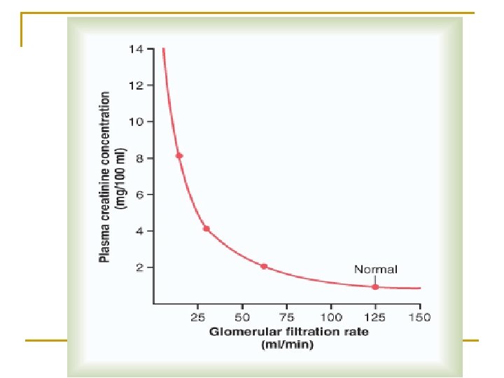

Effect of reducing GFR by 50 % on serum creatinine concentration and creatinine excretion rate

Plasma creatinine can be used to estimate changes in GFR

Filtration in systemic capillary beds VS Glomerular Filtration n n filtration across the systemic capillary bed of capillaries is only 20 L/day ; 17 L is reabsorbed by veins and 3 L by lymphatics (kidneys are not included). filtration through the glomerular capillaries is 180 L/day; i. e. , 9 times more than the systemic filtration. Why?
![GFR=Kf. [(PGC-PBS)-( GC- BS)]= Kf * Peff (use Ohm’s law again) • The](http://slidetodoc.com/presentation_image_h/238237c72a7d1325496f1cefc3a81a70/image-78.jpg "GFR=Kf. [(PGC-PBS)-( GC- BS)]= Kf * Peff (use Ohm’s law again) • The")
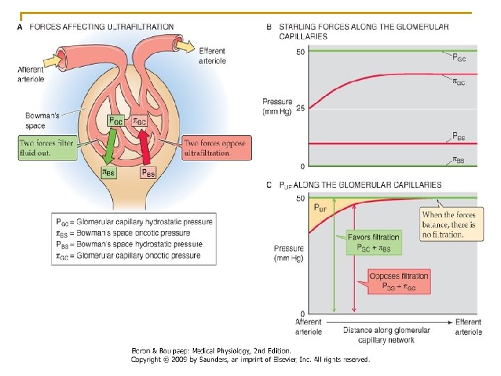
GFR=Kf. [(PGC-PBS)-( GC- BS)]= Kf * Peff (use Ohm’s law again) • The driving force is the summation of Starling forces which are 2 forces inside and only one force outside. • The inside ones are: 1. Capillary hydrostatic pressure (PGC). 2. Colloid capillary pressure ( GC) provided by albumin and globulin mostly by albumin. • The outside ones are: 1. PBS; it is 18 mm. Hg (encapsulated organs) and it will oppose filtration ( in most tissue interstitial pressure is subatmospheric or –ve)

Dirving Forces affecting Filtration n 1. 2. n 1. Favoring Filtration: Hydrostatic Pressure in the Glomerular capillaries. (PGC) =60 mm. Hg. . the highest in our body Oncotic (Colloid) Pressure of the filtrate in the Bowman’s capsule. ( BS)=32 mm. Hg Opposing Filtration: Hydrostatic Pressure in the Bowman’s capsule. (PBS )=18 mm. Hg
")
I. Glomerular Hydrostatic Pressure: n n n The difference between 20 L/day (systemic capillaries) and the 180 L/day (GFR) is either due to increased Peff or increased Kf or both. The PGC here is 60 mm. Hg as opposed to 15 -30 mm. Hg in systemic capill, or 7 -10 mm. Hg in pulmonary capillaries…. . WHY? If we look at systemic capillaries they have an arterial end a venous end but in the glomerulus they have both arteriolar ends afferent and efferent arterioles. This makes the pressure in the glomerular capillaries the highest (60 mm. Hg).

Arteriolar diameter effect on GFR: n n n Afferent dilatation means an increase in the blood coming to the capillaries so increased PGC and GFR. Constriction of efferent arteriole increases PGC to a limit. If it goes over this limit filtration will decrease as no more blood entering the capillaries. To regulate PGC you either control the afferent arteriolar dilatation or the efferent arteriolar constriction or both.
 Pressure: In the systemic capillaries the stays 28 mm Hg")
II. Glomerular Capillaries Oncotic(colloid) Pressure: In the systemic capillaries the stays 28 mm Hg at both the arterial and venous ends. WHY? Because what is filtered is only 0. 5% from the whole incoming fluid, so it does not affect the concentration of proteins much. n But filtration in the kidneys is 20% so it must have an effect so GC that becomes 36 mm Hg at the efferent end (avg. 32 mm. Hg). n
 : Bowman’s Space contains protein free glomerular filtrate; i.")
III. Interstitial forces (Bowman’s Space) : Bowman’s Space contains protein free glomerular filtrate; i. e, too small BS. So in the kidneys Starling forces have been reduced to 3 forces instead of 4 forces n Hydrostatic Pressure (P) of Bowman’s space is 18 mm. Hg due to the fluids filtered. n Net Driving forces favoring filtration= 60 – (32 + 18 ) = 10 mm. Hg n Knowing that P = 10 mm. Hg and GFR is 125 then Kf will equal =12. 5 ml/min. mm. Hg n (125=10*KF) n

Renal Kf Basement membrane is negatively charged n In nephrotic syndrome, loss of negative charge causes albumin loss and edema (Alb MW is slightly less than 70 K but still not filtered bcs it is negatively charged. When basement membrane losses its negative charge …now albumin can filter leading to hypoalbuminemia. ). n (Remember the four cause of hypoalbuminemia : malnutrition, malabsorption, malproduction, and increased loss from the kidney). n Hypoalbuminemia →↑GFR.

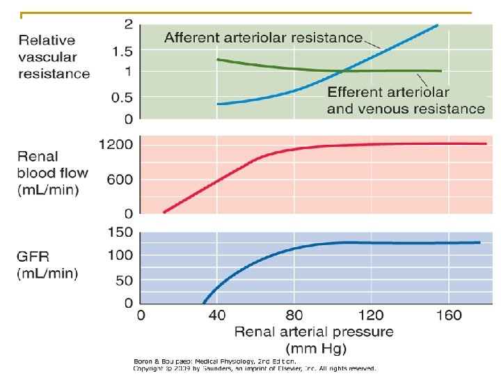
Renal Autoregulation n n Autoregulation of GFR…see the figure after 3 slides GFR is fluctuates slightly in relation to changes in arterial blood pressure but this translates in a large increase in urine output… why is that? GFR = 125 ml/min and UOP is only = 1 ml/min = 1. 5 L/day which means 124 ml/min is reabsorbed (99. 4% of the filtered water is reabsorbed and only 0. 6% is excreted) so a little change in GFR changed the urine output a lot. Therefore, GFR must be regulated and this is achieved mainly by the renal vascular system (glomerular capillary hydrostatic pressure) and this is controlled by afferent and efferent arterioles by the following mechanism:

Tubuloglomerular Feedback n 1. 2. n n In the distal tubule the afferent arteriole touches a few cells in its wall…these DT cells are sensors (macula densa). They sense the content of Na +, K+, Ca++. When the amount of these electrolytes reach macula densa is small → 2 messages are sent. The first message: dilatation of the afferent arteriole so the blood flow increases to the capillaries. (Myogenic Response) The second message: is to the granular cell in the afferent arteriole to secret rennin (hormonal response). Rennin leaves the kidney and goes to the circulation where it cleaves 4 amino acids for a 14 -aa small peptide produced by the liver and called angiotensinogen forming the decapeptide angiotensin-I. In the lungs the ACEs (angiotensin converting enzymes) convert AI to AII (octapeptide). In bleeding we have to protect the kidneys by keeping normal GFR and at the same time conserve water. Angiotensin-II can do this…through

Tubuloglomerular Feedback n n n First: constriction of efferent arteriole leading to increased GFR and at the same time the pressure in the peritubular capillaries decreases giving a better chance for reabsorbing to get the minimal urine output which is 0. 5 L/day. Second: angiotensin acts directly on the adrenal cortex to secret aldosterone that enhances the reabsorption of Na+ from the distal tubule and sodium bring with it water. Third: angiotensin itself act directly to enhance sodium reabsorption in the proximal tubule.
 • Normally changes as a function of GFR, not")
Bowman’s Capsule hydrostatic Pressure (PB) • Normally changes as a function of GFR, not a physiological regulator of GFR • Tubular Obstruction kidney stones tubular necrosis • Urinary tract obstruction Prostate hypertrophy/cancer 89
 • Arterial Plasma Oncotic Pressure (")
Factors Influencing Glomerular Capillary Oncotic Pressure ( G) • Arterial Plasma Oncotic Pressure ( A) A G • Filtration Fraction (FF) FF G FF = GFR / Renal plasma flow = 125 / 650 ~ 0. 2 (or 20%) 90

Increase in colloid osmotic pressure in plasma flowing through glomerular capillary 91

Net Filtration Pressure Decreases Along the Glomerulus Because of Increasing Glomerular Colloid Osmotic Pressure PG = 60 G = 28 Net Filtration Pressure PG = 60 14 G =32 6 G = 36 PB = 18 92
 • Plasma Protein Concentration Arterial Plasma")
Factors Influencing Glomerular Capillary Oncotic Pressure ( G) • Plasma Protein Concentration Arterial Plasma Oncotic Pressure ( A) A G • Filtration Fraction (FF) FF G FF= GFR / Renal plasma flow 93

Measuring GFR • The concept of Clearance…make sure you understand this concept fully…it is the most important concept in renal physiology…it tells you how the nephron or any segment of the nephron handles any substance…for instance we use the clearance to measure GFR, RPF etc Given the concentration of a substance in the plasma and the amount of the substance excreted in the urine per minute, you will compute the plasma clearance rate. Measurement of GFR = Cinulin Inulin is a starch-like polymer of fructose (MW 5000). Inulin is a glomerular marker Ccreatinine…instead of inulin we use creatinine… we either use t. GFR or e. GFR (“t” stands for true and “e” stands for estimated) 94

Use of Clearance to Measure GFR For a substance that is freely filtered, but not reabsorbed or secreted (inulin, 125 I-iothalamate, creatinine), renal clearance is equal to GFR amount filtered = amount excreted GFR x Pin = GFR = Uin x V Pin

Creatinine MW 114 The typical reference ranges are: Men 0. 7 to 1. 2 mg/d. L (60 -110 μmol/l), for women: 0. 5 to 1. 0 mg/d. L (about 45 -90 μmol/l While a baseline serum creatinine of 2. 0 mg/d. L (150 μmol/l) may indicate normal kidney function in a male body builder, a serum creatinine of 0. 7 mg/d. L (60 μmol/l) can indicate significan renal disease in a frail old woman. In the United States, creatinine is typically reported in mg/d. L, while in Canada and Europe μmol/litre may be used. 1 mg/d. L of creatinine is 88. 4 μmol/l 1.

Plasma Clearance Let us practice calculating plasma clearance using the clearance equation. In all your calculations, assume that the urine production rate (V) is 2 ml/min. Let’s start with the substance inulin (not insulin!). If after a dose of inulin, your urine has 30 mg/ml and your plasma has 0. 5 mg/ml of this substance, what is the inulin clearance rate? If you got 120 ml/min, you are correct! Unit 1 - Objective 9
")
Clearances of Different Substances Substance glucose albumin sodium urea inulin creatinine PAH Clearance (ml/min) 0 0 0. 9 70 125 140 600

Plasma Clearance Test your ability to conduct further calculations by calculating the clearance rate for the following substances: Substance Urine concentration Plasma concentration Urea 7. 0 mg/ml 0. 2 mg/ml Glucose 0. 0 mg/ml 1. 0 mg/ml Penicillin 298 mg/ml 0. 7 mg/ml Urine production rate is (2 ml/min). The clearance rate for each of the above substances will be: Urea = 70 ml/min; Glucose = 0 ml/min; Penicillin = 851 ml/min!!!. Were you able to get the right answers? If not, go back and restudy the clearance process. Unit 1 - Objective 9

• Since Inulin is an exogenous substance it is only used for research purposes and not as a clinical test. • We need an endogenous substance: Creatinine: Comes from high energy bound, muscle phosphocreatinine (PC). • Is muscle protein • Small molecule (MW is 114)) • Its concentration does not fluctuate from day to day in plasma • Freely filtered, not reabsorbed but SLIGHTLY SECRETED To convert mol /l of creatinine to mg/dl, divide by 88. 4. To convert mg/dl of creatinine to mol/l, multiply by 88. 4 Creatinine in the urine comes from 90% filtered and 10% through secretion. This has the potential to overestimates GFR by 10%. But in actuality it does not…why? In fact, it does overestimate GFR in end-stage renal failure…again WHY? Look for the answer in both cases

Through this equation: Creatinin Clearance=Ccr = GFR = UCr * V P Cr Last point to describe which is the To answer the question I asked earlier: 10% of Cr in urine is secreted which overestimates the GFR. But it was found that 10% of Cr in plasma is bounded to protein… it is the total Cr we measure and not only the free portion. (so, both factors cancel each others). Ccr is good estimation of GFR.
 • Is the determinant of GFR snd is subject to")
Glomerular Hydrostatic Pressure (PG) • Is the determinant of GFR snd is subject to physiological control • Factors that influence PG - arterial pressure (effect is buffered by autoregulation) - afferent arteriolar resistance - efferent arteriolar resistance 102
 Normal kidney 60")
Autoregulation of Glomerular Hydrostatic Pressure 80 Glomerular Hydrostatic Pressure (mm. Hg) Normal kidney 60 Kidney disease 40 20 0 50 100 150 200 Arterial Pressure (mm. Hg) 103

Autoregulation of renal blood flow and GFR but not urine flow 104

Effect of afferent and efferent arteriolar constriction on glomerular pressure Ra Re PG Blood Flow GFR Ra GFR + PG Renal Blood flow Flow Blood Flow GFR Re GFR + Renal Blood flow Flow 105

Effect of changes in afferent arteriolar or efferent arteriolar resistance. Biphasic Effect 106

G determined by : FF = GFR / RPF G RBF _ GFR RE PG + 107

Summary of Determinants of GFR Kf PB G A FF PG RA RE (as long as GFR Renal Disease, DM, HP GFR Renal stones GFR Decrease RBF G G GFR PG PG GFR GFR RE < 3 -4 x normal) 108
 RBF = P / R P = difference")
Determinants of Renal Blood Flow (RBF) RBF = P / R P = difference between renal artery pressure and renal vein pressure = 100 -4 mm. Hg R = total renal vascular resistance = Ra + Re + Rv = sum of all resistances in kidney vasculature 109

Table 26 -3. Approximate Pressure and Vascular Resistances in the Circulation of Normal Kidney Afferent + efferent contribute to about 70% of the intrarenal vascular resistance (mainly efferent). Pressure mm. Hg % Total Vascular R Beginning 100 End 100 85 Afferent Glomerular capillaries 85 60 60 59 Efferent 59 18 Peritubular Capillaries Interlobar, arcuate and interlolbular veins 18 8 8 4 10 4 Renal vein 4 ≈4 0 Renal Artery Interlobar, arcuate and interlolbular arteries 0 15 25 1 only 1 mm. Hg which means little resistance 43 resistance mainly resides her 110
")
Renal blood flow • High blood flow 1100 ml/min (~22 % of cardiac output) • High blood flow needed for high GFR • Oxygen and nutrients delivered to kidneys normally greatly exceeds their metabolic needs • A large fraction of renal oxygen consumption is related to renal tubular sodium reabsorption 111
")
Blood flow in kidneys and other organs Organ Approx. blood flow (ml/min/g of tissue) A-V O 2 difference (ml/L) Kidney 4. 00 12 -15 (depends on reabsorption of Na+ ) Heart 0. 80 96 Brain 0. 50 48 Skeletal muscle (rest) 0. 05 - Skeletal muscle (max. exercise) 1. 00 - 20 5 Carotid bodies
 A-V difference (Vol %) Oxygen onsumption ml/gm/min tissue weight")
Tissue blood flow (ml/g/min) A-V difference (Vol %) Oxygen onsumption ml/gm/min tissue weight Heart 0. 8 (250÷ 315) 11 0. 086 0. 5 6. 2 0. 033 6 0. 0018 Brain 750/1400 Skeletal Muscle 0. 03 1200÷ 28000 Liver Kidney 0. 6 3. 4 4. 2 1100/262 1. 4 0. 059 (twice the brain) O 2 consumption is directly related to Na + reabsorption. If GFR is high → Na+ reabsorption is high→O 2 consumption is high. When GFR is severely depressed (Acute RF) → decrease need for O 2 and this is how renal cells can tolerate ischemia. In this case 20 -25% of RBF is enough to supply the basic metabolic needs of the renal cells Carotid bodies 20 0. 58/0. 029 0. 5 0. 1 most active cells in our body (twice the kidney)

Renal oxygen consumption and sodium reabsorption 114
 115")
Control of GFR and renal blood flow • Neurohumoral • Local (Intrinsic) 115

Renal Blood Flow n We expect Flow to be a function of Pressure Gradient n We find Flow to be constant n Therefore: Vascular Resistance changes in response to a change in Blood Pressure

Control of GFR and renal blood flow 1. Sympathetic Nervous System /catecholamines RA + R E GFR + RBF e. g. severe hemorrhage. Under normal conditions Sympathetic tone have little influence on RBF. Sympathetic system may not influence RBF under normal circumstances, but in severe sympathetic stimulation it may decrease RBF significantly 2. Angiotensin II RE GFR + RBF (prevents a decrease in GFR) e. g. low sodium diet, volume depletion 117

Control of GFR and renal blood flow 3. Prostaglandins RA + R E GFR + RBF Blockade of prostaglandin synthesis → ↓ GFR This is usually important only when there are other disturbances that are already tending to lower GFR. If Aspirin is administered which suppresses PGs then a severe decrease in GFR might occur. e. g. nonsteroidal antiinflammatory drugs NDAID in a volume depleted patient, or a patient with heart failure, cirrhosis, etc 118
 RA +")
Control of GFR and renal blood flow 4. Endothelial-Derived Nitric Oxide (EDRF) RA + R E GFR + RBF • Protects against excessive vasoconstriction • Patients with endothelial dysfunction (e. g. atherosclerosis) may have greater risk for excessive decrease in GFR in response to stimuli such as volume depletion 119

Control of GFR and renal blood flow 5. Endothelin RA + R E GFR + RBF • Hepatorenal syndrome – decreased renal function in cirrhosis or liver disease? • Acute renal failure (e. g. contrast media nephropathy)? • Hypertensive patients with chronic renal failure? Endothelin antagonists may be useful in these conditions 120

Summary of neurohumoral control of GFR and renal blood flow Effect on GFR Effect on RBF Sympathetic activity Catecholamines Angiotensin II EDRF (NO) Endothelin Prostaglandins increase decrease no change 121

Local Control of GFR and renal blood flow 7. Autoregulation of GFR and Renal Blood Flow • Myogenic Mechanism • Macula Densa Feedback (tubuloglomerular feedback) • Angiotensin II ( contributes to GFR but not RBF autoregulation) 122
 100 80 Glomerular Filtration Rate Renal")
Renal Autoregulation 120 Renal Artery Pressure (mm. Hg) 100 80 Glomerular Filtration Rate Renal Blood Flow 0 1 2 3 Time (min) 4 5 123

Importance of Autoregulation Arterial Pressure GFR Reabsorption Urine Volume 1 - Poor Autoregulation + no change in tubular reabsorption 100 125 124 120 150 124 1. 0 26. 0 = 37. 4 L/day! 2 - Good Autoregulation + no change in tubular reabsorption 120 130 124 6. 0 3 Good Autoregulation+adaptive increase in tubular reabsorption 120 130 128. 8 1. 2 124

Myogenic Mechanism Arterial Pressure Stretch of Blood Vessel Blood Flow and GFR Vascular Resistance Cell Ca++ Entry Intracell. Ca++ 125

Structure of the juxtaglomerular apparatus: macula densa 126

Macula Densa Feedback GFR Distal Na. Cl Delivery Macula Densa Na. Cl Reabsorption (macula densa feedback) Afferent Arteriolar Resistance 127

Macula Densa Feedback Proximal Na. Cl Reabsorption Distal Na. Cl Delivery Macula Densa Na. Cl Reabsorption (macula densa feedback) Afferent Arteriolar Resistance (Dilatation) GFR 128

Regulation of GFR by AII GFR Macula Densa Na. Cl Blood Pressure Renin AII Efferent Arteriolar Resistance 129
 1200 800")
Ang II Blockade Impairs GFR Autoregulation 1600 Renal Blood Flow ( ml/min) 1200 800 Normal Ang II Blockade 400 0 Glomerular Filtration Rate (ml/min) 120 80 40 0 0 50 100 150 Arterial Pressure (mm. Hg) 200 130

Macula densa feedback mechanism for regulating GFR 131

Other Factors That Influence GFR • Fever, pyrogens: increase GFR • Glucorticoids: increase GFR • Aging: decreases GFR 10% / decade after 40 yrs • Hyperglycemia: increases GFR (diabetes mellitus) • Dietary protein: high protein increases GFR low protein decreases GFR 132

Protein Ingestion Amino Acids Tubular Amino Acid Reabs. Proximal Nacl Reabs. Macula Densa Na. Cl (macula densa feedback) Afferent Arteriolar Resist. GFR 133
- Slides: 133