The Nephron The epithelium of the visceral and


- Slides: 18

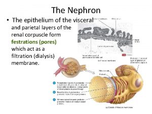
The Nephron • The epithelium of the visceral and parietal layers of the renal corpuscle form festrations (pores) which act as a filtration (dialysis) membrane.

The Nephron • Fluid filtered from the glomerular capillaries enters Bowman’s space, (the space between the two layers of the glomerular capsule), which is the lumen of the urinary tube.

The Nephron • Blood plasma is filtered through the glomerular capillaries into the glomerular capsule. – Filtered fluid passes into the renal tubule, which has three main sections: • the proximal convoluted tubule (PCT) • the loop of Henle • the distal convoluted tubule (DCT)

The Nephron • The distal convoluted tubules of several nephrons empty into a single collecting duct. – Collecting ducts unite and converge into several hundred large papillary ducts which drain into the minor calyces, major calyces, renal pelvis, and ureters.

The Nephron • The first part of the loop of Henle (the descending limb) dips into the renal medulla. It then makes a hairpin turn and returns to the renal cortex as the ascending limb. – The descending limb of the loop of Henle is composed of a simple squamous epithelium. – The ascending limb of the loop may be either “thin” (composed of a simple squamous epithelium) or “thick” (composed of simple cuboidal to low columnar cells). • Some nephrons contain both thick and thin ascending limbs.

The Nephron • Based on the length of the loop of Henle and the presence of thin segments in the ascending limb, nephrons can be sorted into two populations: cortical and juxtamedullary. • Nephrons with long loops of Henle enable the kidneys to create a concentration gradient in the renal medulla and to excrete very dilute or very concentrated urine.

The Nephron • Cortical nephrons make up about 80– 85% of the 1 million microscopic nephrons that comprise each kidney. – Their renal corpuscles are located in the outer portion of the cortex, with short loops of Henle that penetrate only a small way into the medulla. – The ascending limbs of their loops of Henle consist of only a thick segment, lacking any thin portions. – Nephrons with short loops receive their blood supply from peritubular capillaries that arise from efferent arterioles.

The Nephron

The Nephron • The other 15– 20% of the nephrons are juxtamedullary nephrons. – Their renal corpuscles lie deep in the cortex, close to the medulla, and they have long loops of Henle that extend into the deepest region of the medulla. – The ascending limbs of their loops of Henle consist of both thin and thick segments. – Nephrons with long loops receive their blood supply from the vasa recta that arise from peritubular capillaries before becoming peritubular venules.

Juxtamedullary Nephron

The Nephron • In each nephron, the final part of the ascending limb of the loop of Henle makes contact with the afferent arteriole serving that renal corpuscle. Because the columnar tubule cells in this region are crowded together, they are known as the macula densa. § Alongside the macula densa, the wall of the afferent arteriole contains modified smooth muscle fibers called juxtaglomerular (JG) cells. • Together with the macula densa, they constitute the juxtaglomerular apparatus (JGA).

The Nephron • As we will shortly see, the JGA helps regulate blood pressure within the kidneys.

Renal Physiology • The 3 basic functions performed by nephrons and collecting ducts are: – Glomerular filtration - pressure forces filtration of waste -laden blood in the glomerulus. The glomerular filtration rate (GFR) is the amount of filtrate formed in all the renal corpuscles of both kidneys each minute. – Tubular reabsorption – the process of returning important substances from the filtrate back to the body. – Tubular secretion – the movement of waste materials from the body to the filtrate.

Glomerular Filtration • Glomerular filtration is the formation of a protein-free filtrate (ultrafiltrate) of plasma across the glomerular membrane. – Only a portion of the blood plasma delivered to the kidney via the renal artery is filtered. – Plasma which escapes filtration, along with its protein and cellular elements, exits the renal corpuscle via the efferent arteriole, perfuses the tubular capillary beds, and is eventually collected in the renal venous system.

Glomerular Filtration • In the average adult, the body’s entire extracellular fluid volume is filtered about 12 times per day. l Since we cannot afford to lose that amount of fluid, the vast majority must be reclaimed, with just a small portion being excreted in the urine.

Glomerular Filtration Amount in 180 L of filtrate (/day) Amount returned to blood/d (Reabsorbed) Amount in Urine (/day) 3 L 180 L 178 -179 L 1 -2 L Protein (active) 200 g 2 g 1. 9 g 0. 1 g Glucose (active) 3 g 162 g 0 g Urea (passive) 1 g 54 g 24 g 30 g 0. 03 g 1. 6 g Total Amount in Plasma Water (passive) Creatinine (about 1/2) 0 g 1. 6 g (all filtered) (none reabsorbed) The daily composition of plasma, filtrate, and urine are compared.

Glomerular Filtration • Filtration is controlled by 2 opposing hydrostatic forces and 2 opposing osmotic forces at the glomerular membrane called Starling forces. – Blood hydrostatic pressure (55 mm. Hg) is the main force that “pushes” water and solutes through the filtration membrane (promotes filtration). – Capsular hydrostatic pressure (15 mm. Hg) is exerted against the filtration membrane by fluid in the capsular space (opposes filtration).

Glomerular Filtration • Starling forces continued – Blood osmotic (oncotic) pressure (30 mm. Hg) is the pressure of plasma proteins “pulling” on water (opposes filtration). – Normally very little protein escapes through the filtration membrane making capsular oncotic pressure a negligible force except in certain disease states. • Net Filtration = Blood Hydrostatic Pressure – Blood Osmotic Pressure – Capsular Hydrostatic Pressure – Net Filtration = 55 -30 -15 = 10 mm. Hg
 Efron of a nephron
 Pleura parietal visceral
 Peritoneum
 Path of food from mouth to anus
 What is the peritoneum
 Applied anatomy of mediastinum
 Myogenic autoregulation
 Nephron diagram and function
 Kidney tubular secretion
 Epiploic foramen
 Heart membranes
 Hypothalamus ans
 Sympathetic
 Visceral leishmaniasis
 Pleural cavity
 Lung surface anatomy
 Taxonomy
 What is visceral mass in molluscs
 Visceral afferent vs efferent
