Dynamin Monique Krger http www google deimgres imgurlhttp3


- Slides: 22
Dynamin - Monique Krüger - http: //www. google. de/imgres? imgurl=http%3 A%2 F%2 Fstatic 1. squarespace. com%2 Fstatic%2 F 52 e 79 cb 5 e 4 b 0 b 77 fb 0239620%2 Ft%2 F 52 ef 8 d 1 be 4 b 07 fbe 88868 c 13%2 F 1391430942719%2 FBDLPxtal_map_filament. png&imgrefurl=http%3 A%2 F%2 Fwww. thelowlab. org%2 Fresearch 1%2 F&h=354&w=1000&tbnid=PLAYf. DDOc. Us 9 M%3 A&docid=mv 0_0 jn. STC_R 0 M&ei=A 9 I 3 Vs. KZKYKhy. AOltqrg. Ag&tbm=isch&iact=rc&uact=3&dur=12632&page=1&start=0&ndsp=15&ve d=0 CD 8 Qr. QMw. Cmo. VCh. MIgqf. Cgt. Tyy. AIVgh. By. Ch 0 lmwos
Table of contents • Cell / organell division • Dynamin superfamily • Dynamin structure • Dynamin as mechanoenzymes • Dynamin function • Arabidopsis genome • DRP 1 – DRP 5
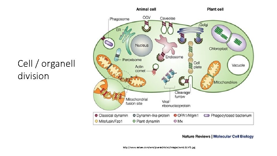
Cell / organell division http: //www. nature. com/nrm/journal/v 5/n 2/images/nrm 1313 -f 1. jpg
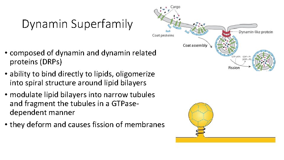
Dynamin Superfamily • composed of dynamin and dynamin related proteins (DRPs) • ability to bind directly to lipids, oligomerize into spiral structure around lipid bilayers • modulate lipid bilayers into narrow tubules and fragment the tubules in a GTPasedependent manner • they deform and causes fission of membranes
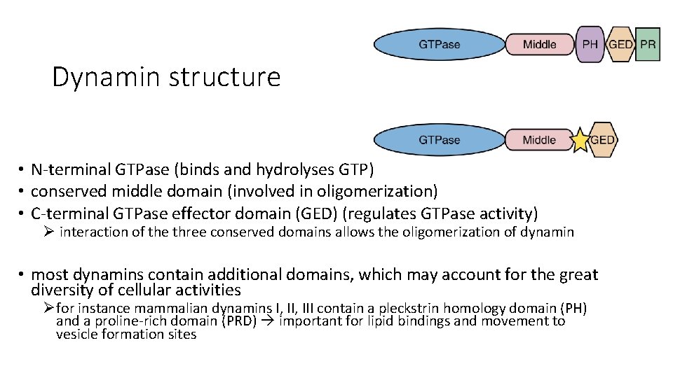
Dynamin structure • N-terminal GTPase (binds and hydrolyses GTP) • conserved middle domain (involved in oligomerization) • C-terminal GTPase effector domain (GED) (regulates GTPase activity) Ø interaction of the three conserved domains allows the oligomerization of dynamin • most dynamins contain additional domains, which may account for the great diversity of cellular activities Øfor instance mammalian dynamins I, III contain a pleckstrin homology domain (PH) and a proline-rich domain (PRD) important for lipid bindings and movement to vesicle formation sites
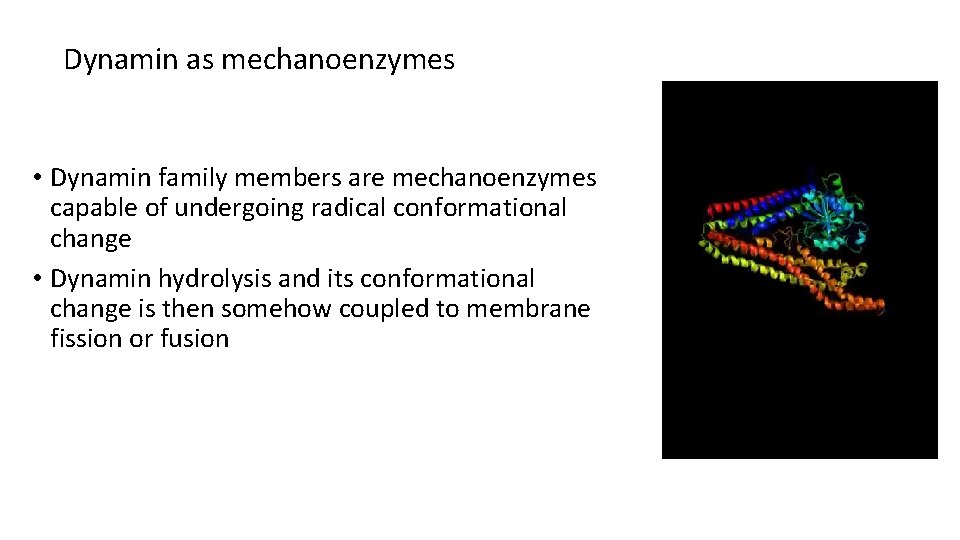
Dynamin as mechanoenzymes • Dynamin family members are mechanoenzymes capable of undergoing radical conformational change • Dynamin hydrolysis and its conformational change is then somehow coupled to membrane fission or fusion
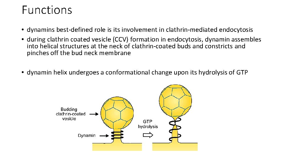
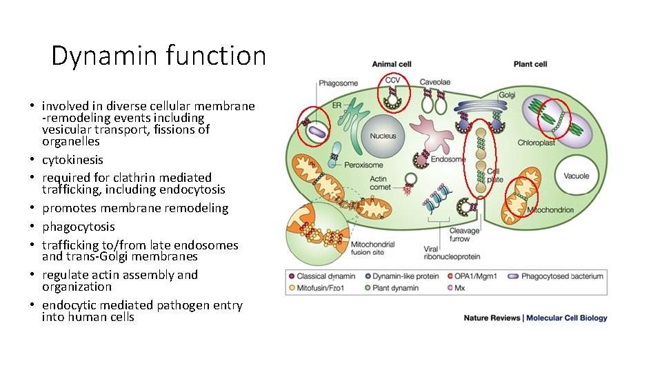
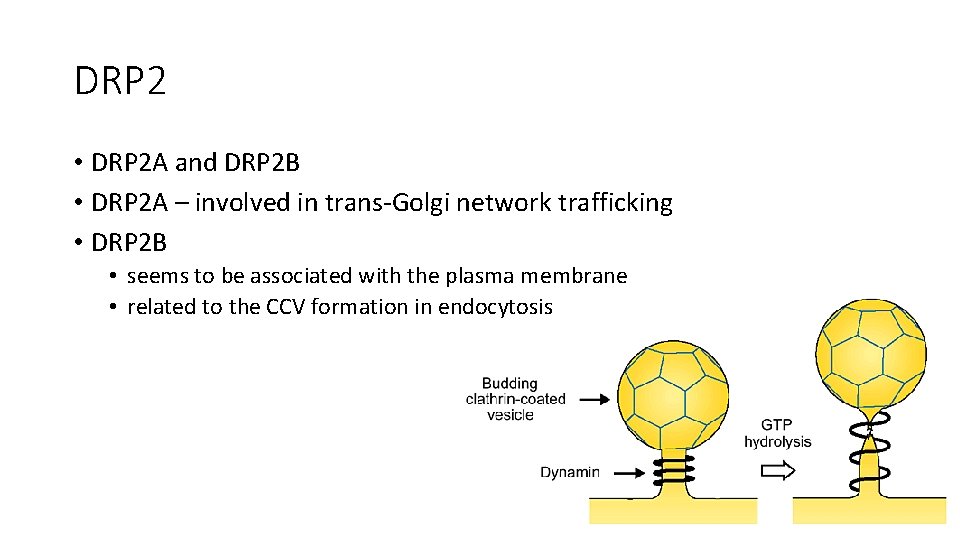
Functions • dynamins best-defined role is its involvement in clathrin-mediated endocytosis • during clathrin coated vesicle (CCV) formation in endocytosis, dynamin assembles into helical structures at the neck of clathrin-coated buds and constricts and pinches off the bud neck membrane • dynamin helix undergoes a conformational change upon its hydrolysis of GTP
Dynamin function • involved in diverse cellular membrane -remodeling events including vesicular transport, fissions of organelles • cytokinesis • required for clathrin mediated trafficking, including endocytosis • promotes membrane remodeling • phagocytosis • trafficking to/from late endosomes and trans-Golgi membranes • regulate actin assembly and organization • endocytic mediated pathogen entry into human cells
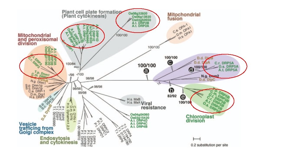
Arabidopsis genome • has 16 dynamin related proteins • grouped into 6 subfamilies (DRP 1 -DRP 6)
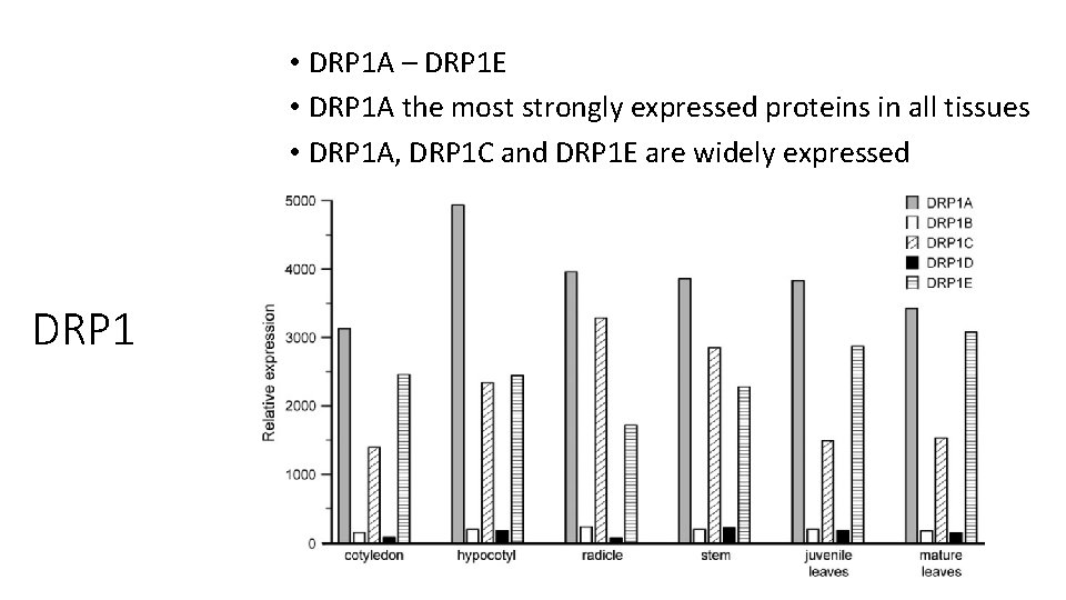
DRP 1 • grouped in DRP 1 A - E • DRP 1 A, C and E accumulate in cell plate during cytokinesis • DRP 1 s constrict vesicles and tubules in the cell plate • DRP 1 A is related to the CCV formation side in endocytosis • DRP 1 A were identified by studying rsw 9, a null mutant • tissue samples were chosen from seedlings (cotyledon, hypocotyl and radicle), leaves (immature and mature) and stems
• DRP 1 A – DRP 1 E • DRP 1 A the most strongly expressed proteins in all tissues • DRP 1 A, DRP 1 C and DRP 1 E are widely expressed DRP 1
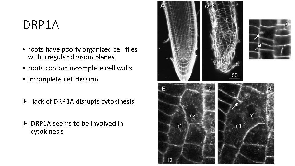
DRP 1 A • roots have poorly organized cell files with irregular division planes • roots contain incomplete cell walls • incomplete cell division Ø lack of DRP 1 A disrupts cytokinesis Ø DRP 1 A seems to be involved in cytokinesis
DRP 2 • DRP 2 A and DRP 2 B • DRP 2 A – involved in trans-Golgi network trafficking • DRP 2 B • seems to be associated with the plasma membrane • related to the CCV formation in endocytosis
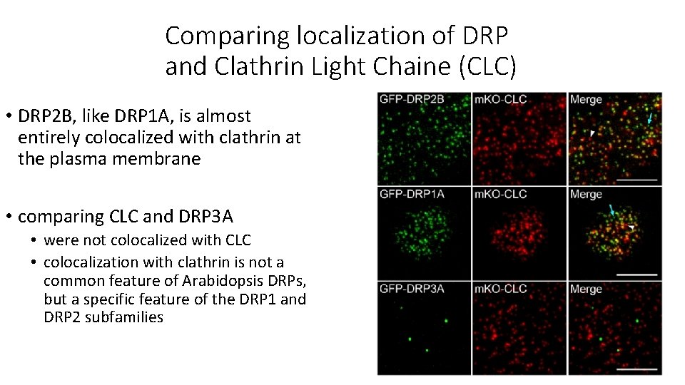
Comparing localization of DRP and Clathrin Light Chaine (CLC) • DRP 2 B, like DRP 1 A, is almost entirely colocalized with clathrin at the plasma membrane • comparing CLC and DRP 3 A • were not colocalized with CLC • colocalization with clathrin is not a common feature of Arabidopsis DRPs, but a specific feature of the DRP 1 and DRP 2 subfamilies
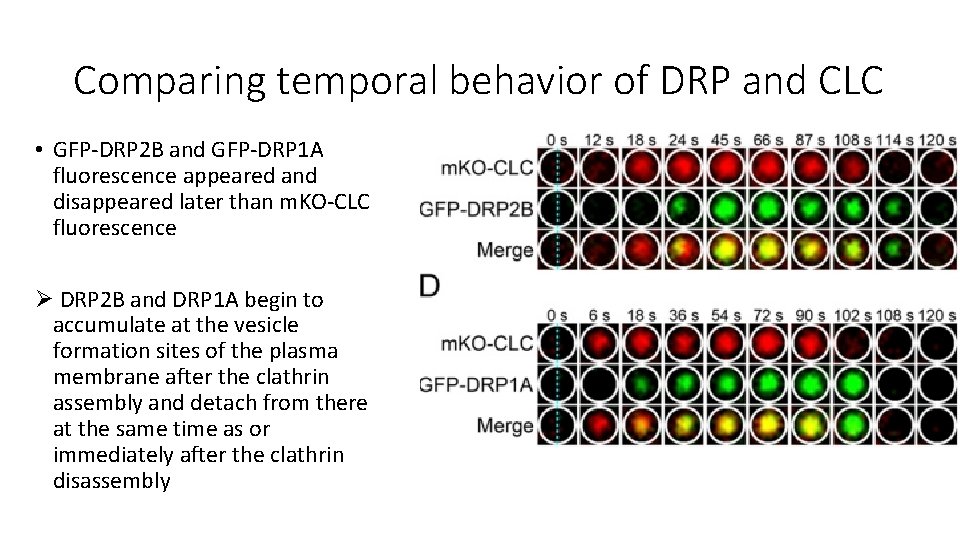
Comparing temporal behavior of DRP and CLC • GFP-DRP 2 B and GFP-DRP 1 A fluorescence appeared and disappeared later than m. KO-CLC fluorescence Ø DRP 2 B and DRP 1 A begin to accumulate at the vesicle formation sites of the plasma membrane after the clathrin assembly and detach from there at the same time as or immediately after the clathrin disassembly
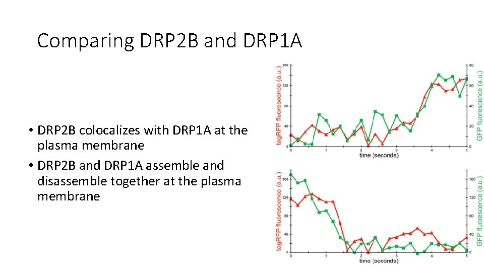
Comparing DRP 2 B and DRP 1 A • DRP 2 B colocalizes with DRP 1 A at the plasma membrane • DRP 2 B and DRP 1 A assemble and disassemble together at the plasma membrane
Results DRP 2 B and DRP 1 A • same localization • appear and disappear shortly after CLC fluorescence • assemble and disassemble together at the plasma membrane Ø DRP 2 B and DRP 1 A interact with each other Ø suggesting that they form a molecular complex on the CCV formation site
DRP 3 • includes DRP 3 A and DRP 3 B • localized to peroxisomes and mitochondria • involved in fission of organelles
DRP 5 B • involved in division of peroxisomes and chloroplasts • DRP 5 B is the only DRP besides DRP 3 A and DRP 3 B that is known to play a direct role in organelle division
DRP 5 A • DRP 5 A is involved in cytokinesis instead of organell division • signals were detected in meristematic tissues • expression is limited to dividing cells
Thanks for your attention!