DNA and Replication 1 History of DNA 2


 “Legs")


 Guanine (G) A or G •")


 = Thymine (30%) •")


 • S phase during interphase of the cell cycle •")


 Enzyme Helicase unwinds and separates the 2 DNA strands")
 Primase gathers nucleotides and brings them into the replication")
 The enzyme DNA Polymerase matches free nucleotides with the")


 The enzyme ligase connects any “breaks” in the new")


 radiation damage the DNA in our")
- Slides: 49
DNA and Replication 1
History of DNA 2
History of DNA • Early scientists thought protein was the cell’s hereditary material because it was more complex than DNA • Proteins were composed of 20 different amino acids in long polypeptide chains 3
Transformation • Fred Griffith worked with virulent S and nonvirulent R strain pneumonia bacteria • He found that R strain could become virulent when it took in DNA from heat-killed S strain • Study suggested that DNA was probably the genetic material 4
Griffith Experiment 5
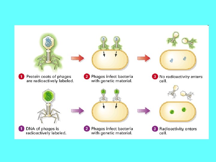
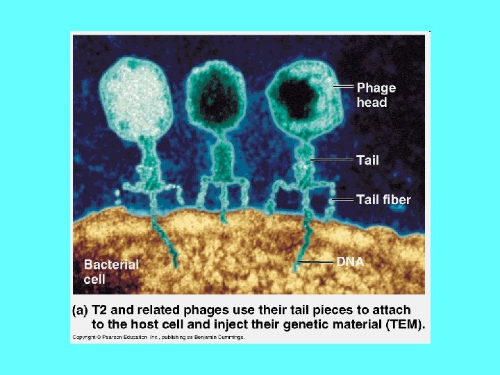
History of DNA • Viruses are made of DNA in a protein “coat” • Experiments on viruses by Hershey & Chase proved that DNA was the cell’s genetic material Radioactive DNA was injected into bacteria! 6
DNA Structure • Rosalind Franklin took diffraction x-ray photographs of DNA crystals • In the 1950’s, Watson & Crick built the first model (double helix) of DNA using Franklin’s x -rays 9
Rosalind Franklin 10
Watson and Crick We’re big fat thieves!
Video: History of DNA Discovery Questions to Ponder… 1. Who was Linus Pauling and why was his DNA model incorrect? 2. How did Rosalind Franklin “help” Watson and Crick to develop their model of DNA? . . Downloaded VideosThe Secret of Life -- Discovery of DNA Structure. avi
DNA Structure 13
The Basics • DNA is a type of Nucleic Acid • DNA: Deoxyribonucleic Acid • Made of monomers called nucleotides • Function: to store genetic information
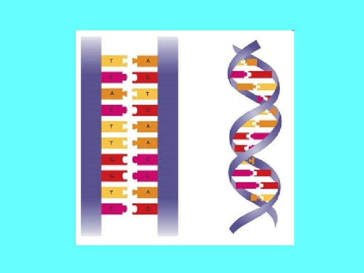
DNA • Two strands coiled called a double helix • Sides made of a sugar Deoxyribose bonded to phosphate (PO 4) groups • Center made of nitrogen bases bonded together by weak hydrogen bonds 15
DNA Double Helix “Rungs of ladder” Nitrogenous Base (A, T, G or C) “Legs of ladder” Phosphate & Sugar Backbone 16
DNA • Double helix is formed by nucleotides linked to one another • Nucleotide made of: 1. Phosphate group 2. 5 -carbon sugar 3. Nitrogenous base 18
DNA Nucleotide Phosphate Group O O=P-O O 5 CH 2 O N C 1 C 4 Sugar (deoxyribose) C 3 C 2 Nitrogenous base (A, G, C, or T) 19
DNA Strands • The part of a strand that ends with a phosphate group is called the 5 prime (5’) end • The part of a strand that ends with a sugar is called the 3 prime (3’) end 20
Antiparallel Strands • One strand of DNA goes from 5’ to 3’ • The other strand is opposite in direction going 3’ to 5’ Strand 1: Strand 2: 5’ to 3’ 3’ to 5’
Nitrogenous Bases • Double ring PURINES Adenine (A) Guanine (G) A or G • Single ring PYRIMIDINES Thymine (T) Cytosine (C) T or C 22
Base-Pairings • Purines only pair with Pyrimidines • Hydrogen bonds connect the bases 3 H-bonds G C 23
The process of specific bases bonding together to form the rungs of the ladder is called Complementary Base Pairing
Chargaff’s Rule • Adenine must pair with Thymine • Guanine must pair with Cytosine T A G C 25
Discovery of DNA Structure • Erwin Chargaff showed the amounts of the four bases on DNA ( A, T, C, G) • In a body or somatic cell: A = 30. 3% T = 30. 3% G = 19. 5% C = 19. 9% 26
Question: • If there is 30% Adenine, Adenine how much Cytosine is present? 27
Answer: • There would be 20% Cytosine • Adenine (30%) = Thymine (30%) • Guanine (20%) = Cytosine (20%) • Therefore, 60% A-T and 40% C-G 28
Question Write out the sequence of a strand complementary to the following strand… TTAGCATGG
Answer Original Strand: Complementary Strand: TTAGCATGG AATCGTACC
DNA Replication 31
Replication Facts • DNA has to be copied before a cell divides • DNA is copied during the S or synthesis phase of interphase • New cells will need identical DNA strands 32
Synthesis Phase (S phase) • S phase during interphase of the cell cycle • Nucleus of eukaryotes S DNA replication takes place in the S phase G 1 interphase G 2 Mitosis -prophase -metaphase -anaphase -telophase 33
DNA Replication • Begins at Origins of Replication • Two strands open forming Replication Forks (Y-shaped region) • New strands grow at the forks 5’ Parental DNA Molecule 3’ 3’ Replication Fork 34 5’
DNA Replication • As the 2 DNA strands open at the origin, Replication Bubbles form • Prokaryotes (bacteria) have a single bubble • Eukaryotic chromosomes have MANY bubbles Bubbles 35
Steps of DNA Replication 1) Enzyme Helicase unwinds and separates the 2 DNA strands by breaking the weak hydrogen bonds copyright cmassengale 36
Steps of DNA Replication 2) Primase gathers nucleotides and brings them into the replication fork. A “primer” is created to start the new strand. 37
Steps of DNA Replication 3) The enzyme DNA Polymerase matches free nucleotides with the correct base pairs on the template (parent) strands. 38
copyright cmassengale 39
copyright cmassengale 40
Steps of DNA Replication 4) The enzyme ligase connects any “breaks” in the new strands and the 2 new strands rewind back together. The Big Question: Why are there “breaks” in the new strands at all? ! 41
• DNA polymerase can only add nucleotides to the 3’ end of the DNA • This causes the NEW strand to be built in a 5’ to 3’ direction 5’ 3’ Nucleotide DNA Polymerase Direction of Replication RNA Primer 42 5’
Remember the Strands are Antiparallel 43
Making New DNA Strands • The Leading Strand is built into the replication fork copyright cmassengale 44
• The Lagging Strand is built in short sections in the opposite direction (out of the fork) • This causes the “breaks” in the strand. copyright cmassengale 45
Joining of Fragments of DNA • The enzyme Ligase joins the sections together to make one strand DNA ligase 5’ 3’ 3’ 5’ Lagging Strand 46
A Quick Video to Help You Remember • . . Downloaded VideosDNA Replication Process. avi
Proofreading New DNA • DNA polymerase makes about 1 in 10, 000 base pairing errors • Enzymes proofread and correct these mistakes • The new error rate for DNA that has been proofread is 1 in 1 billion base pairing errors 48
DNA Damage & Repair Chemicals & ultraviolet (UV) radiation damage the DNA in our body cells Types of Repair: 1) Excision repair – when a repair enzyme removes damaged DNA 2) DNA polymerase and ligase work together to replace and bond the new nucleotides together 49