The Molecule of Life DNA is the molecule


 • Draw the DNA")
 1. Worked with two strains of")

 1. Repeated Griffith’s experiment, but added enzymes to heat-killed bacteria")

 1. Provided further evidence that DNA is")


 molecules and phosphates. This")


 Each time DNA is copied,")


 DNA codon: cytosine-adenine (CCA")


= a major component of ribosomes; also helps bond amino acids")
= helps transfer amino acids to the corresponding m. RNA")
 If the m. RNA codon is CUU, that would translate to the")

 chain, amino")


 43 =")

- Slides: 79
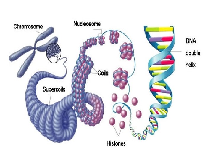
The Molecule of Life
DNA is the molecule of life. All living things contain DNA is found in the nucleus of cells. DNA contains genetic codes that determine physical features.
DNA stands for deoxyribonucleic acid. It may sound gibberish, but the name actually tells us two things: - DNA contains deoxyribose (a 5 -carbon sugar) - DNA is a nucleic acid (molecule made up of nucleotides) Nucleotide
Nucleotides consist of a sugar molecule attached to a nitrogen base and a phosphate group. Phosphate Nitrogen Base Sugar Nucleotides have 3 parts: 1) Sugar 2) Nitrogen Base 3) Phosphate
The Nitrogen Bases There are 4 possible different nitrogen bases: -adenine -guanine These 4 different bases -cytosine allow for genetic -thymine diversity
Base-pairing rules DNA is a double stranded molecule – the two strands are connected by the nitrogen bases. Adenine can only pair with thymine (and vice versa). Guanine can only pair with cytosine (and vice versa). C T G A
Purines vs. Pyrimidines The molecular structure of the 4 bases fall under two categories: 1) Purines- double ring structures - adenine and guanine are purines 2) Pyrimidines- single ring structures - thymine and cytosine are pyrimidines Purines always bond with pyrimidines (as per the base pairing rules). HINT: Think OPPOSITES- The BIGGER word is the smaller molecule; the smaller word is the BIGGER molecule.
On Your Desk (or a blank spot on your packet) • Draw the DNA Structure – Make sure to Label: – Phosphate – Deoxyribose Sugar – Adenine, Thymine, Guanine, Cytosine
I. Scientists Discovering DNA A. Frederick Griffith (1928) 1. Worked with two strains of a pneumonia bacterium, one pathogenic and one harmless 2. When he mixed heat-killed remains of the pathogenic strain with living cells of the harmless strain, some living cells became pathogenic 3. Transformation – one strain of bacteria is changed into another strain; some “factor” was transferred from one strain to the other
Griffith’s Experiment
B. Oswald Avery (1944) 1. Repeated Griffith’s experiment, but added enzymes to heat-killed bacteria that would destroy carbs, proteins, lipids, & RNA. 2. When mixed with harmless bacteria, transformation still occurred (mice died) 3. Repeated and added enzymes that destroyed DNA, and when mixed with harmless bacteria, no transformation occurred (mice lived!) 4. Proved DNA was the transformation “factor”, meaning DNA stores & transmits genetic info.
Avery’s Experiment
C. Alfred Hershey & Martha Chase (1952) 1. Provided further evidence that DNA is genetic material by using bacteriophages (phages), viruses that infect bacteria 2. Used a phage known as T 2 which infects E. coli 3. Radioactive Isotopes – used to tag DNA and protein • Sulfur to tag protein • Phosphorus to tab DNA 4. DNA was proven to be the genetic material of viruses versus protein
Chargaff’s Rule Chargaff's rules states that DNA from any cell should have a 1: 1 ratio of pyrimidine and purine bases (as per the base pairing rules). • In other words, the amount of guanine is equal to cytosine and the amount of adenine is equal to thymine. Erwin Chargaff developed Chargaff’s rules through careful experimentation. His discoveries helped Watson and Crick develop their model of the double helix.
Chargaff’s Rule Example: If a DNA molecule contains 28% cytosine, we can figure out how much guanine, thymine, and adenine are present in the molecule. Cytosine 28% Guanine 28% Adenine 22% According to How much of the Chargaff’s rule, DNA have we how much accounted for so guanine is far? present? Thymine 22% 28% + 28% = 56% This tells us that 44% of the molecule must be made up of adenine and thymine.
Watson and Crick The shape of DNA is very complex. In 1953, James Watson and Francis Crick determined the shape of DNA based on X-ray diffraction.
The Structure of DNA: Discovery Though Watson and Crick tend to get most of the credit for discovering the structure of DNA, their discovery was more of a puzzle completion. • The work and information of other scientists was used to determine the structure of DNA, namely Rosalind Franklin.
The Structure of DNA: Discovery Rosalind Franklin was a very methodical and esteemed scientist. • Franklin was offered a position at Kings College in London to help Maurice Wilkins perfect X-ray crystallography. • Franklin and Wilkins allegedly did not get along. • Wilkins showed Watson and Crick Rosalind’s work, namely her famous “Photo 51”, which confirmed the helical structure of DNA.
Scientists! Recap: • Somewhere in your packet write a recap of the scientists we just learned: • Griffith- showed that bacteria caused disease through mice • Chargaff- rule of complementary bases • Franklin- discovered shape of DNA using x -ray crystallography • Watson and Crick- credited for discovering shape of DNA
II. Prokaryotic vs. Eukaryotic DNA Replication A. Prokaryotic 1. Have 1, circular DNA molecule 2. Replication begins at one point, 2 replication forks form, and replication occurs in opposite directions until the 2 forks meet on opposite sides of the DNA loop.
DNA- The Double Helix Watson and Crick discovered that the shape of DNA was a double helix. Double= 2 strands of nucleotides Helix= twisted Picture a twisted ladder or staircase.
The Double Helix If DNA is straightened out and flattened, it looks like a ladder.
The sides of the ladder are composed of sugar (deoxyribose) molecules and phosphates. This is called the “sugarphosphate backbone”. The nitrogen bases make up the rungs (steps) of the ladder.
The two strands of DNA are connected to each other at the bases. The bases bond together using hydrogen bonds. Adenine and thymine have two hydrogen bonds. Guanine and cytosine have three hydrogen bonds.
Hydrogen bonds are the weakest type of bond. You might think that DNA should be strongly held together- but it does need to unzip- and quite often! DNA comes apart during DNA Replication.
DNA Replication Recall that DNA is found in the nucleus of all cells. In order to make more cells (which you are constantly doing), you must make a copy of DNA first! DNA Replication occurs during the synthesis phase of the cell cycle (before the cell actually divides).
DNA Replication- Step 1 The first step required in order for DNA to make a copy of itself is to break those hydrogen bonds between the bases. An enzyme called DNA helicase breaks the hydrogen bonds and unzips the original parent DNA molecule.
DNA Replication- Step 2 Once the DNA strands are unzipped, the nucleotides are exposed. 1 The second step involves another enzyme called DNA Polymerase. This enzyme reads the DNA and determines which NEW nucleotides to add to the parent strand. 1 2 3
Replication Forks DNA is a very long molecule that must be tightly coiled and packed into our cells. Replication fork If the enzymes had to go from one end of DNA all the way to the other, it would take too long! 2 3 Replication forks form at multiple points in the DNA to speed up replication.
Two replication forks make replication “bubbles”.
5’ and 3’ Since DNA is a 3 -Dimensional molecule made of linked nucleotides, it really doesn’t have a “left” or “right”; “up” or “down”. If we have to refer to DNA’s direction we use 5’ and 3’ (5 prime and 3 prime). Recall that deoxyribose is a 5 -carbon sugar. These numbers (5, 3) are in respect to the position on the 5 -carbon sugar.
Antiparallel DNA molecules are antiparallel- meaning the two strands run parallel to one another, but in different directions. (It always looks like one strand is up-side down relative to the other).
During DNA replication, DNA polymerase READS the parent molecule in the 3’ 5’ direction. New DNA is synthesized in the 5’ 3’ direction (opposite). (How to Remember? When you READ a book you would read chapters 3 to 5)
Leading and Lagging Daughter Strands DNA Polymerase moves from 3’ to 5’ One new strand will move continuously TOWARD the replication fork- this is known as the leading strand. Because the strands are anti-parallel, the other strand will move AWAY from the replication fork- this is the lagging strand.
Leading and Lagging Daughter Strands The leading strand has continuous replication - it goes along with the replication fork. The lagging strand has discontinuous replication- it moves against the replication fork.
Lagging Strand Since the lagging strand is traveling away from the fork, as the fork continues to open up, the lagging strand needs to jump backwards to adjust (discontinuous). Okazaki fragments are the short segments of new DNA on the lagging strand. Replication fork
Final Product- DNA Replication The final product of DNA replication is two molecules of DNA (4 strands total since each molecule is double stranded). However, it would not be appropriate to call the molecules “new”.
DNA Replication is semi-conservative (semi= half; conserve= to save) Each time DNA is copied, the original DNA molecule is saved. DNA is never destroyed during replication! Each new molecule consists of one parental strand, and one (new) daughter strand.
Self Check Quiz 1. The letters D. N. A. stand for ______________. Deoxyribonucleic acid Double helix 2. DNA is shaped like a _______. thymine 3. The four nitrogen bases are: adenine, ______, cytosine guanine _____, ______. thymine 4. Adenine always bonds with ______. guanine 5. Cytosine always bonds with ______. 6. DNA is important because it determines your physical traits ________. semi conservative 7. DNA replication is _______- _________. 8. DNA replicates (circle one) [before | after] cell division. 9. DNA replicates using specific [enzymes | carbohydrates]. 10. Thymine and cytosine are [purines | pyrimidines]. 11. Nitrogen bases are paired together using [hydrogen | covalent] bonds.
Why DNA is important: DNA is important because it holds the “recipe” for making proteins. Your entire body is made out proteins! DNA is your personalized instruction manual and yours is unique to you (though everyone in this room shares about 99% of the same DNA, that’s what makes us human!)
DNA is very important; it controls the workings of the cell. However, it is trapped inside the nucleus. (Like a mob boss in jail? ) In order to get all of its instructions to the rest of the cell, DNA relies on its trusty sidekick. .
End of Day 1 • Next Time: DNA Models and Review
DNA Model Please only participate if you are able to work together and ensure that every piece makes it back as you found it, in the bag and in the box. If you are feeling squirrely and will lose pieces, you can watch the group and still participate or draw the diagram in your notes. No horseplay, points will be taken off for lost pieces
Ribonucleic Acid R. N. A. is also a nucleic acid- it is made out of linked nucleotides (like DNA). Recall that nucleotides are made of a sugar, phosphate, and nitrogen base.
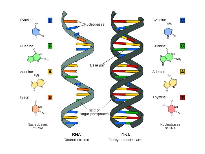
DNA vs. RNA and DNA are very similar, but there are some differences. First of all, DNA is double stranded, and RNA is single stranded. This means that RNA is SMALLER than DNA.
RNA contains 4 nitrogen bases: adenine, guanine, cytosine and URACIL. *Thymine is NOT present in RNA. Uracil is complementary to adenine in DNA. It essentially takes the place of thymine.
The last major difference between DNA and RNA is that RNA contains the 5 -carbon sugar ribose. (Recall DNA contains deoxyribose). Ribose has one more oxygen atom than deoxyribose. Ribose Deoxyribose
Recap RNA is single stranded, so it is smaller than DNA. This means it can leave the nucleus (which DNA cannot). RNA contains the sugar ribose. RNA has 4 bases: A, T, C, and U. The base pairing rules are as follows: C pairs with G pairs with A pairs with U pairs with G C U A NO thymine in RNA
3 Types of RNA’s job is to help DNA make proteins. DNA must deliver its code to the remainder of the cell- it relies on 3 molecules: 1) Messenger RNA (m. RNA) 2) Transfer RNA (t. RNA) 3) Ribosomal RNA (r. RNA)
Messenger RNA m. RNA is complementary to the original strand of DNA. m. RNA is first created in the nucleus and then travels to the ribosomes out in the cytoplasm. m. RNA uses the DNA’s code (or message) to make proteins! Example) DNA Strand: G G C T T A m. RNA strand: C C G A A U
Proteins Recall that proteins are made up of smaller parts called amino acids. Another word for protein is “peptide”. Individual DNA codes are called “codons”. The codons correspond to specific amino acids. m. RNA also has codons, which are complementary to DNA codons.
Codons consist of groups of 3 nucleotides called triplets. Example) DNA codon: cytosine-adenine (CCA for short) A C C Each codon codes for one amino acid. This is where we need RNA’s help.
DNA Template Strand: A C G T T A m. RNA strand: U G C A A U G C C C G G m. RNA is always complementary to the template DNA strand. How many codons are there? What does the other DNA strand look like?
Three DNA codons are transcribed into three m. RNA codons are specific to amino acids. This is the beginning step of PROTEIN SYNTHESIS. Protein= well, protein synthesis= to make
DNA Template Strand: A C G T T A G C C m. RNA strand: U G C A A U C G G 1) UGC 2) AAU 3) CGG Which three amino acids do these m. RNA codons code for?
Amino Acids Though there are only 20 different amino acids, they are sequenced differently and come in different shapes to make for thousands of different proteins.
Protein Synthesis • A two part process in which DNA is decoded into corresponding proteins • The first process is known as transcription • The second process is translation • Occurs in the nucleus and cytoplasm
Transcription is the first part of protein synthesis. During transcription, m. RNA is created by transcribing the DNA’s code. Transcription occurs in the nucleus. (That’s where the DNA is!)
Transcription During transcription, the enzyme RNA polymerase temporarily unzips DNA and adds complementary RNA nucleotides to the growing m. RNA strand.
Transcription Recall that m. RNA is the messenger. It copies DNA’s code (or “message”; “instructions”) and it is now responsible for delivering this message to the rest of the cell. Once the m. RNA strand is completed, it leaves the nucleus (exits via nuclear pores). Transcription is complete. (No protein yet. . . next stop, the ribosomes!)
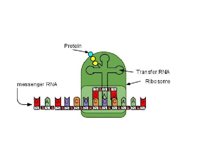
Translation is the final step of protein synthesis- it involves ALL THREE types of RNA (m. RNA, t. RNA and r. RNA). • Translation is a process in which the m. RNA that was manufactured during transcription is translated into an amino acid sequence (proteins) • occurs in the cytoplasm, on the ribosomes
Ribosomal RNA (r. RNA)= a major component of ribosomes; also helps bond amino acids together to make polypeptides (proteins)! The goal of the 3 types of RNA is to work together to make proteins using the DNA’s instructions!
Translation Transfer RNA (t. RNA)= helps transfer amino acids to the corresponding m. RNA codons (t. RNA is always complementary to the m. RNA strand) m. RNA codons: U G C A t. RNA anticodons: A C G U A U C G G U A G C C t. RNA bases are referred to as “anti-codons” because they are complementary to m. RNA codons. m. RNA strand (codons) t. RNA strand (anti-codons U G A C C A A U C G G G U U A G C C
LEUCINE Example) If the m. RNA codon is CUU, that would translate to the amino acid leucine. The t. RNA molecule that will deliver leucine to the ribosome has the anticodon GAA. ANTICODON Once the amino acid is delivered, the t. RNA releases itself from the ribosome, and leaves to find another amino acid to add to the growing protein chain.
Translation t. RNA transfers amino acids to the ribosome. The amino acids are attached to the t. RNA via a specialized enzyme called t. RNA synthetase. Analogy: t. RNA is a librarian; m. RNA codons are the book codes; amino acids are the books. t. RNA reads the m. RNA and fetches the appropriate amino acid.
Label the following: m. RNA, t. RNA, ribosome, codons, anti-codons, peptide (protein) chain, amino acid
V. Translation – taking m. RNA, “reading” it to bring the correct amino acids and connect them to make a protein. 1. m. RNA is made from a single strand of DNA in the nucleus (transcription). 2. m. RNA is carried from the nucleus to the cytoplasm. 3. m. RNA goes to a ribosome (r. RNA). The ribosome attaches to the start codon.
Mutations - changes in DNA sequence I. Gene Mutations: result from changes in a single gene A. Point Mutations: affect 1 DNA nucleotide 1. Also called substitutions, as 1 DNA nucleotide is substituted for another
B. Frameshift Mutations: shift the “reading frame” of the genetic message Insertion Deletion
II. Chromosomal Mutations: changes in the # or structure of chromosomes A. Deletion: loss of all or part of a chromosome B. Duplication: segment of a chromosome is repeated C. Inversion: part of a chromosome becomes oriented in reverse directions D. Translocation: part of 1 chromosome breaks off and attaches to another chromosome
Chromosomal Mutations
Puttin’ it all together DNA codes for proteins Proteins are made during protein synthesis Transcription occurs in the nucleus. The final product of transcription is an m. RNA strand. Translation occurs on the ribosomes. The final product of translation is a protein.
Codons There are 64 possible codons: 3 bases in a codon (triplets) 43 = 64 4 possible bases (A, T, C or G) 64 codons for 20 amino acids. Can more than one codon specify the same amino acid?
Start and Stop Signals Proteins are made up of a very specific sequence of amino acids. DNA contains “start” and “stop” codons so that the cell knows where to start decoding proteins and where to stop. The start codon= AUG (codes for the amino acid methionine) Stop codons= UAA, UAG, UGA