Cells of the Immune System and Antigen Recognition


- Slides: 29

Cells of the Immune System and Antigen Recognition Jennifer Nyland, Ph. D Office: Bldg#1, Room B 10 Phone: 733 -1586 Email: jnyland@uscmed. sc. edu

Teaching objectives • To review the role of immune cells in protection from different types of pathogens • To discuss the types of cells involved in immune responses • To describe the nature of specificity in adaptive immune responses • To understand the role of lymphocyte recirculation in immune responses

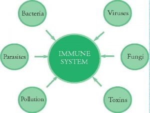
Overview of the immune system • Purpose: – Protection from pathogens • Intracellular (viruses, some bacteria and parasites) • Extracellular (most bacteria, fungi, and parasites) – Eliminate modified or altered “self” • Cancer or transformed cells • Sites of action: – Extracellular – Intracellular

Overview- extracellular pathogens • Ab are primary defense – Neutralization – Opsonization – Complement activation

Overview- intracellular pathogens • Cell-mediated responses are primary defense – Ab are ineffective – Two scenarios: • Pathogen in cytosol – Cytotoxic T cell (CD 8) • Pathogen in vesicles – Th 1 (CD 4) releases cytokines – Activates macrophages

Cells of the immune system Immune system Myeloid cells Lymphoid cells Granulocytic Monocytic T cells B cells Neutrophils Basophils Eosinophils Macrophages Kupffer cells Dendritic cells Helper cells Suppressor cells Cytotoxic cells Plasma cells NK cells

Development of the immune system Stem cell T cell Granulocyte Myeloid progenitor Lymphoid progenitor NK cell Mast cell B cell Monocyte Macrophage Dendritic cell Plasma Cell

Cells of the immune system Eosinophil Lymphocyte (T, B, NK) Plasma cell Basophil Granular Agranular (35% in circulation) Monocyte Neutrophil Dendritic cell

Phagocytosis and Intracellular killing Neutrophils and Macrophages

Phagocytes – neutrophils (PMNs) • Characteristic nucleus, cytoplasm • Granules • CD 66 membrane marker protein Neutrophil Geimsa stain Source: www. dpd. cdc. gov

Characteristics of neutrophil granules Primary granules Secondary granules Azurophilic; young neutrophils Specific for mature neutrophils Contain: cationic proteins, lysozyme, defensins, elastase and Contain: Lysozyme, NADPH oxidase components and myeloperoxidase Lactoferrin and B 12 -binding protein

Phagocytes – macrophages Macrophage Source: Dr. Peter Darben, Queensland University of Technology, used with permission • Characteristic nucleus • lysosomes • CD 14 membrane marker protein

Non-specific killer cells NK cells Eosinophils

Natural killer (NK) cells • Also known as large granular lymphocytes (LGL) • Kill virus-infected or transformed cells • Identified by the CD 56+/CD 16+/CD 3 • Activated by IL-2 and IFN-γ to become LAK cells

Eosinophils • Characteristic bi-lobed nucleus • Cytoplasmic granules, stain with acidic dyes (eosin) – Major basic protein (MBP) – Potent toxin for helminths Source: Bristol Biomedical Image Archive, used with permission • Kill parasitic worms

Mast cells Source: Wikimedia • Characteristic cytoplasmic granules • Responsible for burst release of preformed cytokines, chemokines, histamine • Role in immunity against parasites

Cells of the immune system: innate • Phagocytes – Monocytes/macrophages – PMNs/neutrophils • • NK cells Basophils and mast cells Eosinophils Platelets

Cells of the immune system: APC • Cells that link the innate and adaptive arms – Antigen presenting cells (APCs) • Heterogenous population with role in innate immunity and activation of Th cells • Rich in MHC class II molecules (lec 11 -12) – Examples • • Dendritic cells Macrophages B cells Others (Mast cells)

Cells of adaptive immune response T cells and B cells

Cells of the immune system: adaptive • Lymphocytes – B cells • Plasma cells (Ab producing) – T cells • Cytotoxic (CTL) • Helper (Th) – – Th 1 Th 2 Th 17 T-reg

Major distinguishing markers Marker B cell CTL T-helper Antigen R BCR (surface Ig) TCR CD 3 -- + + CD 4 -- -- + CD 8 -- + -- CD 19/ CD 20 + -- -- CD 40 + -- --

Specificity of adaptive immune response • Resides with Ag R on T and B cells • TCR and BCR – both specific for only ONE antigenic determinant • TCR is monovalent • BCR is divalent T cell TCR Ag Ag B cell BCR Ag

Specificity of adaptive immune response • Each B and T cell has receptor that is unique for a particular antigenic determinant on Ag • Vast array of different Ag. R in both T and B cell populations • How are the receptors generated? – Instructionist hypothesis • Does not account for self vs non-self – Clonal selection hypothesis • Ag. R pre-formed on B and T cells and Ag selects the clones with the correct receptor

Four principles of clonal selection Hθ 1. Each lymphocyte has a SINGLE type of Ag. R 2. Interaction between foreign molecule and Ag. R with high affinity leads to activation 3. Differentiated effector cell derived from activated lymphocyte with have the same Ag. R as parental lymphocyte (clones) 4. Lymphocytes bearing Ag. R for self molecules are deleted early in lymphoid development and are absent from repertoire

Specificity of adaptive immune response • Clonal selection Hθ can explain many features of immune response – Specificity – Signal required for activation – Lag in adaptive immune response – Discrimination between self and non-self

Development of the immune system Bone Marrow Tissues Thymus Stem cell Granulocyte T cell Myeloid progenitor Lymphoid progenitor NK cell Mast cell B cell Monocyte 2° Lymphoid Macrophage Dendritic cell Plasma Cell

Lymphocyte recirculation • Relatively few lymphocytes with a specific Ag. R – 1/10, 000 to 1/100, 000 • Chances for successful encounter enhanced by circulating lymphocytes – 1 -2% recirculate every hour

Lymphocyte recirculation • Lymphocytes enter 2° lymphoid organs via high endothelial venules (HEVs) • Ag is transported to lymph nodes via APC • Upon activation, lymphocytes travel to tissues Bone marrow Thymus T cell B cell Virgin lymphocytes B cell Primed lymphocytes B cell Monocyte B cell Spleen and lymph nodes T cell DC APC Tissues

Lymphocyte recirculation • After activation, new receptors (homing R ) are expressed to direct to tissues • R on lymphocytes recognize CAMs on endothelial cells • Chemokines at infection help attract activated lymphocytes Bone marrow Thymus T cell B cell Virgin lymphocytes B cell Primed lymphocytes B cell Monocyte B cell Spleen and lymph nodes T cell DC APC Tissues
 O antigen and h antigen
 Primary immune response and secondary immune response
 Immune cells meaning
 Mbg 453
 Immune effector cells
 Chapter 35 immune system and disease
 Primary vs secondary immune response
 Lesson 12 blood and immune system
 Lesson 12 blood and immune system
 Malt tonsils
 Paranasal sinuses development
 Red blood cells and white blood cells difference
 Similarities between plant and animal cells venn diagram
 Masses of cells form and steal nutrients from healthy cells
 What is the third line of defense in the immune system
 Flow chart of wbc
 Third line of defense immune system
 Second line of defense immune system
 Ap bio immune system
 Immune system lymph nodes
 Overreactions of the immune system
 Lymphatic vs immune system
 Phagocitize
 Defination of tuberculosis
 What are the first line of defense
 What is the main function of the immune system
 Thymus
 Immune complex
 Thalassemia facies
 1what's the purpose of the body's immune system?
