Immune cells Immune cells Classification of immune cells


 natural killer cell")

 l. Increased organelles l. Enhanced phagocytic")


 PRRs recognizes conserved molecular structures on microorganisms These molecular structures")

 u lipopolysaccharide (LPS, gram-positive bacteria) u")

 l Thirteen receptors identified in mice and humans")
 Ig. G Fc receptor (FcγR) 2) complement receptor (CR 1)")
 Oxygen-dependent killing mechanisms 2) Oxygen-independent killing mechanisms")
 Oxygen-dependent killing mechanisms During phagocytosis, the respiratory burst occurs A. reactive oxygen intermediates(ROIs)")
 Oxygen-independent killing mechanisms Lysozyme Defensins: a group of cationic peptides hydrolytic enzymes")


 pcovered with long membrane extensions puptake, processing and presentation of")

 cell p Large, granular lymphocytes with cytotoxic activity p No")
 cell p. The first line of defense against Ø virus")


")

 l The exact nature of ARs on NK cells is not")
 KIR 2 DL, KIR 3 DL l")


 is phagocytic cell important in the body’s defense")
- Slides: 46
Immune cells
Immune cells
Classification of immune cells p. Cells of the innate immune system monocyte/macrophage neutrophil eosinophil basophil dendritic cell (DC) natural killer cell (NK cell) p. Cells of the adaptive immune system B lymphocyte T lymphocyte
Cells mediating innate immunity monocyte/macrophage neutrophil eosinophil basophil dendritic cell (DC) natural killer cell (NK cell)
I. monocyte/macrophage Origin and development of macrophages
monocyte macrophage l. Enlarged cell size (5~10 -fold) l. Increased organelles l. Enhanced phagocytic ability
Macrophages in different tissues have various names
Functions of macrophage 1. Ingestion and digestion of pathogens
How do Macrophages Recognize Pathogens?
Pattern Recognition Receptors (PRRs) PRRs recognizes conserved molecular structures on microorganisms These molecular structures are not expressed by host tissues Mannose receptor PRRs Scavenger receptor Toll-like receptor (TLR)
Mannose receptor
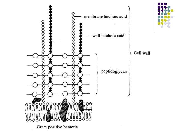
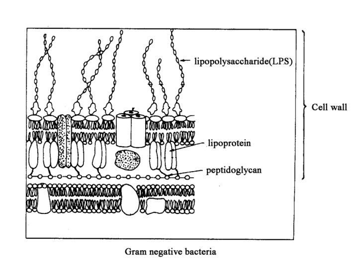
Scavenger receptor recognize u teichoic acid (gram-negative bacteria) u lipopolysaccharide (LPS, gram-positive bacteria) u phosphatidyl serine (apoptotic host cells)
Scavenger receptor Phagocytosis of apoptotic cells
Toll-like receptor (TLR) l Thirteen receptors identified in mice and humans
Opsonic receptor 1) Ig. G Fc receptor (FcγR) 2) complement receptor (CR 1)
Mechanisms of killing phagocytosed pathogens 1) Oxygen-dependent killing mechanisms 2) Oxygen-independent killing mechanisms
1) Oxygen-dependent killing mechanisms During phagocytosis, the respiratory burst occurs A. reactive oxygen intermediates(ROIs) NADH/NADPH oxidase O 2— (superoxide anion) . OH (hydroxyl radicals) H 2 O 2 (hydrogen peroxide) B. reactive nitrogen intermediates(RNIs) i. NOS (induced nitric oxide synthetase) L-arginine + O 2 + NADPH NO (nitric oxide) + L-citrulline + NADP NO reacts with O 2— forming peroxynitrite
2) Oxygen-independent killing mechanisms Lysozyme Defensins: a group of cationic peptides hydrolytic enzymes
Functions of macrophage 2. Antigen processing and presentation
Functions of macrophage 3. Secretion of factors
Granulocytes Neutrophil Eosinophil Basophil
II. Neutrophil p. Constitute 50%– 70% of the circulating WBC p. A life span of only 2 -3 days p. Rapid responders to pathogen invasion sensitive to chemoattractants the first to arrive at a site of inflammation
The process of neutrophil extravasation The steps in transendothelial migration and how it is directed are still largely unknown
II. Neutrophil p. Potent phagocytic activity ØPrimary granules: a type of lysosome contain peroxidase, lysozyme, and hydrolytic enzymes ØSecondary granules: contain collagenase, lactoferrin, and lysozyme p. Exhibit a larger respiratory burst than macrophages ØProduce more ROIs and RNIs ØNeutrophils are more likely than macrophages to kill ingested pathogens
III. Eosinophil p. Constitute 1%– 3% of the circulating WBC p. Contents of eosinophilic granules may damage the parasite membrane p. Help against parasites
IV. Basophil p< 1% of the circulating WBC p. Nonphagocytic granulocytes p. Pharmacologically active substances Øhistamine, leukotriene etc. p. Involved in allergic responses
V. Mast cell p. In mucosae and connective tissues p. Not in circulation p. Functionally indistinguishable from basophil
VI. Dendritic cell (DC) pcovered with long membrane extensions puptake, processing and presentation of antigens pexpress high levels of class II MHC molecules and costimulatory molecules
Interaction of DC and T cell
VII. Natural killer (NK) cell p Large, granular lymphocytes with cytotoxic activity p No specific antigen receptor CD 16 (Ig. G Fc receptor) and CD 56: typical NK cell markers p No thymic development Nude Mice Ølack a thymus Øno T cells Øhave functional NK-cell populations
VII. Natural killer (NK) cell p. The first line of defense against Ø virus infected cells Ø tumor cells p Cytolytic mechanisms of NK cell Ø Fas. L-Fas pathway Ø perforin/granzyme pathway Ø antibody-dependent cell-mediated cytotoxicity (ADCC)
The Fas. L-Fas pathway
The perforin/granzyme pathway l The most common mechanism
The perforin/granzyme pathway
Antibody-dependent cellmediated cytotoxicity (ADCC)
The mechanism by which NK cells recognize target cells l NK cells express two different categories of receptors Activation receptors The cytoplasmic tails contain the immunoreceptor tyrosine-based activation motif (ITAM) deliver activation signals Inhibition receptors The cytoplasmic tails contain the immunoreceptor tyrosine-based inhibition motif (ITIM) deliver inhibition signals
Activation receptors (ARs) l The exact nature of ARs on NK cells is not completely clear l Some of the candidate activation receptors l The ligands : are not known May be abnormal patterns of glycosylation on the surface of target cells
Inhibitory receptors l Killer immunoglobulin-like receptor (KIR) KIR 2 DL, KIR 3 DL l Killer lectin-like receptor CD 94/NKG 2 A l Ligand: MHC class I molecules
Opposing Signal Model of Cytotoxicity By NK Cells
VIII. NK T cell l Some of the characteristics of both T cells and NK cells Express T cell receptors (TCRs) Recognition of CD 1 -presented antigens Express NK-specific markers Kill tumor and virus-infected cells
What to remember l Cell function monocyte/macrophage neutrophil eosinophil basophil/mast cell dendritic cell NKT cell
Study question l What are functions of macrophages? 1. Ingestion and digestion of pathogens 2. Antigen processing and presentation 3. Secretion of factors
Study question l a. b. c. Explain why each of the following statements is false. Activation of macrophages increases their expression of class I MHC molecules, making the cells present antigen more effectively. All lymphoid cells have antigen-specific receptors on their membrane. Follicular dendritic cells can process and present antigen to T lymphocytes.
Study question l l l (Eosinophil) is phagocytic cell important in the body’s defense against parasitic organisms (Neutrophil) is generally first cell to arrive at site of inflammation (Basophil) is nonphagocytic granulocytic cell that release various pharmacologically active substances (Basophil) is white blood cell that migrate into the tissues and play an important role in the development of allergies (B cell) is an antigen-presenting cell that arises from the same precursor as a T cell but not the same as a macrophage