Antigen presenting cells T cells B cells primary

Antigen presenting cells, T cells, B cells, primary and secondary immune organs, mucosal immune system Martin Liška

Antigen presenting cells

Antigen presenting cells • A. Professional - Expression of MHC class I and II gp. - Monocytes, macrophages, dendritic cells and B cells • B. Non-professional - Expression of MHC class I gp. ; expression of MHC class II gp. when induced by pro-inflammatory cytokines - fibroblasts, endothelial and epithelial cells
) in")
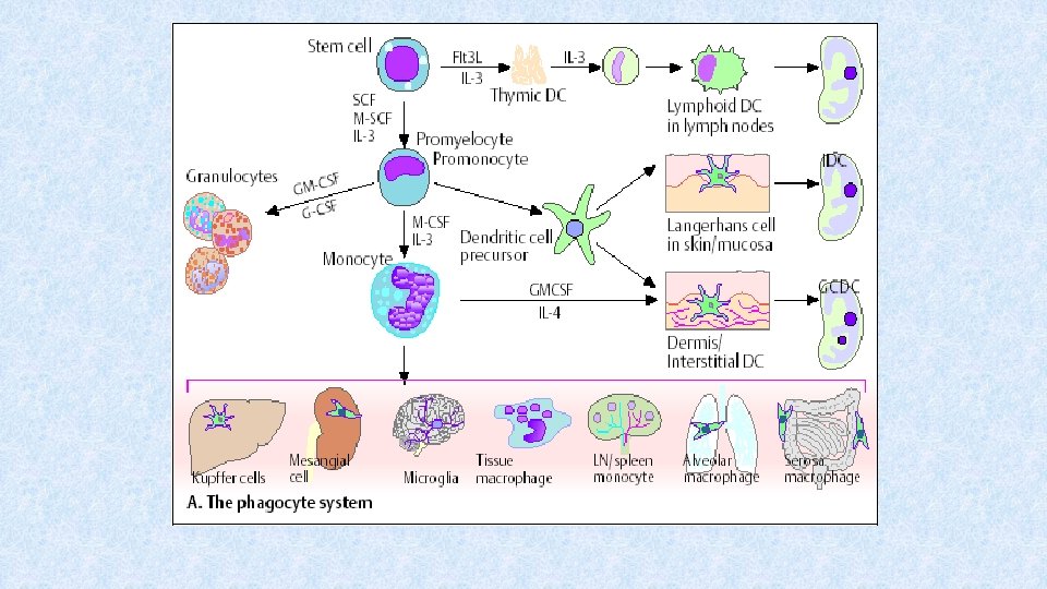
Monocytes • Differentiate from myeloid precursor (differentiated form hematopoietic stem cell (CD 34+)) in bone marrow • Continuously released from bone marrow to periphery • 7% in blood circulation; the rest is located in bone marrow or spleen – the proportion is influenced by cytokines and bacterial products • Monocytes adhere to vessel wall by using b 1 -integrins binding to VCAM-1 on endothelial cells

Monocytes • Then, monocytes move among vessel endothelial cells to tissues • In tissues, they differentiate into different subtypes of tissue macrophages (Kupffer cells, microglia, alveolar macrophages etc. )

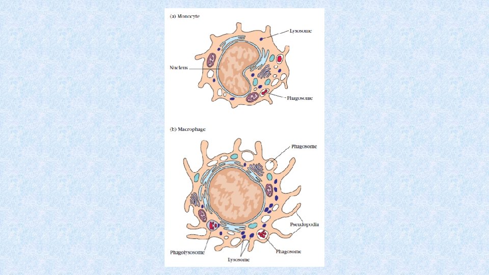
Macrophages • Terminal stage of differentiation of monocytemacrophage cell line • Mature monocytes are released to peripheral blood stream, then catched in organs and differentiate to tissue macrophages • There are different subpopulations of macrophages in different tissues

Subtypes of macrophages • Kupffer cells – liver • Alveolar macrophages - lungs • Microglia – CNS • Osteoclasts – bone • Histiocytes – connective tissue • Mesangial cells - kidney
 – produced by")
Cytokines influencing the development of macrophages • SCF (Stem Cell Factor) – produced by stromal cells → development and maintenance of hematopoietic stem cell function • GM-CSF (Granulocyte-Monocyte Colony Stimulating Factor) – produced by bone marrow stromal cells, lymphocytes → stimulation of monocyte differentiation • M-CSF (Monocyte Colony Stimulating Factor) – produced by stromal cells, lymohocytes, endothelial and epithelial cells → stimulation of monocytes differentiation • IL-3 – produced by lymphocytes → monocyte (and other cells) differentiation

Macrophage surface markers • MHC class I and II gp. • CD 35 - CR 1 receptor for C 3 b fragment of complement system • Fc receptor for Ig. G • CD 14 – receptor binding bacterial lipopolysaccharides • Receptors recognizing apoptotic cells
 – presentation of Ag")
Macrophages - function • Proffesional antigen presenting cells (APC) – presentation of Ag at an early stage of the specific immune response • Phagocytes – uptake of microbes and old or damaged own cells • Completely functional when activated by cytokines produced by T cells (IFN-g) • Production of cytokines, enzymes, complement system components, microbicidal, cytotoxic and tumoricidal substances, bioactive lipids (PG, PC, TX, LT)

Cytokines produced by macrophages • IL-1 a and b – stimulation of T and B cells, activation of the other macrophages • IL- 6 – endogenous pyrogen, activator acute phase proteins synthesis in liver • TNFa – similar function to IL-1

Cytokines produced by macrophages • IL-8 – secreted by activated macrophages, induce chemotaxis of neutrophils, T cells • IL-12 – induction of Th 1 response, supression Th 2 response • IFN-a – induction of production of enzymes inhibiting virus replication, increases expression of MHC class I gp. by host cell, activation of NK cells, T cells and the other macrophages
 • Differentiate from myeloid or lymphoid precursor • DCs are the")
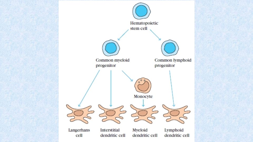
Dendritic cells (DCs) • Differentiate from myeloid or lymphoid precursor • DCs are the most important APCs • Migration of DCs precursors to tissues is influenced by chemokines (RANTES, MCP-1, MIP 1 a, MIP-1 b) • DCs are dispersed in all tissues and organs
 • Upon contact with antigens, they mature and migrate to secondary")
Dendritic cells (DCs) • Upon contact with antigens, they mature and migrate to secondary immune organs where induce antigen-specific immune response • Expression of MHC class I and II gp. , co-stimulatory ligands and adhesive molecules necessary for activation of naïve T cells • DCs have numerous processes enabling contact with up to 3000 T cells • In lymph nodes, DCs increasing expression of: • MHC class I and II gp. + co-stimulatory molecules (CD 80, CD 86)

Types of DCs • Langerhans cells – from myeloid line, in epidermis • Interstitial DCs – from myeloid line, in dermis and most of organs • Lymphoid (plasmocytoid) DCs – form lymphoid line, in blood and secondary immune organs • Follicular dendritic cells (FDCs) – presentation of antigen to B cells during secondary immune response

Surface markers of DCs • Receptors for Ig. G, Ig. E and C 3 b – enable internalization of immune complexes • Pattern Recognition Receptors - TLR and CD 14 (= receptor for LPSs) • Receptors recognizing apoptotic cells • Molecules important for presentation of antigen - mature DCs exprime MHC class I and II gp. and co-stimulatory molecules CD 80 and CD 86

Function of DCs • DCs are the most important APCs • DCs can be easily infected by viruses → viral proteins processed in a complex with MHC class I gp. → activation of Tc cells • DCs are able to internalize extracellular viral particles → viral peptides presented in a complex with MHC class II gp. → activation of Th 2 cells → help to B cells → production of anti-viral antibodies • DCs can also be activated by apoptotic cells

B cells

B cells • B cells are responsible for specific antibody-mediated immune response • B cells recognize native antigen by using BCR (B cell receptor) = surface bound Ig. M (or Ig. D) • Such B cell is stimulated to proliferation and differentiation to effector (plasma) cells producing huge amount of antibodies of the same specifity like BCR • Part of stimulated B cells differentiate to memory cells

Surface markers of B cells • CD 19 - typical B cell surface marker • CD 20 – expressed on the surface of Ig-positive B cells • Ig. M, Ig. D - BCR • MHC class I and II gp. - antigen presenting molecules • CD 40 – co-stimulatory receptor

Differentiation of B cell differentiation begins in bone marrow and completes in secondary immune organs after contant with an antigen Hematopoietic stem cell B cell progenitor → start of recombination processes → many clones of B cells with distinct BCR Pre B cell → expression of pre-B receptor (H(m) chain and surrogate L chain) Immature B cell → expression of surface Ig. M (BCR) elimination of autoreactive clones Mature B cell → expression of surface Ig. M or Ig. D (BCR)

BCR • BCR consists of surface immunoglobulin (Ig. M, Ig. D – H chains are membrane-bound; recognition of Ag) and associated signalling molecules (Iga and Igb) which are associated with cytoplasmic protein tyrosin-kinases (PTK) Src heavy chain light chain • If Ag binds simultaneously to 2 or more BCRs → PTKs draw closer → mutual phosphorylation of PTKs → phosphorylation of other cytoplasmic proteins → changes in gene transcription → proliferation, differentiation of B cell → secretion of antibodies cytoplasmic membrane B cell

Elimination of autoreactive clones of B cells • Recombination processes could give arise the clones of B cells who bear autoreactive BCRs and produce autoreactive antibodies • Majority of autoreactive B cells is eliminated at stage of immature B cell – if they bind autoantigen by their BCR with a sufficient afinity, then they receive signal leading to apoptosis • If some autoreactive clones pass through elimination proces, then it does not manifest – lack of suitable TH cells, many autoantigens are cryptic, or they are in low concentrations → ignored

Meeting of B cell with an antigen in secondary immune organ antigen presenting cell mitosis differentiation plasma cells antibodies memory B cells

Function of B cells • Antibody response • Immunological memory • Presentation of antigen – B cells are able to pick-up exogenous antigens by using BCR (typically allergens or toxins), to internalize them by endocytosis and to present them with MHC class II gp. to TH cells

Primary immune organs

Primary immune organs • Bone marrow, thymus • Sites of inception, maturation and differentiation of immunocompetent cells • Immature lymphocytes get there their antigenic specifity

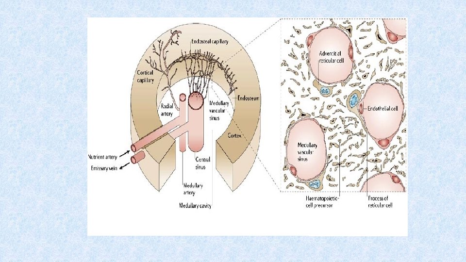
Bone marrow • Site of haematopoiesis in postnatal period • Localized in flat bones (pelvis, scapula, sternum, ribs) or in cancellous material of epiphysis of long bones • Consists of cells, extracellular matrix and blood vessels • Blood cells differentiate from hematopoietic stem cell in regions bordered by vascular sinuses • Sinuses lined with endothelial cells producing cytokines • Outer wall of sinuse bordered by reticular cells
 Haematopoiesis starts in yolk sac, then it moves to fetal liver")
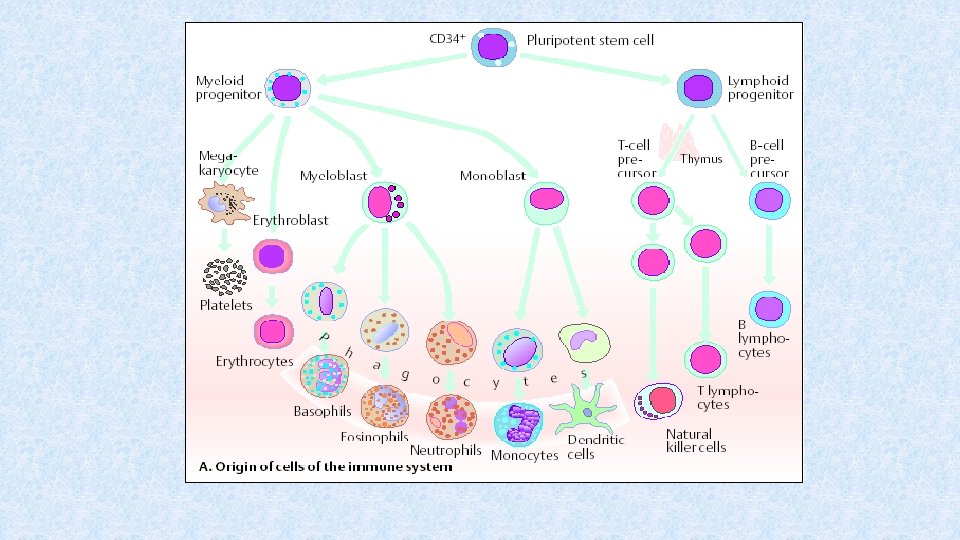
Immune cells (immunocytes) Haematopoiesis starts in yolk sac, then it moves to fetal liver and spleen (3 th to 7 th month of gestation) The main site of postnatal haematopoiesis is bone marrow All blood cells differentiate from hematopoietic stem cell (HSC)(CD 34+). HSC proliferates and maintains throughout whole life Haematopoiesis is controlled by cytokines secreted by stromal cells, activovated TH cells and macrophages
 – is")
Haematopoiesis in bone marrow • Differentiation from HSCs (CD 34+ CD 45+) – is regulated by membrane interactions between HSCs and stromal cells + cytokines (SCF, IL-3, thrombopoietin, erythropoietin) • Differentiation from less differentiated precursors to more differentiated stages • Haematopoiesis react to momentary needs of macroorganism

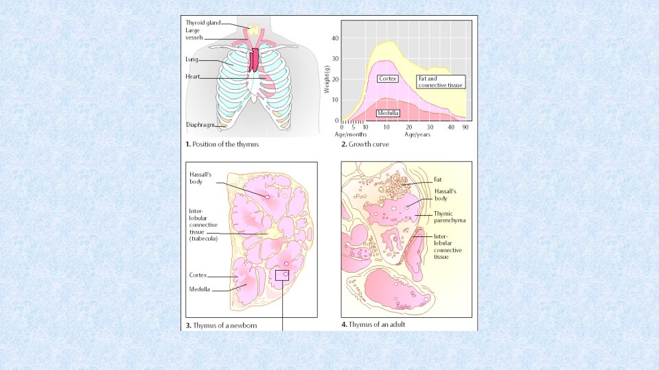
Thymus • Site of T cell differentiation • 2 lobes consisting of cortex (cluster of lymphocytes = rapidly proliferating cells) and medulla (mature cells, Hassall’s corpuscles) separated by corticomedulary junction • In cortexu and corticomedullary junction, there are macrophages and dendritic cells influencing the differentiation of thymocytes
 secreted by thymic")
Thymus • Differentiation is regulated by thymic hormones (thymulin, thymopoietin, thymosin) secreted by thymic epithelial cells and the other growth factors (SCF, IL-7) • Proliferation and differentiation of T cells in thymus → development of TCR → negative and positive selection of T cells • Thymus – induction of tolerance of own antigens

Secondary immune organs

Secondary immune organs • „Meeting-point“ of immunocompetent cells and antigens • Proliferation of immunocompetent T and B cells • Terminal differentiation of lymphocytes to effector cells

Secondary immune organs • Spleen – in comparison to lymph nodes, spleen filtrates the blood and picks-up antigens • Lymph nodes – filtration of lymph, uptake of antigens • MALT (Mucosa Associated Lymphoid Tissue) – diffuse and organized lymphoid tissue, uptake of antigens passing through mucous membranes, their presentation and induction of immune response

Spleen and lymph nodes • Passage of the majority of circulating lymphoid cells • Uptake of microbial antigens from blood and lymph • Lobular structure with trabeculae • Stabilization by reticular connective tissue

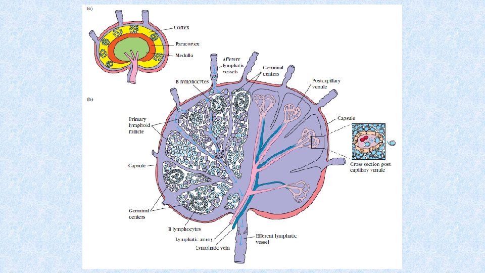
Lymph nodes • Located along lymphatic vessels • Drainage of the skin and the other surface tissues - cervical, axillary, inguinal l. n. • Drainage of mucous membranes and internal organs - mesenterial, mediastinal, paraaortic l. n. • Convex side of lymph node – entrance of afferent lymphatic vessels (subcapsular sinus) • Hilum – entrance of afferent and efferent blood vessels, exit of efferent lymphatic vessels • Node is surrounded by fibrous capsule which forms trabeculae containing vessels and nerves

Lymph node - cortex • Contains primary and secondary follicles – outer portion more B cells, deeper layers more T cells • Paracortical region - T cells and accesory cells (macrophages, dendritic cells) • Dendritic cells enter to lymph nodes with processed antigen → antigen presentation to T cells OR antigen alone enters the lymph node → uptake by APCs → antigen processing → antigen presentation

Lymph nodes - medulla • Medullary cords surround medullary sinuses that run to hilum • B and T cells migrate from follicles and paracortical region to medulla together with plasma cells → efferent vessels → migration to tissues and organs • T and B cells - effector cells, a portion differentiates into memory cells • Efferent lymphatic vessels → ductus thoracicus → vena subclavia (blood circulation)

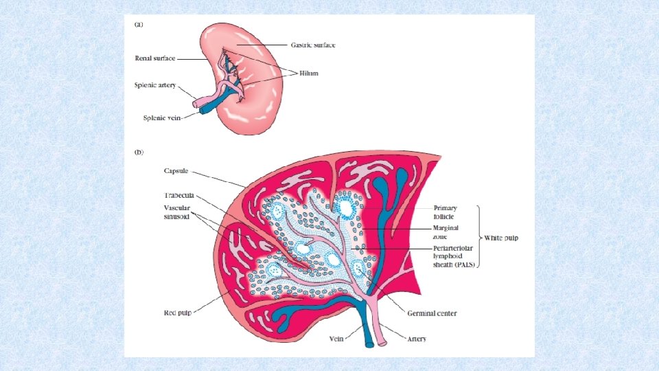
Spleen • Oval organ with lobular structure • Located in the left upper quadrant of abdomen • Consists of red and white pulp

Spleen - red pulp • Splenic sinuses between arteries and veins → filtration of blood • Marcophages remove from the blood old erythrocytes and leukocytes and microbial antigens • Elimination of immune complexes bound to erythrocytes

Spleen – white pulp • Lymphoid tissue located in periarteriolar lymphoid sheats and lymphoid follicles • In lymphoid follicles more B cells, in periarteriolar lymphoid sheats more T cells • Primary and secondary lymphoid follicles • Secondary follicles – germinal centre (isotype switching, somatic hypermutations → affinity maturation of B cells´ response)

Spleen - function • Filtration of blood – removal of old or damaged blood cells • Removal of immune complexes • Immune functions – specific cell-mediated and humoral immune response (affinity maturation, somatic hypermutations, isotype switching, response to T-independent antigens) – in case of absence or loss of spleen, vaccination against Pneumococci, Haemophilus, Neisseria is necessary • Reservoir of erythrocytes, lymphocytes and the other blood elements
")
Mucosa Associated Lymphoid Tissue (MALT)

Function and structure of mucosal and skin immune system • Mucosa and skin are in permanent contact with the environment, concentration of cca 80% immunocompetent cells • Skin – barrier against mechanical, physical and chemical damage and penetration of microorganism, in human cca 1. 5 m 2 • Mucosal immune system – barrier against penetration of pathogenic micoorganisms, prevention of induction of selfdamaging immune reactions against pathogens or harmless antigens from environment, cca 400 m 2

Mucosal immune system • Mucous membrane of oral and nasal cavity, respiratory, gastrointestinal and urogenital tract, eye, ear etc. • Non-immunological defense mechanisms: mucociliary clearance, flow of the air and fluids, secretions of exocrinal glands with bactericidal effects (fatty acids, lysozyme, pepsin, defensins), low p. H in stomach and urine
 BALT (Bronchus Associated")
Structure of mucosal immune system • MALT (Mucosa Associated Lymphoid Tissue) BALT (Bronchus Associated Lymphoid Tissue) GALT (Gut Associated Lymphoid Tissue) • o-MALT (organized) – lymphoid follicles in submucosa; Waldeyer’s tonsilar ring, appendix, Peyer’s patches • d-MALT (diffuse) – leukocytes spread in lamina propria mucosae (T and B cells, macrophages, neutrophils, eosinophils, mast cells)

Intraepithelial T cells • Located mainly in villi of small intestine • Mostly bear TCRgd and co-receptor CD 8 • Produce TGFb (healing of mucous membrane) • Suppress undesirable immune reactions against food allergens (→ oral tolerance)

Humoral mechanisms of mucosal immune system s Ig. A • Secretory immunoglobulin A • The most important mucosal immunoglobulin, also in breast milk • transcytosis – Ig. A is transported through epithelium by using transport Fc receptor (poly-Igreceptor) → in luminal surface, Ig. A is cleaved-off together with portion of transport Fc-receptor (secretory component) which protects s. Ig. A from gut proteases • Neutralization of antigens attacking mucous membrane; Ig. A does not activate complement system; Ig. A binds Fc receptors of phagocytes; Ig. A- containing immune complexes can be catched in Peyer’s patches and then induce immune response

Induction of mucosal immune reaction Oral tolerance * the majority of orally administered antigens induces suppression of specific immune response (a size of antigen is crucial) * Treg cells (regulatory) – production of IL-10 Induction of mucosal immune reaction M cells - specialized enterocytes ensuring transportation of Ag (endocytosis) - in close contact with lymphocytes and APCs Immunization in mucosa stimulates Th 2 and Th 3 cells and production of Ig. A

Mucosal barrier of the gut
- Slides: 59