Cells and organisation of living organisms Hevi Diyowati


- Slides: 28

Cells and organisation of living organisms Hevi Diyowati

Learning Objectives • State that living organisms are made of cells. • Identify and describe the structure of a plant cell (palisade cell) and an animal cell (liver cell), as seen under a light microscope. • Describe the differences in structure between typical animal and plant cells. • Relate the structures seen under the light microscope in the plant cell and in the animal cell to their functions.

Cells Every living thing on Earth is made up of cells. Cells keep living things organized. Some organisms, like bacteria, are only as big as a single cell. In an organism as complex as a human, there is no way you we could do everything we do and be just a single cell. There a lot of cells work in our body. Cells contain so many organelles with it’s own function and shape. Page 3

Animal cell • A animal cell ENDOPLASMIC RETICULUM (ER) Rough ER Smooth ER Nuclear envelope Nucleolus NUCLEUS Chromatin Flagelium Plasma membrane Centrosome CYTOSKELETON Microfilaments Intermediate filaments Ribosomes Microtubules Microvilli Golgi apparatus Peroxisome Figure 6. 9 Mitochondrion Lysosome In animal cells but not plant cells: Lysosomes Centrioles Flagella (in some plant sperm)

Plant cell • A plant cell Nuclear envelope Nucleolus Chromatin NUCLEUS Centrosome Rough endoplasmic reticulum Smooth endoplasmic reticulum Ribosomes (small brwon dots) Central vacuole Tonoplast Golgi apparatus Microfilaments Intermediate filaments CYTOSKELETON Microtubules Mitochondrion Peroxisome Plasma membrane Chloroplast Cell wall Plasmodesmata Wall of adjacent cell Figure 6. 9 In plant cells but not animal cells: Chloroplasts Central vacuole and tonoplast Cell wall Plasmodesmata

Cytoplasm : Place where many chemical reactions take place • semifluid, “factory area” • Major Elements ▫ Cytosol – semitransparent, largely water ▫ Organelles – “little organs”, specialized compartments, specific functions

The Nucleus: Genetic Library of the Cell • The nucleus – Contains most of the genes in the eukaryotic cell • The nuclear envelope – Encloses the nucleus, separating its contents from the cytoplasm Nucleus 1 µm Nucleus Nucleolus Chromatin Nuclear envelope: Inner membrane Outer membrane Nuclear pore Pore complex Rough ER Surface of nuclear envelope. 1 µm Ribosome 0. 25 µm Close-up of nuclear envelope Figure 6. 10 Pore complexes (TEM). Nuclear lamina (TEM).

Ribosomes: Protein Factories in the Cell Ribosomes Cytosol ER Free ribosomes Endoplasmic reticulum (ER) Bound ribosomes Large subunit 0. 5 µm TEM showing ER and ribosomes Figure 6. 11 – Carry out protein synthesis Small subunit Diagram of a ribosome

The Endoplasmic Reticulum: Biosynthetic Factory There are two distinct regions of ER Smooth ER, which lacks ribosomes (SER) The smooth ER ◦ Synthesizes lipids ◦ Metabolizes carbohydrates ◦ Stores calcium ◦ Detoxifies poison Rough ER, which contains ribosomes (RER) The rough ER ◦ Has bound ribosomes ◦ Produces proteins and membranes, which are distributed by transport vesicles Figure 6. 12 Smooth ER Nuclear envelope Rough ER ER lumen Cisternae Ribosomes Transport vesicle Smooth ER Transitional ER Rough ER 200 µm

The Golgi Apparatus: Shipping and Receiving Center • Functions of the Golgi apparatus include – Modification of the products of the rough ER; packaging. – Manufacture of certain macromolecules

Lysosomes: Digestive Compartments • A lysosome – Is a membranous sac of hydrolytic enzymes – Can digest all kinds of macromolecules Nucleus 1 µm Lysosome contains active hydrolytic enzymes Food vacuole fuses with lysosome Hydrolytic enzymes digest food particles Digestive enzymes Lysosome Plasma membrane Digestion Food vacuole (a) Phagocytosis: lysosome digesting food

Vacuoles: Diverse Maintenance Compartments • A plant or fungal cell – May have one or several vacuoles • Central vacuoles – Are found in plant cells – Hold reserves of important organic compounds and water Central vacuole Cytosol Tonoplast Nucleus Central vacuole Cell wall Chloroplast 5 µm

Mitochondria Are the sites of cellular respiration • Mitochondria are enclosed by two membranes – A smooth outer membrane – An inner membrane folded into cristae Mitochondrion Intermembrane space Outer membrane Free ribosomes in the mitochondrial matrix Inner membrane Cristae Matrix Figure 6. 17 Mitochondrial DNA 100 µm

Chloroplasts: Capture of Light Energy • The chloroplast – Contains chlorophyll • Chloroplasts – Found only in plants, are the sites of photosynthesis – Are found in leaves and other green organs of plants and in algae • Chloroplast structure includes – Thylakoids, membranous sacs – Stroma, the internal fluid

• Chloroplasts Chloroplast Ribosomes Stroma Chloroplast DNA Inner and outer membranes Granum Figure 6. 18 1 µm Thylakoid

Animal cell

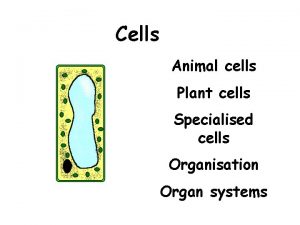
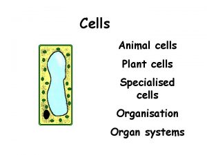
Plant cell

Plant cell & Animal Cell The Differences Between Animal & Plant Cell The Part of Cell Animal Cell Plant Cell Morpholo gy Physiolog y Reproducti on Size Relatively smaller Relatively larger Shape Irregular Regular Cell wall No Present Vacuole Small or absent Large Nucleus position Centre Near cell wall Food store Glycogen Starch Chloroplast No Present Sentrosom Present No

Organisation of Living Organisms • • • We must have many different, and many different kinds of cells. One of the main functions of cells is to organize the body. We have brain cells, stomach cells, bone cells, and many other types of cells. They all do special jobs to help our bodies function the way they are supposed to. Although the cells in our bodies do many different jobs, they all contain similar parts called organelles. In the same way there are different kinds of cells inside us, different organisms have different types of cells. Trees have different cells than us and so do dogs and birds. Each of those cells is different in some way. Every cell has a special job to do.

Specialised Cells Plant cells may become specialised to carry out particular functions. The cells are organised into plant tissues and plant tissues are organised into plant organs, (roots, stems, leaves and flowers). Plant tissue Main function Parenchyma A packing and supporting tissue. Epidermis Provides a waterproof, microorganism proof, outer layer to the plant. Xylem Transports water and salts from roots to leaves. Phloem Carries sugars and other foods from the leaves to where they are needed in the plant.

The drawings below illustrate how some plant cells/tissues are adapted in structure to perform particular functions. Epidermal cells (Surface layer of plant). Thick waterproof, microbe-proof cuticle. Prevents dehydration and infection. Xylem vessels Phloem sieve tube Rings and spirals of impermeable, strong lignin for support and strength. Parenchyma cells (The main ‘packing’ tissue of the plant, also give support). Air spaces Cells packed tightly together Cells become empty tubes as end walls break down. Allows transport of water and salts upwards. End wall forms a sieve plate which allows transport of dissolved foods up or down.

Structure of a leaf: Waxy cuticle – waterproof layer (reduces water loss from surface) ue la cu r s tis Veins (strands of xylem and phloem from midrib) as v f eo m or a dl n xylem bu F phloem Leaf stalk (petiole) stem Stomata – Midrib (vascular tissue i. e. pores on phloem and xylem) underside of leaf through which carbon dioxide, oxygen and water vapour are exchanged

Section through a leaf. Upper side of leaf waxy cuticle to prevent water loss Upper epidermis – transparent, no chloroplasts nucleus vacuole cytoplasm Palisade cell – many chloroplasts for much photosynthesis Chloroplast – membranes covered with chlorophyll xylem – transports water Spongy mesophyll cells – fewer chloroplasts Air space phloem – transports food lower epidermis Guard cells Stoma lower side of leaf

Level of Organisation • A tissue is a group of cells, of similar structure, which perform a particular function • An organ is a functional unit made up of several different tissues, the individual functions of which contribute to the functions of the organ as a whole. • Organ system Organs work together form organ system which have particular task • Organisms, Organ systems work to together in organisms

A Hierarchy of Biological Organization • The hierarchy of life. Extends through many levels of biological organization 1 The biosphere Figure 1. 3 • From the biosphere to organisms

A Hierarchy of Biological Organization • From cells to molecules 9 Organelles 1 µm Cell 8 Cells Atoms 10 µm 7 Tissues 50 µm 6 Organs and organ systems Figure 1. 3 10 Molecules

A Closer Look at Ecosystems • Each organism – Interacts with its environment • Both organism and environment – Are affected by the interactions between them • The dynamics of any ecosystem include two major processes – Cycling of nutrients, in which materials acquired by plants eventually return to the soil – The flow of energy from sunlight to producers to consumers

Page 28
 Venn diagram living and nonliving things
 Single celled and multi celled organisms
 Competitive interaction
 5 levels of organisms
 Onodi cells and haller cells
 Chlorocruorin
 Venn diagram animal and plant cells
 Masses of cells form and steal nutrients from healthy cells
 Eucariote
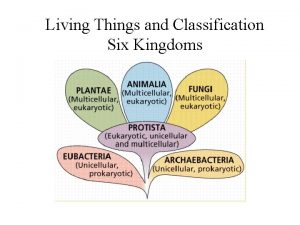
 Kingdoms of living things
 Which principle
 Are all living things based on the metric system
 Fungi cell
 Apkbs
Apkbs Living organisms differ in
 Two different organisms living together
 Archaebacteria vs eubacteria
 What is the smallest unit of living organisms
 Living organisms
 Diffrent types of fossils
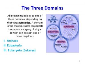
 The three domains of life
 Similar
 Two different organisms living together
 Guinea worm
 Small living creatures tom 21
 Unicellular living organisms
 Remains imprints or traces of once-living organisms
 Mitochondria information
 Regulation of tubular reabsorption
