Case Studies in Sports Dermatology Kent Scheff MD




- Slides: 58
Case Studies in Sports Dermatology Kent Scheff MD University of Michigan Primary Care Sports Medicine Fellow
Disclosures • None
Objectives • Be able diagnose common infections encountered in the training room. • Choose appropriate therapy for the above infections • Be able to propose an appropriate time for athletes to return to competition following infection • Propose methods for reducing further infection and recurrence.
Case #1 Too Close for Comfort
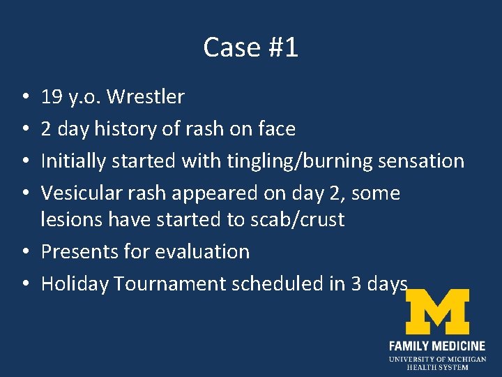
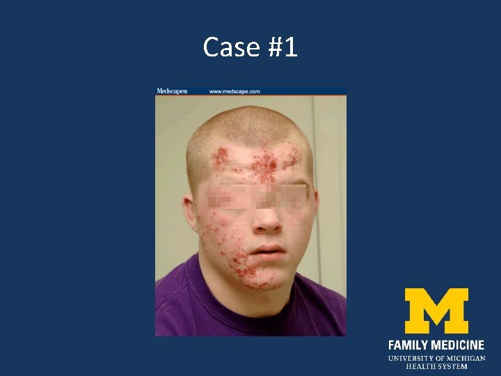
Case #1 19 y. o. Wrestler 2 day history of rash on face Initially started with tingling/burning sensation Vesicular rash appeared on day 2, some lesions have started to scab/crust • Presents for evaluation • Holiday Tournament scheduled in 3 days • •
Case #1
Herpes Gladiatorum • Caused by Herpes Simplex Virus 1 • Prominent among wrestlers and in other sports with skin to skin contact • Accounts for 39% of skin infections in the NCAA injury surveillance of wrestlers • Incubation is 2 -20 days however, most lesions appear after 3 -5 days of exposure
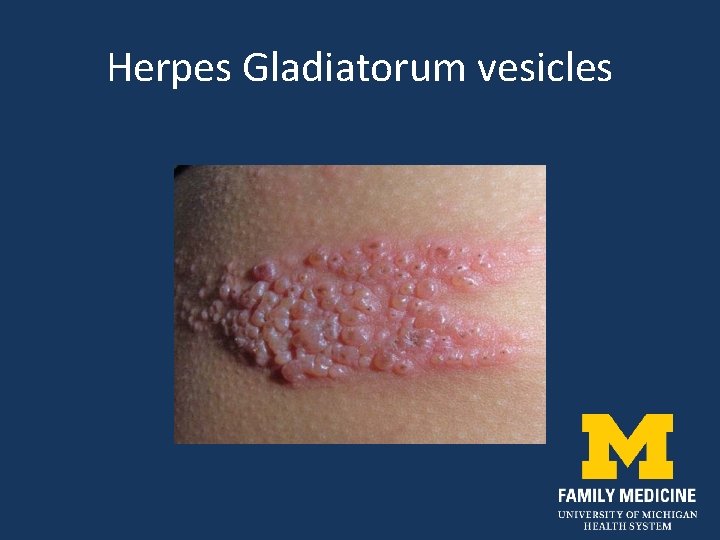
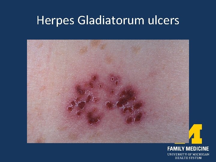
Herpes Gladiatorum • Initial presentation is seen on head, neck, face (right side) ears, torso or upper extremities • Primary infections may present with constitutional symptoms along with burning, tingling or stinging at the site • Vesicles then form on an erythematous base • Vesicles ulcers plaques
Herpes Gladiatorum vesicles
Herpes Gladiatorum ulcers
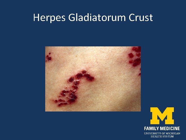
Herpes Gladiatorum Crust
Diagnostic Methods • Gold standard is Viral Culture remembering that maximum sensitivity is achieved in 2 -7 days. • Sample base of a disrupted vesicle during active lesion • HSV PCR test is more sensitive and faster though more expensive • Serology Ig. M 20 -25 days, Ig. G 6 -8 weeks after exposure
Treatment • Early treatment is key; virus exponentially replicates within the first 48 hours • Acyclovir 400 mg TID • Valacyclovir 1 gm BID 7 -10 days • Famciclovir 500 mg Bid • Recurrence: identical dosing for 5 days
Return to Play • Free of systemic symptoms • No new lesions for 72 hours • All lesions must be dry with a firm adherent crust • Appropriate treatment with a systemic antiviral for 120 hours prior to competition • Active lesions are not to be covered to allow competition
Prevention • Early diagnosis and quarantine of affected athletes • Athlete education “no sharing” of towels • Disinfection of commonly used items, mats, exercise equipment • Antiviral prophylaxis has been shown to decrease acquisition and spread of Herpes Gladiatorum in wrestlers • Valacyclovir 500 mg to 1 gm daily
Case #2 Training Room Challenge
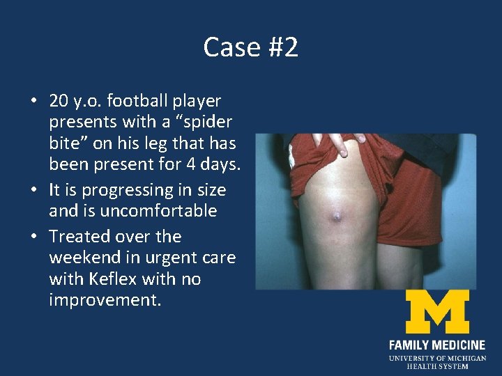
Case #2 • 20 y. o. football player presents with a “spider bite” on his leg that has been present for 4 days. • It is progressing in size and is uncomfortable • Treated over the weekend in urgent care with Keflex with no improvement.
Community Acquired MRSA • Most S. aureus are beta-lactamase producing, resistant to penicillin, but susceptible to cephalosporins and the penicillinase-resistant penicillins (nafcillin, oxacillin, dicloxicillin), or Blactam-B-lactamase inhibitor combinations (e. g. , amoxicillinclavulanate)
CA-MRSA • Increased prevalence of S. aureus that have the mec. A gene that produces the penicillin-binding protein PBP 2 a • Confers resistance to all currently FDAapproved B-lactams
CA-MRSA • Study of University affiliated ERs found that 76% of skin/soft tissue infections caused by S. aureus – 59% of total due to MRSA – 77. 8% of S. aureus were MRSA – 99% of MRSA were CA-MRSA
CA-MRSA • Median age of HA-MRSA: 68 yrs • Median age of CA-MRSA: 23 yrs • 2006 survey of Texas Athletic Trainers found that 32% had treated MRSA infections in their athletes
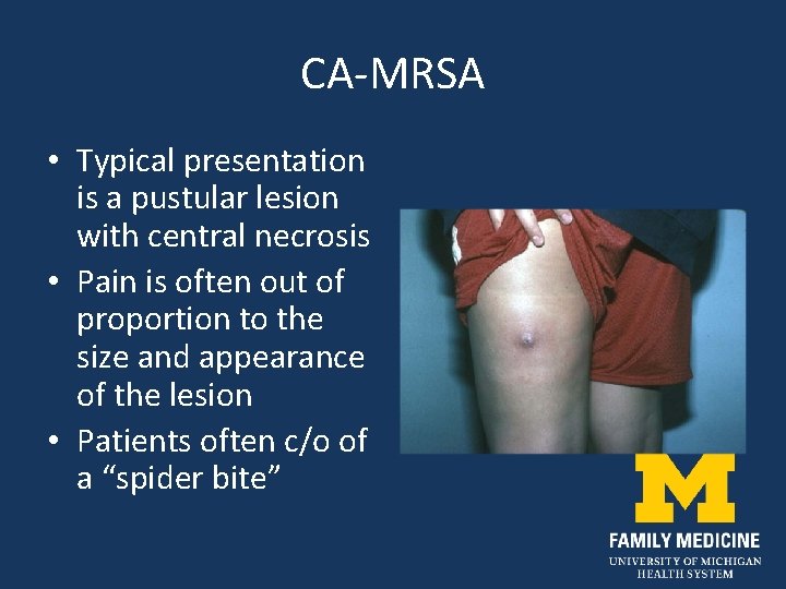
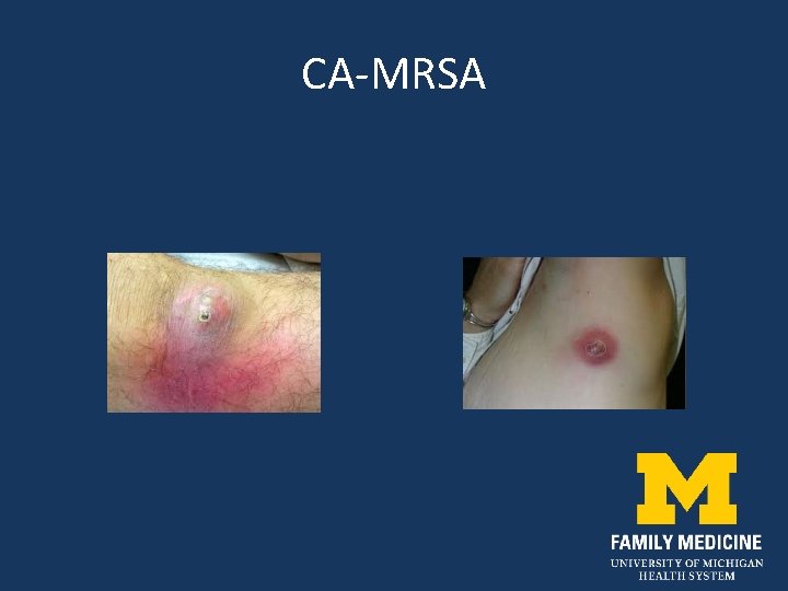
CA-MRSA • Typical presentation is a pustular lesion with central necrosis • Pain is often out of proportion to the size and appearance of the lesion • Patients often c/o of a “spider bite”
CA-MRSA
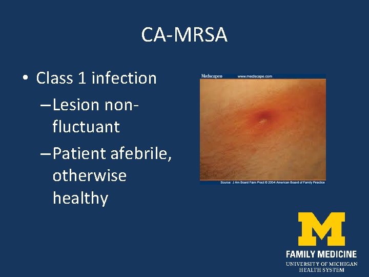
CA-MRSA • Class 1 infection – Lesion nonfluctuant – Patient afebrile, otherwise healthy
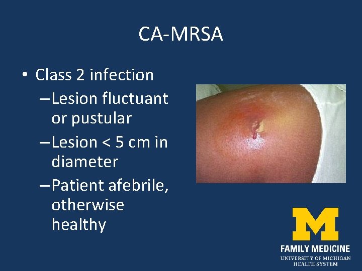
CA-MRSA • Class 2 infection – Lesion fluctuant or pustular – Lesion < 5 cm in diameter – Patient afebrile, otherwise healthy
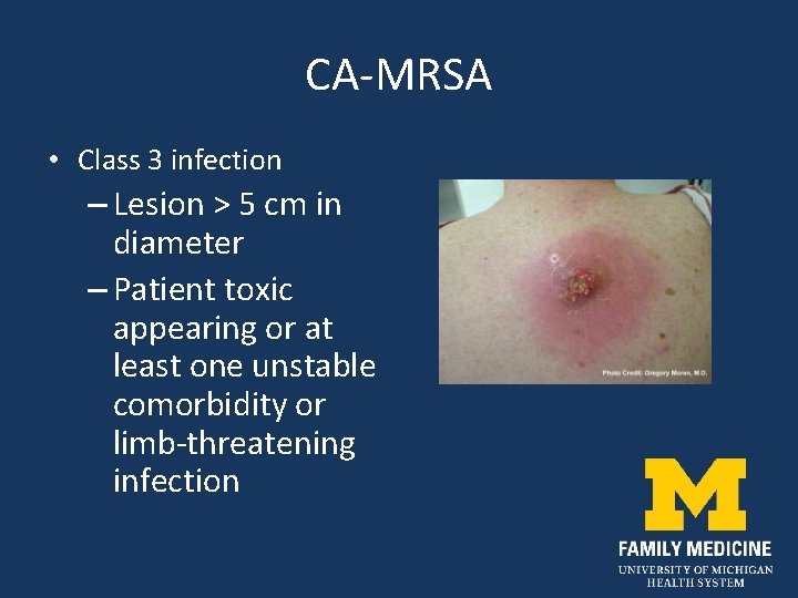
CA-MRSA • Class 3 infection – Lesion > 5 cm in diameter – Patient toxic appearing or at least one unstable comorbidity or limb-threatening infection
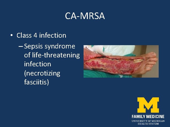
CA-MRSA • Class 4 infection – Sepsis syndrome of life-threatening infection (necrotizing fasciitis)
CA-MRSA Key Point Incision and Drainage of the Lesion! (if possible)
CA-MRSA • Incision and drainage alone without antibiotics has 90% cure rate • Use 11 blade to create a wide opening • Explore wound for loculations and pack • Frequent follow-up for approximately 2 weeks Rajendran et al. Antimicrob Agents Chemotherapy 2007; 51: 4044 -48
CA-MRSA treatment • Class 1 infection – If no drainable abscess, prescribe first or second generation cephalosporin, semi-synthetic penicillin, macrolide, or clindamycin – Follow-up in 1 -2 days to ensure response
CA-MRSA • Class 2 infection – I&D the lesion – Frequent follow-up – If not healing within 7 days, empirically treat with TMP-SMX DS 2 tabs BID – Clindamycin and tetracycline are alternatives, but have slightly less efficacy
CA-MRSA treatment • Class 3 and 4 infections – Admit to hospital – Consult surgery for aggressive debridement – Start vancomycin IV – Consult infectious disease specialist
CA-MRSA Return to Play • Bacterial infections – Treatment for minimum 72 hrs – No new lesions within 48 hrs – No draining or “wet” lesions
CA-MRSA Prevention • Washing/Showering with soap and warm water as soon as possible after competition/practice • Not Sharing soap bars, towels, razors or clothing • Prompt treatment of abrasions, cuts
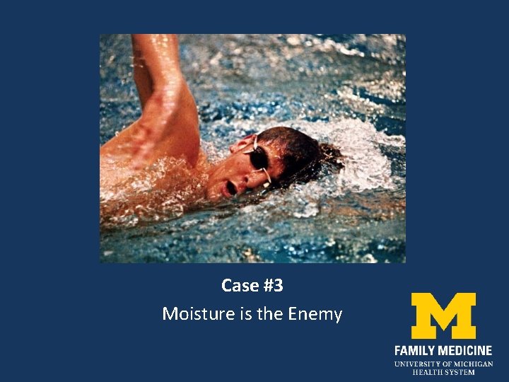
Case #3 Moisture is the Enemy
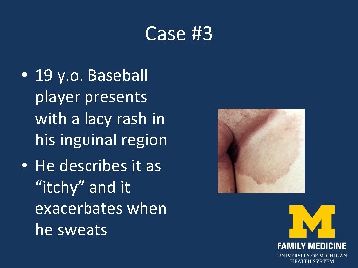
Case #3 • 19 y. o. Baseball player presents with a lacy rash in his inguinal region • He describes it as “itchy” and it exacerbates when he sweats
Tinea Infections • Dermatophytes survive on keratin in the stratum corneum layer of the skin • Warm, moist environments promote fungal growth • Tinea corpora in athletes is primarily spread by skin-to-skin contact • Skin scales containing fungal spores can live outside the host on inanimate surfaces
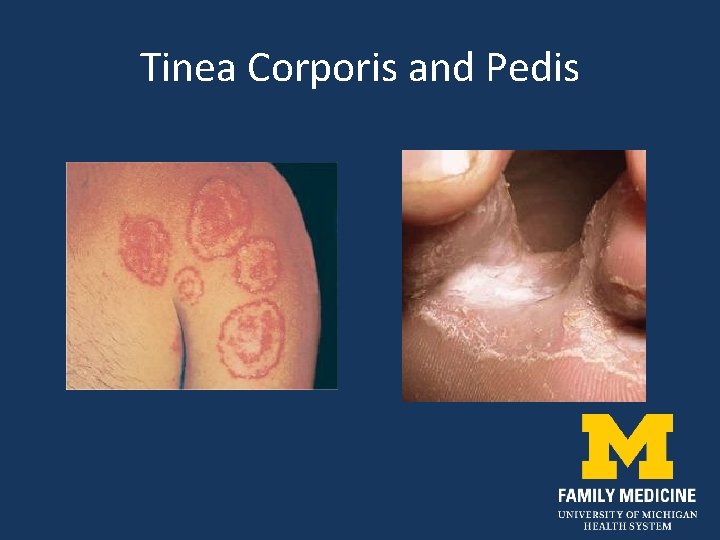
Tinea Corporis and Pedis
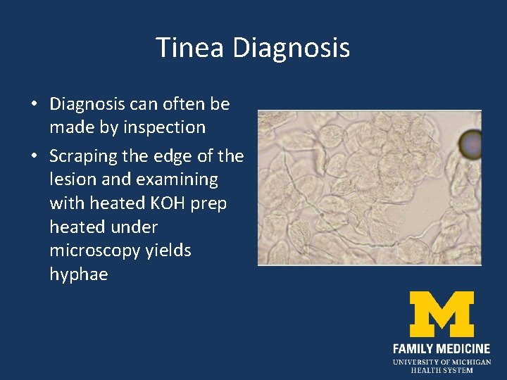
Tinea Diagnosis • Diagnosis can often be made by inspection • Scraping the edge of the lesion and examining with heated KOH prep heated under microscopy yields hyphae
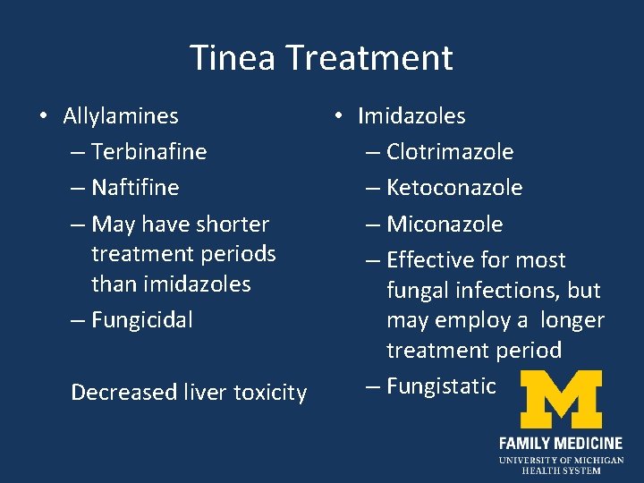
Tinea Treatment • Allylamines – Terbinafine – Naftifine – May have shorter treatment periods than imidazoles – Fungicidal Decreased liver toxicity • Imidazoles – Clotrimazole – Ketoconazole – Miconazole – Effective for most fungal infections, but may employ a longer treatment period – Fungistatic
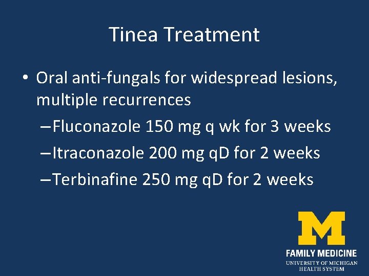
Tinea Treatment • Oral anti-fungals for widespread lesions, multiple recurrences – Fluconazole 150 mg q wk for 3 weeks – Itraconazole 200 mg q. D for 2 weeks – Terbinafine 250 mg q. D for 2 weeks
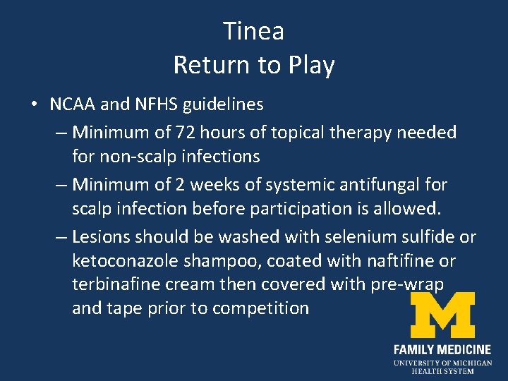
Tinea Return to Play • NCAA and NFHS guidelines – Minimum of 72 hours of topical therapy needed for non-scalp infections – Minimum of 2 weeks of systemic antifungal for scalp infection before participation is allowed. – Lesions should be washed with selenium sulfide or ketoconazole shampoo, coated with naftifine or terbinafine cream then covered with pre-wrap and tape prior to competition
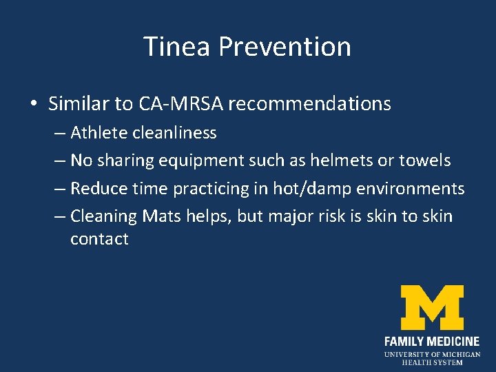
Tinea Prevention • Similar to CA-MRSA recommendations – Athlete cleanliness – No sharing equipment such as helmets or towels – Reduce time practicing in hot/damp environments – Cleaning Mats helps, but major risk is skin to skin contact
Case #4 My Achin’ Feet!
Case #4 • 51 y. o. backpacker • Presents with a blister on his foot • Hiked in rough terrain for 7 miles with a 50 pound pack • Wore wool socks • A recreational runner, but not accustomed to Weight/terrain, etc
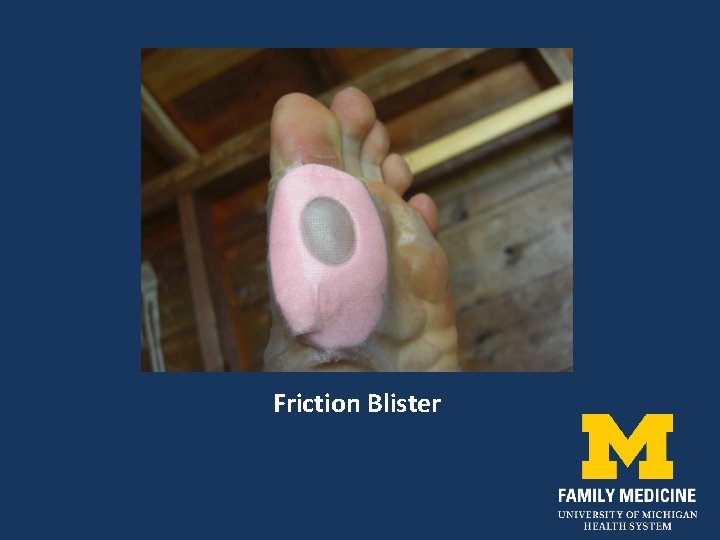
Friction Blister
Friction Blisters • Results from friction forces that separate epidermal cells at the level of the stratum spinosum • Potential space fills with fluid and/or blood
Friction Blisters • Factors promoting blister formation – Moisture increases frictional forces – Heat – Poorly fitting shoes – New activities – Cotton socks
Friction Blisters • Treatment – Decompress large blisters with a sterile needle. – Leave blister roof intact – “Second Skin” or petrolatum, donut pad, should be applied to minimize further friction at the site. – Prevention: acrylic or polyester socks
Friction Blister
Case #5
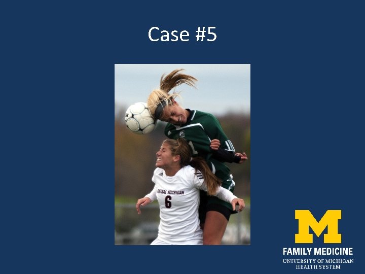
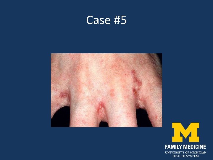
Case #5 • 20 y. o. soccer player presents with a pruritic rash for 2 days between the fingers and in beltline. • 21 y. o. football player presented later that day with similar symptoms and rash distribution • Yet another 21 y. o. soccer player presented with similar rash in the beltline. • What’s going on?
Case #5
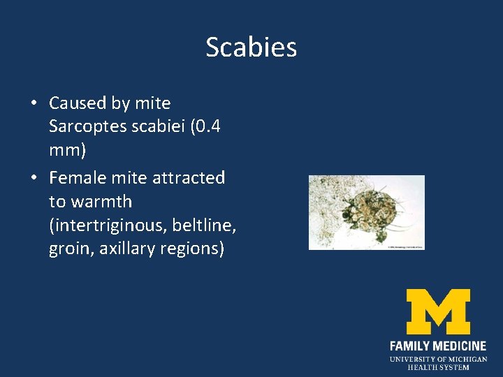
Scabies • Caused by mite Sarcoptes scabiei (0. 4 mm) • Female mite attracted to warmth (intertriginous, beltline, groin, axillary regions)
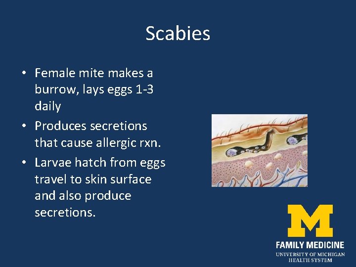
Scabies • Female mite makes a burrow, lays eggs 1 -3 daily • Produces secretions that cause allergic rxn. • Larvae hatch from eggs travel to skin surface and also produce secretions.
Scabies Treatment • Topical application of Permethrin (Elimite cream) from neck to toes at night. • Wash all bedclothing and suspected contaminated clothing in hot water. • Avoid sleeping on roomate’s couch that is infested.
Summary • Reviewed examples of infections and blisters encountered in athletes • Reviewed Diagnosis and treatment • Plans for return to play • Reviewed suggested methods of prevention of infection and re-infection
Bibliography • Pecci M, Comeau D, Chawla V. Skin conditions in the athlete. Am J Sports Med 2009; 37: 406 -17. • Mc. Bride D. Managing community-acquired MRSA lesions: What works? OBG Management 2008; 20: 28 -33. • Cohen P. The skin in the gym. Clinics Derm 2008; 26: 1626. • http: //www. cdc. gov/ncidod/dhqp/ar_mrsa. html • http: //epa. gov/oppad 001/chemregindex. htm • http: //nfhs. org • http: //www. mhsaa. com