Automated Reticulocyte Analysis New Parameters for Anemia Diagnosis


 • Reflects rate")
 • IRF measured as fraction (0. 00")
 Thiazole Orange by Flow Cytometry RBCs Reticulocytes Nucleated Cells IRF")
 • Erythropoietin therapeutic effect: IRF")

 pre-biotinylation 24 hours 6 hours 72 hours")


: Clinical Utility in Medical Practice • Monitor BM or Stem")


- Slides: 27
Automated Reticulocyte Analysis: New Parameters for Anemia Diagnosis and Therapeutic Monitoring & Improved Precision & Laboratory Efficiency Bruce H. Davis, M. D. William Beaumont Hospital Royal Oak, Michigan
Automated Hematology: Desirable New Parameters • CD 4 and CD 8 Lymphocyte Subsets • Reticulocytes with immature reticulocyte maturation (IRF) - alias RMI • Neutrophil activation marker, such as quantitative PMN CD 64 expression • reticulated platelets • Platelet activation markers (eg. CD 62 or CD 41 expression) • Hb F containing RBC enumeration (Kleihauer-Bettke) • Immune activation profile (cytokine/chemokine Rs) • CD 5+ B Cells or light chain+ B cells (CLL, etc. )) • Stem Cell enumeration (CD 34+ cells)
History of Automated Reticulocyte Counting • 1980 -90 Flow cytometric methods – – Tanke: Pyronin Y - Jacobberger: Di. OC(3) Ortho: Acridine Orange - Others: PI, ethidium Br Metzger, Corash: Thioflavin T Lee (BDIS), Davis & Bigelow: Thiazole Orange • 1990: TOA Sysmex R instruments: Auramine O • 1992 -96: Hematology instruments light scatter – Technicon - H 3: Oxazine 750 – Coulter Gen-S, STKS & MAXM: NMB – Abbott Cell Dyn 3500: NMB • 1996: Hematology instruments - fluorescence – Abbott Cell Dyn 4000: thiazole-like dye – Coulter Gen-S: CPO dye
Automated Reticulocyte Counting: Methods Available - 1997 Fluoresence Methods Light Scatter Methods • Thiazole Orange (BD) by Flow Cytometry • CPO dye (Coulter) by flow cytometry • TOA Sysmex R series and SE-Avante by Auramine O • Abbott Cell-Dyn 4000 by CD 4 K 530 • ABX Vega by thiazole orange • Bayer Technicon H 3, Advia by oxazine dye • Coulter STKS/MAXM and Gen-S with new methylene blue (NMB) • Abbott Cell-Dyn 3500 with NMB
Advantages of Automated Reticulocyte Analysis • Amenable to labor efficiencies or robotics –faster analysis per sample –allows for batch analysis or random access • Improved precision of retic counting –superior to visual microscopic counts –greater objectivity • New parameters of erythropoiesis –Immature Reticulocyte Fraction (IRF) –Reticulocyte hemoglobin content
New Parameters with Automated Reticulocyte Analysis • Immature Reticulocyte Fraction (IRF) • Reflects rate of erythropoietic activity • Available on many instruments, methods • Formerly termed reticulocyte maturity index (RMI) • Replaces need for “corrected” reticulocyte count • Reticulocyte MCHC (hypochromic Retics) • Detects early functional iron deficiency in Epo Studies by Brugnaro, d’Onofrio • Available only on Technicon H 3 to date
Reticulocyte Enumeration with Immature Reticulocyte Fraction (IRF) • IRF measured as fraction (0. 00 - 1. 00 range) • Sysmex R: IRF = HFR + MFR (Ref Range: 0. 05 -0. 20) • Thiazole Orange: Cursor at 95% interval (Ref Range: 0. 2 -0. 5) • Report with reticulocyte % and absolute count • Graphic display of retic count vs. IRF • Superimpose refernce ranges for anemia classification • Plotting sequential samples shows erythroid response • Report results with other CBC parameters • Flags for increased reticulocytosis and hypoproliferative response • Automated, random access, discrete testing
Immature Reticulocyte Fraction (IRF) Thiazole Orange by Flow Cytometry RBCs Reticulocytes Nucleated Cells IRF Platelets • IRF = #HFR/#Retics • Data Analysis – exclude nucleated cells (n. RBCs, PMNS, lymphs) – exclude platelets – define IRF region – define retics
Evidence for Pathophysiologic Relevance of Immature Reticulocyte Fraction (IRF) • Erythropoietin therapeutic effect: IRF 1 - 3 days – CD 71 vs TO studies – BJH study, Major et al. • Animal models – in vivo biotinylation studies – CD 71 vs. TO studies • BMT recovery – IRF earliest parameter of engraftment – various methods with demonstrated efficacy
Normal Erythopoiesis • Maturational continuum • EPO effect • blood retic populations – IRF retics (CD 71+) – late retics – stress retics
Reticulocyte Maturation: in vivo biotinylation (K. Ault) pre-biotinylation 24 hours 6 hours 72 hours
Bone Marrow Regeneration Response: Consistent Pattern
Erythroid Parameters with Erythropoietic Response
Evaluation of Erythropoiesis: Bivariate IRF and Retic Count Display
Immature Reticulocyte Fraction (IRF): Clinical Utility in Medical Practice • Monitor BM or Stem Cell Regeneration post-BMT or Chemo. Rx • Monitor Renal Transplant Engraftment (Epo production) • Monitor Neonatal Transfusion Needs • Monitor Anemia Therapy • Monitor EPO Therapy: Renal Failure, AIDS, Infants, MDS • Monitor Bone Marrow Toxic Insults from drugs (eg. AZT) • Prognostic in Anemia of AIDS and Prematurity • Timing for Stem Cell Harvests following Growth Factor or Cytotoxic Drug Therapy • Detection of Aplastic Crisis in Hemolytic Anemias • Diagnosis and monitoring of aplastic anemia • Evaluate Normochromic Anemias of Various Etiologies • Detection of Occult or Compensated Hemorrhage or Hemolysis • Classification of Anemias
Patterns of IRF and Retic counts in Anemia • • • Clinical Condition Retic Ct Aplastic anemia/crisis hypoplastic anemia BM regeneration Chronic disease Iron deficiency Thalassemia Folate/B 12 deficiency Myelodysplasia Hemolytic anemia Blood loss/anoxia Low Low/WNL WNL/high Low/WNL Any level High WNL/high IRF Low High/WNL High WNL/high High
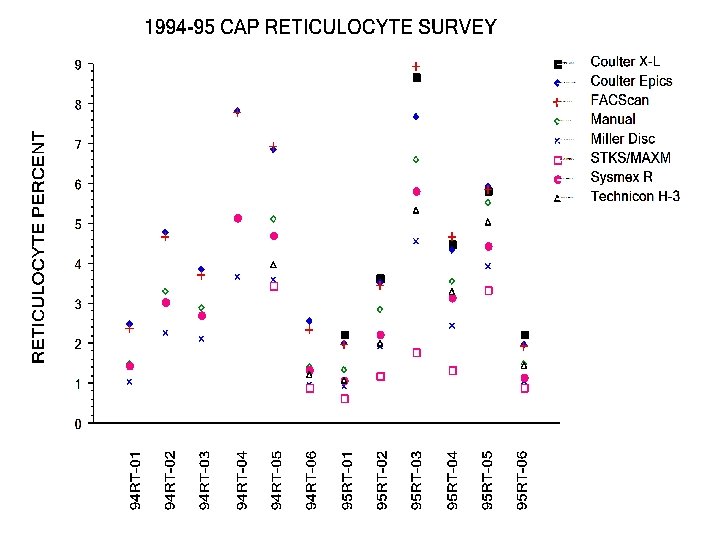
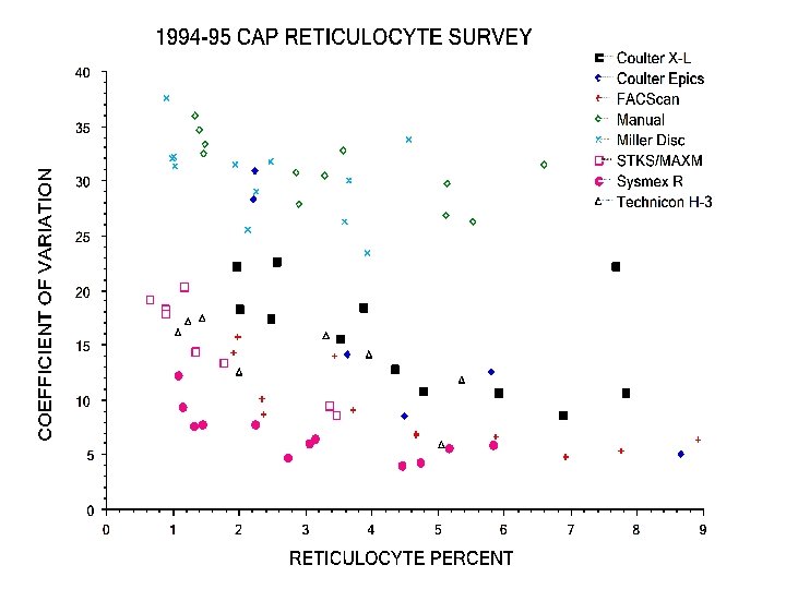
Intermethod Correlation Studies • Single site studies: multiple published – improved precision - CVs <15% – intermethod bias, but “clinically insignificant” • Davis et al: AJCP 102: 468, 1994 – 8 sites, 11 instruments, 310 blood samples – IRF and Retic counts compared • College of American Pathologists’ Reticulocyte RT Survey 1994 -96 – >2, 600 participants – surrogate blood material – Retic % only reported
Retic Counts: Inter-method Correlation
IRF: Inter-method Correlation
Reticulocyte Proficiency Testing College of American Pathologists 1994 -96 Program - surrogate blood material Methodologies Approved for Testing New Methylene Blue visual microscopy Sysmex R series (auramine O) Flow Cytometry - Thiazole Orange Flow Cytometry - Other dyes Coulter STKS/MAXM - NMB Miles Technicon H 3 - Oxazine
CAP Survey 1995 RT-C: Distribution of Methods
CAP RT Survey Experience: No Bias with TO methods secondary to FCM instrument A 1 2 3 4 B 5 Specimen Number 6 A B C D E F Flow Cytometer Instrument G
Known or Potential Interferents • Cellular Elements –Platelet clumps or giant platelets –nucleated cells or fragments • RBC Inclusions –Howell-Jolly bodies –Heinz or Pappenheimer bodies –parasites (malaris, babesia) • Miscellaneous causes –Autoflourescence (drugs, porphyria) –RBC aggregation (paraproteins, cold agglutinins) –coincidence (eg. platelet and RBC) –abnormal RBCs, hemolysis
Controls for Clinical Practice • Commericial Preparations –R&D Systems, Minneapolis, Mn –Streck Lab, Omaha, Ne –Instrument manufacturers • Refrigerated blood samples –short term QC by carry-over comparison –least expensive –will not detect long-term drift • Veterinary blood samples –rabbit –porcine
Reasons for NOT utilizing automated reticulocyte counting • • • Volume does not exceed 3 -5/day Physicians expect “stat” results Waiting for the “next generation” instrument “We’ve always done it this way” Technologists like doing manual counts