AEP Monitor Auditory Evoked Potential Contents 1 2AEP








- Slides: 61
AEP Monitor 麻醉知覺/深度監測儀 (Auditory Evoked Potential) 台大醫學系六年級 全以祖
Contents 1、前言 2、AEP Monitor的理論及臨床應用 3、AEP v. s. BIS 4、WHY AEP?
The patient n The patient has 2 worries: n 1: Will he sleep during the operation? n 2. Will he wake up after the operation?
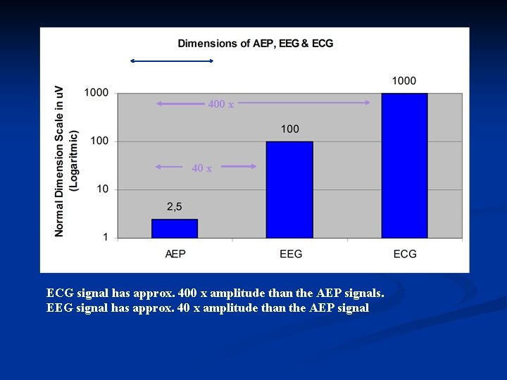
Basic basic n The hearing is the last sense that leaves and the first that returns during anaesthesia. n AEP is just the brain response to a click stimuli through the hearing nerve n AEP is a very weak electrical signal wrapped in the EEG background actvity. n Let’s look at how tiny this signal is.
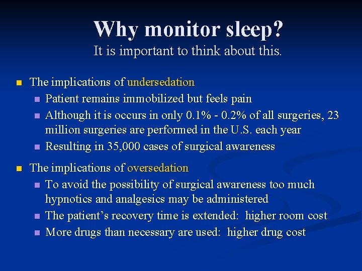
Why monitor sleep? It is important to think about this. n The implications of undersedation n Patient remains immobilized but feels pain n Although it is occurs in only 0. 1% - 0. 2% of all surgeries, 23 million surgeries are performed in the U. S. each year n Resulting in 35, 000 cases of surgical awareness n The implications of oversedation n To avoid the possibility of surgical awareness too much hypnotics and analgesics may be administered n The patient’s recovery time is extended: higher room cost n More drugs than necessary are used: higher drug cost
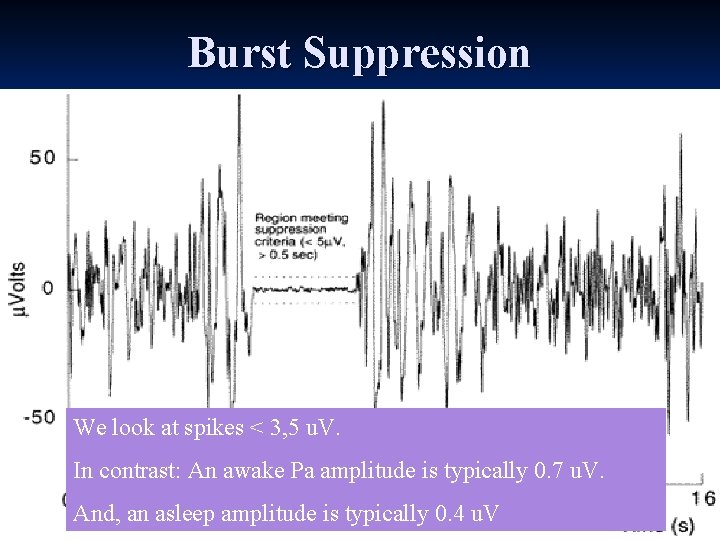
Burst Suppression We look at spikes < 3, 5 u. V. In contrast: An awake Pa amplitude is typically 0. 7 u. V. And, an asleep amplitude is typically 0. 4 u. V
400 x 40 x ECG signal has approx. 400 x amplitude than the AEP signals. EEG signal has approx. 40 x amplitude than the AEP signal
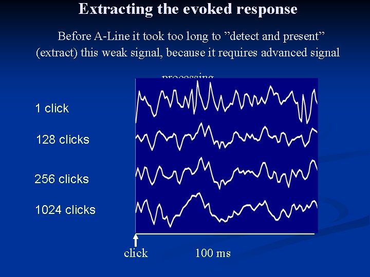
Extracting the evoked response Before A-Line it took too long to ”detect and present” (extract) this weak signal, because it requires advanced signal processing 1 click 128 clicks 256 clicks 1024 clicks click 100 ms
But, lets make this more visible Let’s see what happens when we send a click through the ear.
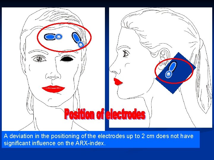
A deviation in the positioning of the electrodes up to 2 cm does not have significant influence on the ARX-index.
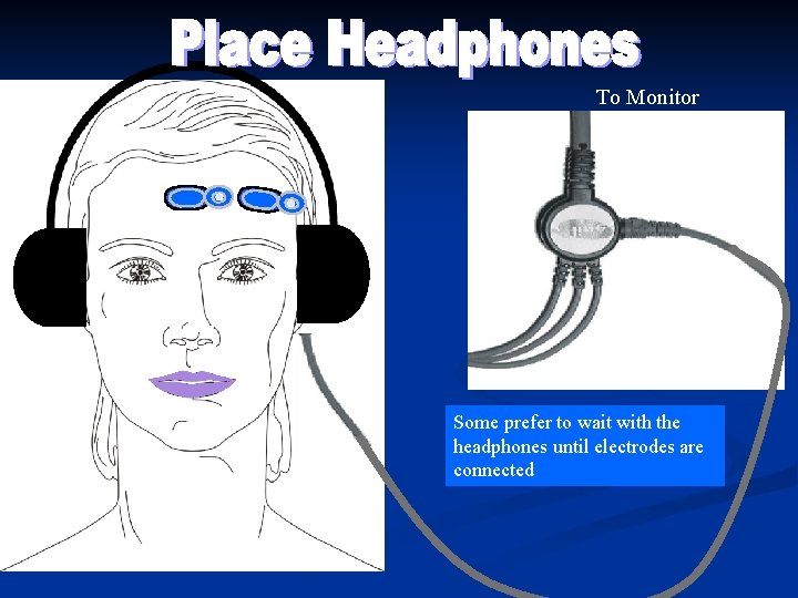
To Monitor Some prefer to wait with the headphones until electrodes are connected
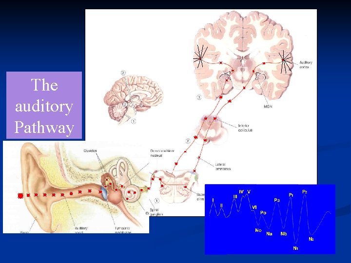
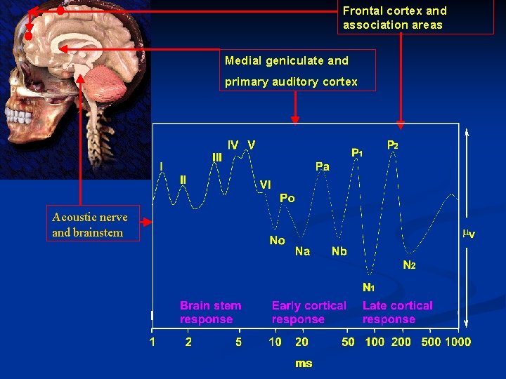
The auditory Pathway 2
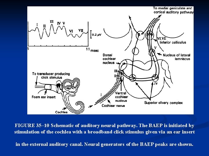
FIGURE 35– 10 Schematic of auditory neural pathway. The BAEP is initiated by stimulation of the cochlea with a broadband click stimulus given via an ear insert in the external auditory canal. Neural generators of the BAEP peaks are shown.
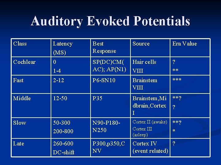
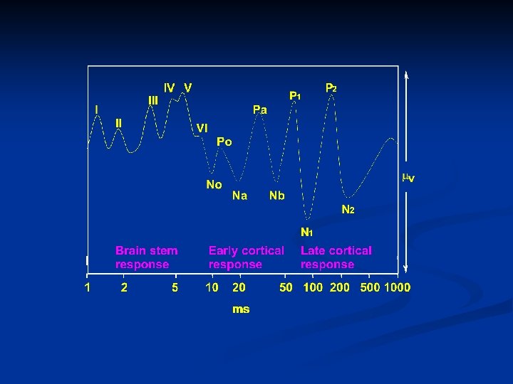
Auditory Evoked Potentials Class Latency (MS) Best Response Source Era Value Cochlear 0 1 -4 SP(DC)CM( AC); AP(N 1) Hair cells VIII ? ** Fast 2 -12 P 6 -SN 10 Brainstem VIII *** Middle 12 -50 P 35 Brainstem, Mi **? dbrain, Cortex ? I Slow 50 -300 200 -800 N 90 -P 180 N 250 Cortex II (awake) Cortex III (asleep) Late 260 -600 DC-shift P 300, p 350, C NV Cortex IV ? (event related) **? *
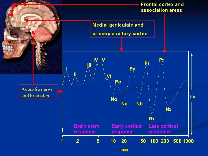
Frontal cortex and association areas Medial geniculate and primary auditory cortex Acoustic nerve and brainstem
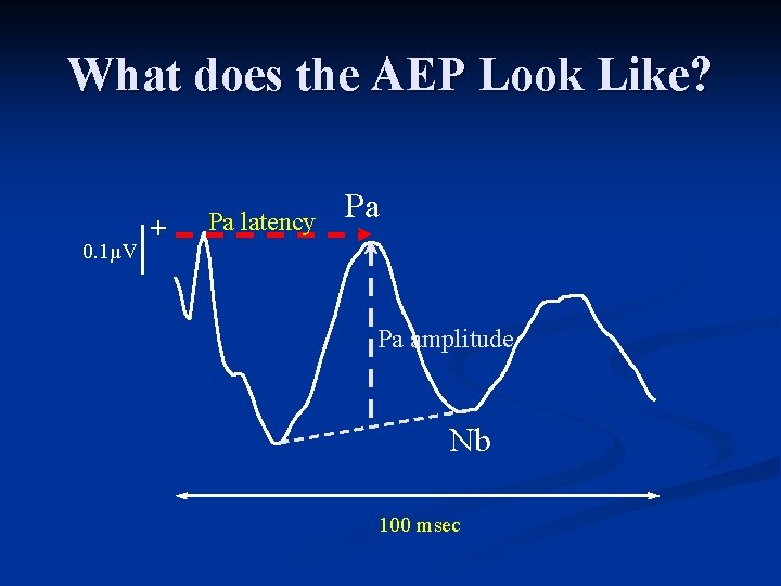
What does the AEP Look Like? 0. 1µV + Pa latency Pa Pa amplitude Nb 100 msec
And, this is what happens
Frontal cortex and association areas Medial geniculate and primary auditory cortex Acoustic nerve and brainstem
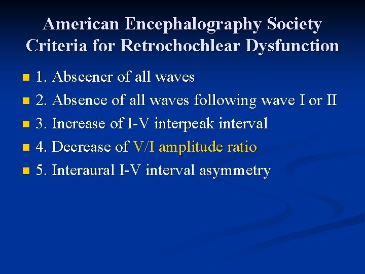
American Encephalography Society Criteria for Retrochochlear Dysfunction 1. Abscencr of all waves n 2. Absence of all waves following wave I or II n 3. Increase of I-V interpeak interval n 4. Decrease of V/I amplitude ratio n 5. Interaural I-V interval asymmetry n
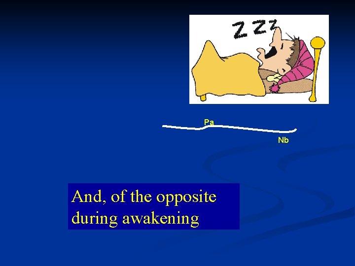
Pa Nb And, of the opposite during awakening
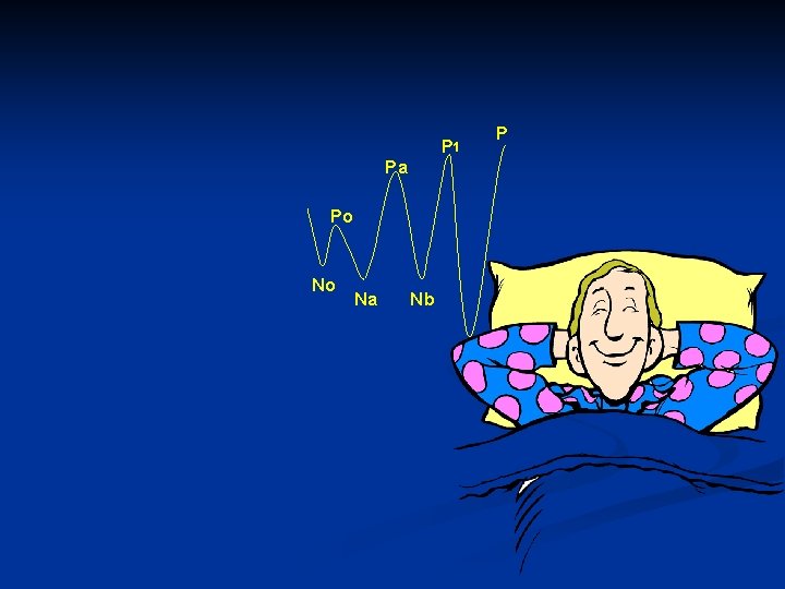
P 1 Pa Po No 7 Na Nb N 1 P
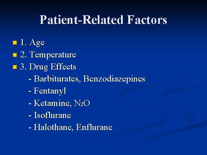
Patient-Related Factors 1. Age n 2. Temperature n 3. Drug Effects - Barbiturates, Benzodiazepines - Fentanyl - Ketamine, N 2 O - Isoflurane - Halothane, Enflurane n
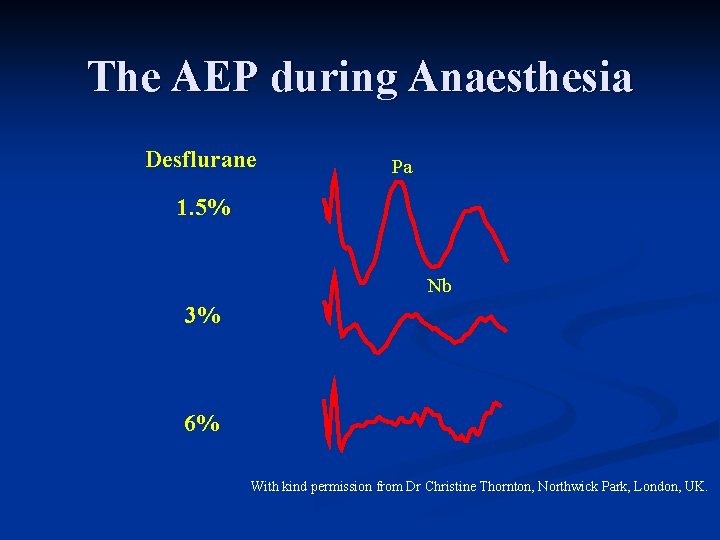
The AEP during Anaesthesia Desflurane Pa 1. 5% Nb 3% 6% With kind permission from Dr Christine Thornton, Northwick Park, London, UK.
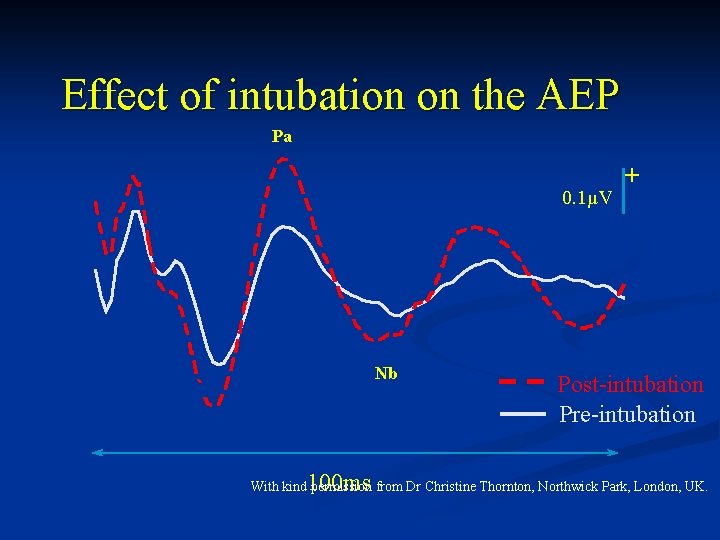
Effect of intubation on the AEP Pa 0. 1µV Nb 100 ms + Post-intubation Pre-intubation With kind permission from Dr Christine Thornton, Northwick Park, London, UK.
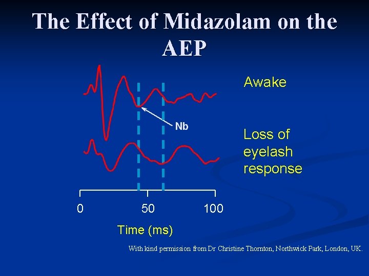
The Effect of Midazolam on the AEP Awake Nb 0 50 Loss of eyelash response 100 Time (ms) With kind permission from Dr Christine Thornton, Northwick Park, London, UK.
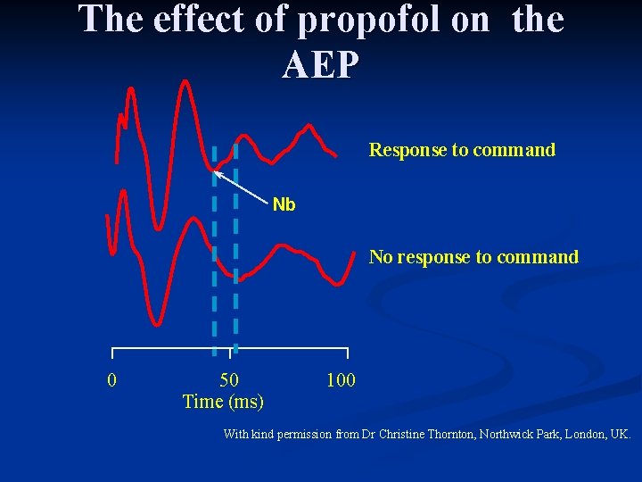
The effect of propofol on the AEP Response to command Nb No response to command 0 50 Time (ms) 100 With kind permission from Dr Christine Thornton, Northwick Park, London, UK.
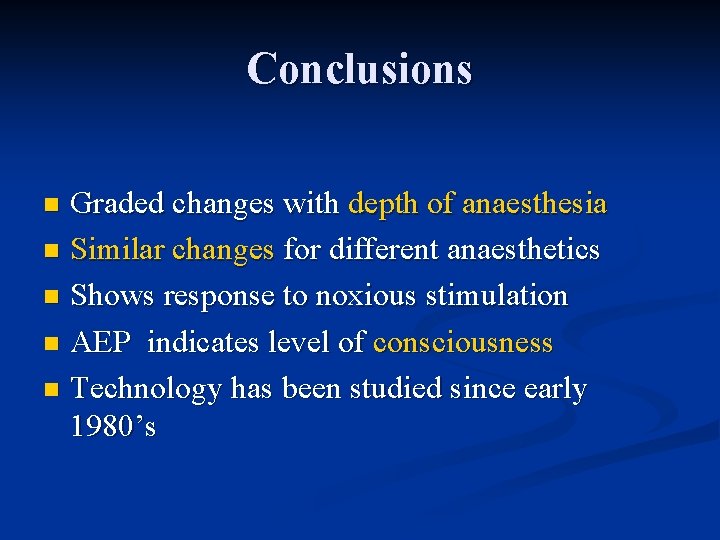
Conclusions Graded changes with depth of anaesthesia n Similar changes for different anaesthetics n Shows response to noxious stimulation n AEP indicates level of consciousness n Technology has been studied since early 1980’s n
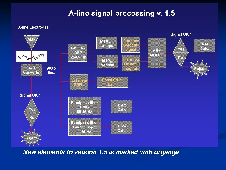
AEP signal processing? How can it be so fast?
New elements to version 1. 5 is marked with organge
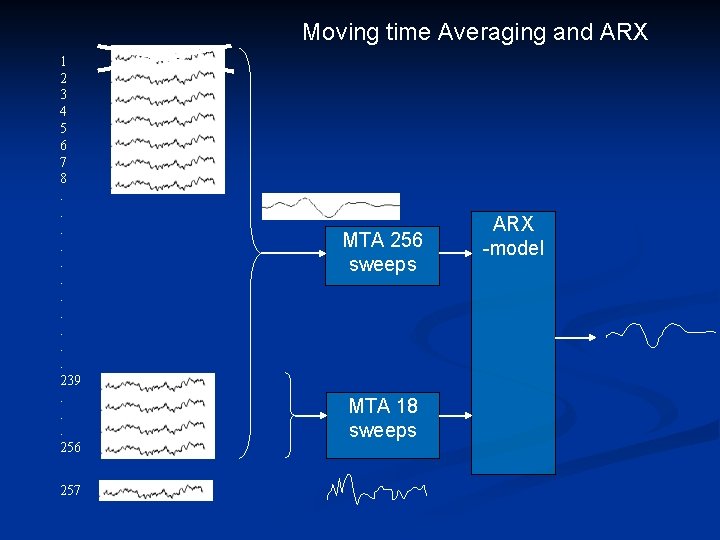
Moving time Averaging and ARX 1 2 3 4 5 6 7 8. . . 239. . . 256 257 MTA 256 sweeps MTA 18 sweeps ARX -model
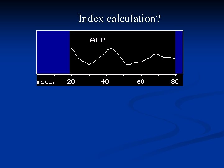
Index calculation?
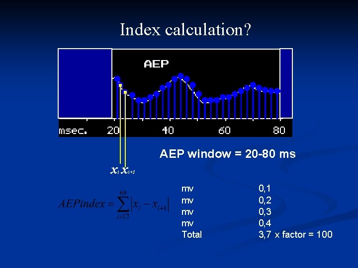
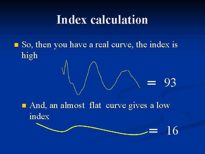
Index calculation? AEP window = 20 -80 ms xx i i+1 mv mv Total 0, 1 0, 2 0, 3 0, 4 3, 7 x factor = 100
Index calculation n So, then you have a real curve, the index is high = 93 n And, an almost flat curve gives a low index = 16
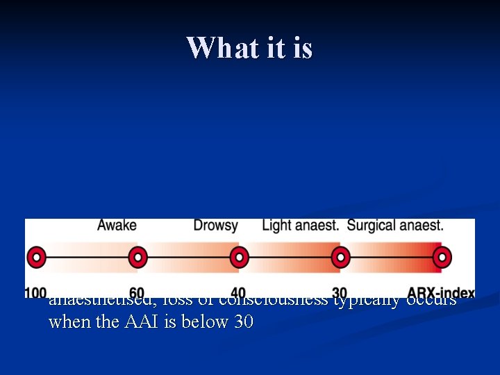
What it is n AAI is typically higher than 60 when the patient is awake and decreases when the patient is anaesthetised; loss of consciousness typically occurs when the AAI is below 30
What we did n n n The click detection is achieved by estimating the Signal to Noise Ratio (SNR). The SNR is defined as the ratio between the measured signal and the noise. The SNR threshold is 1. 45; this means that when the SNR is above 1. 45, the quality of the AEP is satisfactory. On the other hand if the SNR is below 1. 45, the signal quality is low, and a further averaging of the AEP is carried out. Yet if this does not improve the SNR, the symbol LOW AEP or NO AEP will appear on the monitor.
What we did n The symbol NO AEP and flashing AAI will appear in one of the following situations: The Headphones are disconnected or not working properly. n The subject may be hearing impaired. n Too much noise is present. Check grounding of the AAI monitor and the surrounding equipment. n The subject has very high levels of EMG activity, meaning that the AEP is embedded in an excessive amount of noise, impeding the extraction of the AEP. n
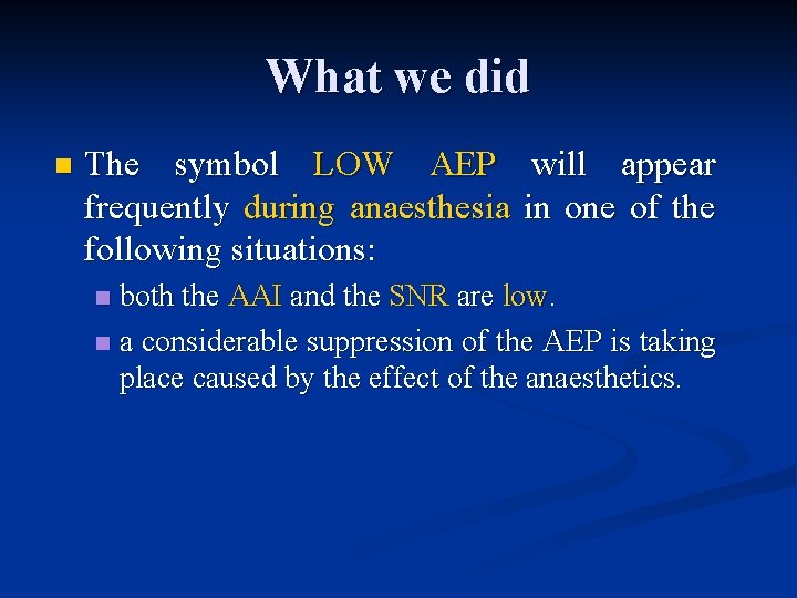
What we did n The symbol LOW AEP will appear frequently during anaesthesia in one of the following situations: both the AAI and the SNR are low. n a considerable suppression of the AEP is taking place caused by the effect of the anaesthetics. n
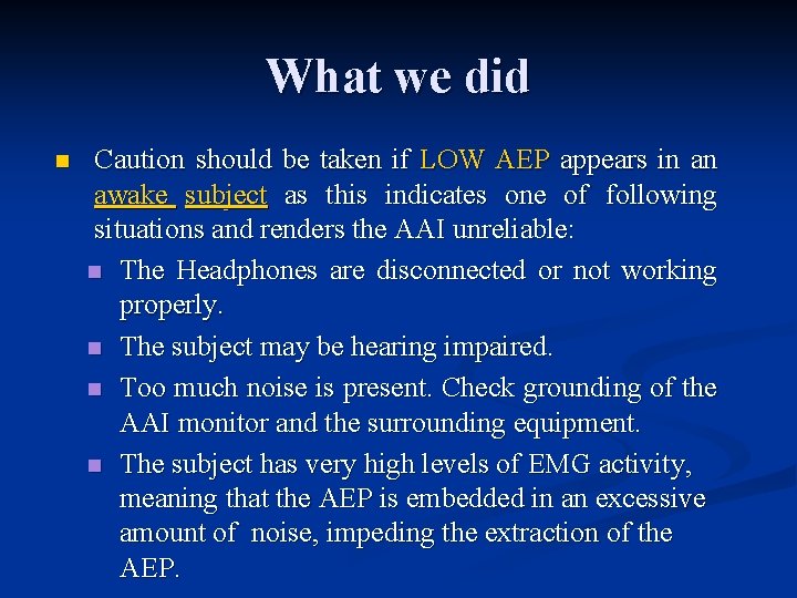
What we did n Caution should be taken if LOW AEP appears in an awake subject as this indicates one of following situations and renders the AAI unreliable: n The Headphones are disconnected or not working properly. n The subject may be hearing impaired. n Too much noise is present. Check grounding of the AAI monitor and the surrounding equipment. n The subject has very high levels of EMG activity, meaning that the AEP is embedded in an excessive amount of noise, impeding the extraction of the AEP.
What we did Ø For both LOW AEP and NO AEP: The response time after the headphones are disconnected until LOW AEP or NO AEP appears in the display has a mean value of 50 seconds and 97 % of the response times will typically be below 2 minutes.
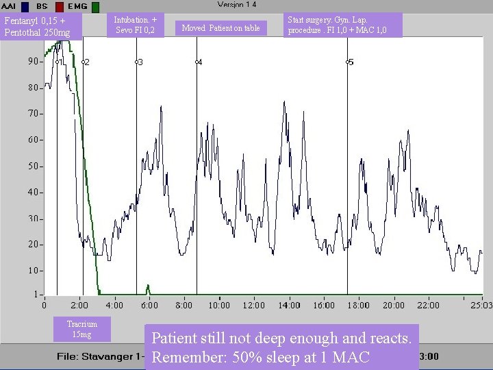
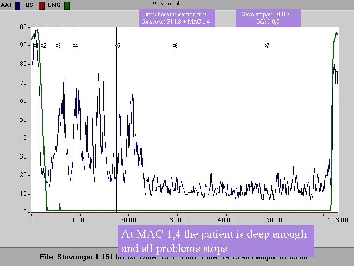
A good case Just to illustrate how important it is. n Customer couldn’t understand why the index was high? n Complained that “something was wrong” n All details captured by our man n After downloading and descriptions the clinicians agreed the anaesthesia was not optimal. n They could actually see things they never n
Fentanyl 0, 15 + Pentothal 250 mg Tracrium 15 mg Intubation. + Sevo FI 0, 2 Moved Patient on table Start surgery. Gyn. Lap. procedure. FI 1, 0 + MAC 1, 0 Index TIVA Intubation dropped with still too induction notsoon. and deep NMB Fentanyl enough and was Maintenance and had Induction started with normal Penthotal Patient was dose was small for to this bereacts. given would Remember: not reached tohave prepare prevented 50% peak intubation sleep effect. this at gas 1 MAC doses patient. moved on Gas table. conc. Dose too low of too low.
Put in trocar (insertion tube for scope) FI 1, 8 + MAC 1, 4 Sevo stopped FI 0, 7 + MAC 0, 9 At MAC 1, 4 the patient is deep enough and all problems stops
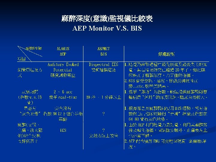
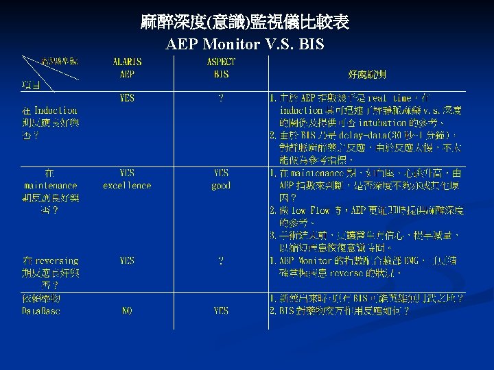
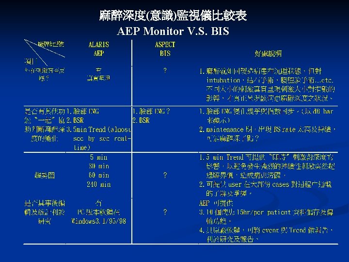
3. AEP Monitor V. S. BIS
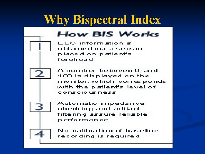
Why Bispectral Index
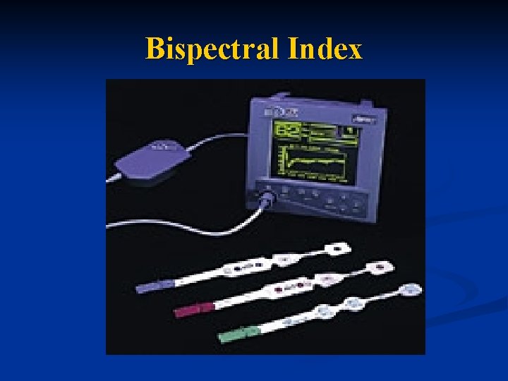
Bispectral Index
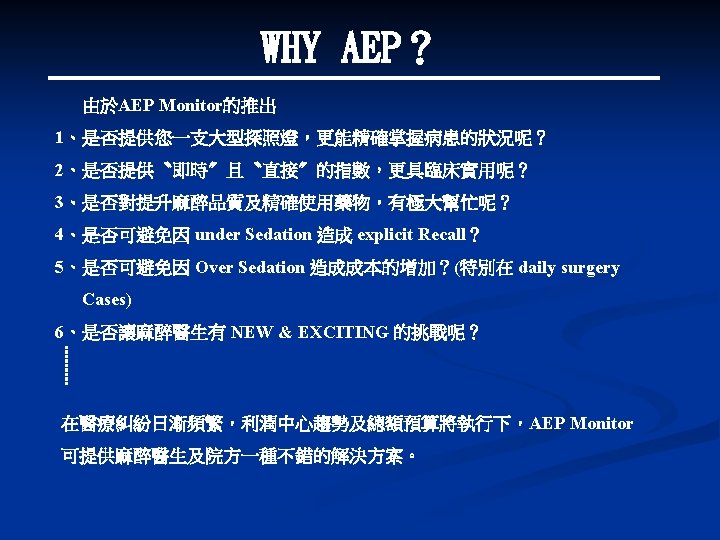
4. WHY AEP?