
Fear Learning The Interworking Circuits that Affect Fear



- Slides: 93
Fear Learning: The Interworking Circuits that Affect Fear By: Cami Brenner
• Usually thought to occur in the amygdala • Long term potentiation leads to a synaptic plasticity • Known as learning • A learned fear can be common in disorders related to stress/fear • Fearful response still exists even when fear is gone Fear Learning
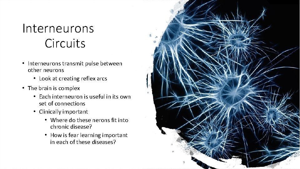
Interneurons Circuits • Interneurons transmit pulse between other neurons • Look at creating reflex arcs • The brain is complex • Each interneuron is useful in its own set of connections • Clinically important • Where do these nerons fit into chronic disease? • How is fear learning important in each of these diseases?
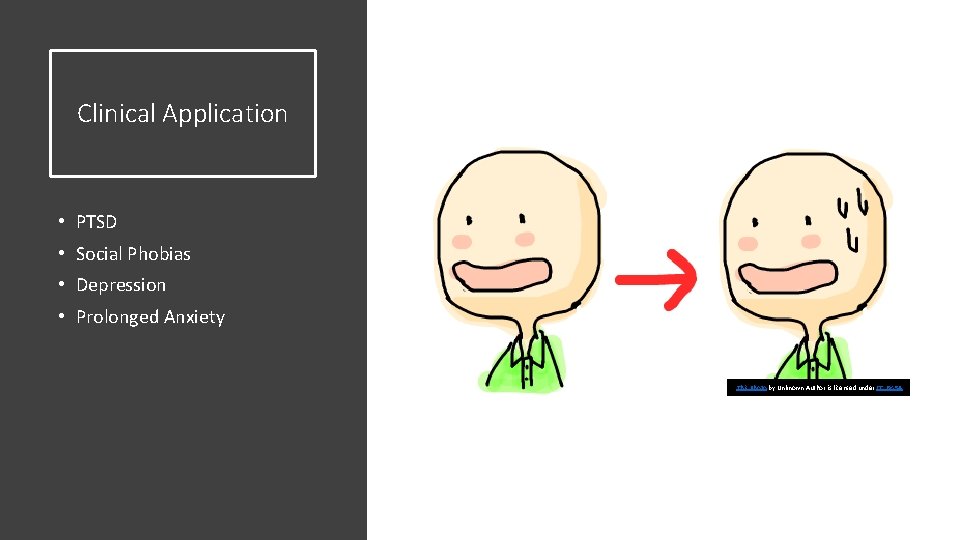
Clinical Application • PTSD • Social Phobias • Depression • Prolonged Anxiety This Photo by Unknown Author is licensed under CC BY-SA
Overarching questions • Can neural gene transfection use interneurons to help treat chronic diseases/disorders? • PTSD • Diagnosis • Will understanding circuitries of neurons help pharmacological approaches to correctly treat these stress disorders? • Can you use these specific neurons to treat these disorders?
Gillet Paper
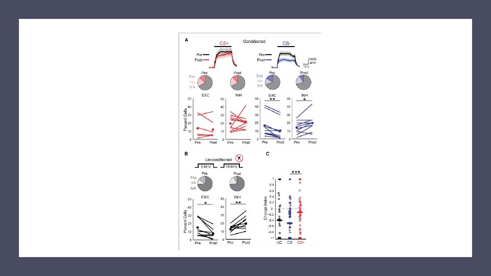
Contributions to Fear Learning • 1 -induction of auditory fear learning • Projections from Auditory cortex to amagdyla • 2 - Fear Conditioning enhances response to conditioned tone • Neurons in auditory cortex show enhanced response to conditioned tones immediately after fear conditioning • 1+2= Cortical Plasticity that contributes to fear learning
Background • -amagdyla: important in associative fear conditioning • Neural circuits (sensory cortex): important for acquisition and expression of learned fear • Removal or inactivation of auditory neural cicruits creates impairment of retrieval of fear memories • This is not understood • Related to complex auditory system? • Related to neural circuitry? • Related to conditioning protocol?
• What is the role of auditory cortex in retrieval of fear memory? • How does fear learning regulate cortical sensory presentations? • How is neural plasticity contributing to these ideas? • Proposal: Changes in cortical sensory representation contributes to memory strength or stimulus discrimination • However, small number of neurons show learning related change in activity Questions
Research • Used optogenetic silencing combined with two photon calcium imaging in mice (during auditory cortex of fear learning) • Optogenic activation of GABAergic interneurons • Two photon calcium for imaging in primary auditory cortex of awake mice • Considers cortical sensory representations before and after discriminative fear learning
• Mice-housed in room with reversed light cycle • Experiments performed during dark period • Mice suited with stainless steel head bar to skull • Muscle that overlies right auditory cortex removed • Small Craniotomy was made (dura intact, 2 x 3 mm) • Sealed with 1. 5% agarose • Injected with virus (10 -16 locations) • Imaging occurred 3 -7 weeks following virus injection Procedures
mice heterozygous for GAD 2 - Cre and Rosa-LSLtd. Tomato Viral Infections AAV 2/9 -syn-GCa. MP 6 s mice heterozygous for PV-Cre or SOM-Cre and Rosa-LSL-td. Tomato AAV 2/9 -syn-FLEX-GCa. MP 6 s
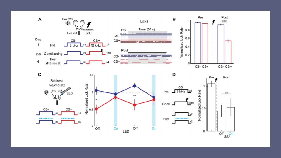
After head bar implant Discriminative Fear Learning: Head Fixed Mice Trained to Obtain Water at a Lick Port • Dehydrated to no more than 80% of free drinking weight • Handled daily • Habituated to head fixation in sound attenuating enclosure (5 -14 days) • Learning measured used licks per second (fear induced suppression of licking) • CS+ is a mouse that is presented with a tone and with a tail shock CS- presented with the tone • Ran over 4 days • Day 1: four preconditioning trials • Day 2/3: conditioning with 10 trials • Day 4: Combined with optogenetic led illumination • Retrieval, habituation
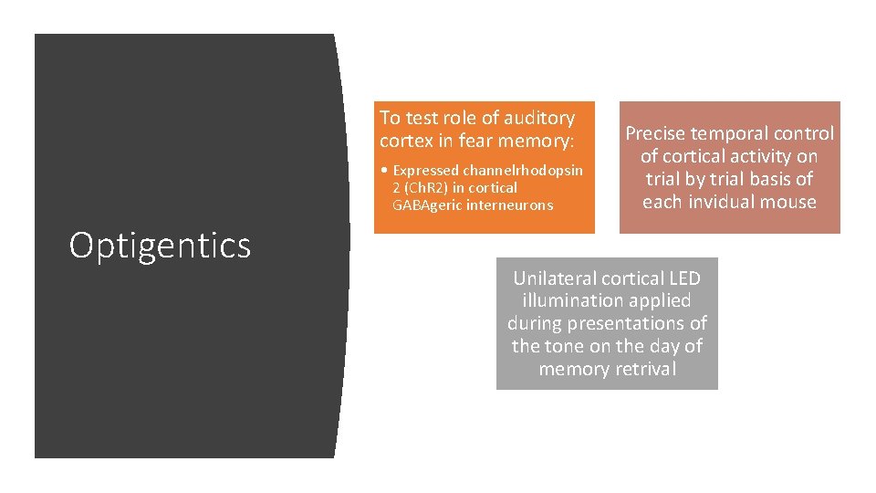
To test role of auditory cortex in fear memory: • Expressed channelrhodopsin 2 (Ch. R 2) in cortical GABAgeric interneurons Optigentics Precise temporal control of cortical activity on trial by trial basis of each invidual mouse Unilateral cortical LED illumination applied during presentations of the tone on the day of memory retrival
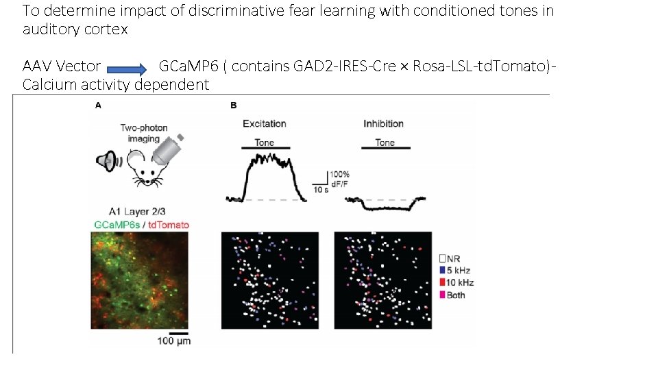
To determine impact of discriminative fear learning with conditioned tones in auditory cortex AAV Vector GCa. MP 6 ( contains GAD 2 -IRES-Cre × Rosa-LSL-td. Tomato)Calcium activity dependent
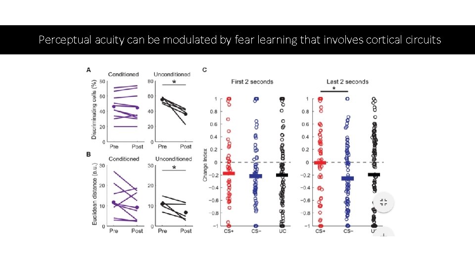
Perceptual acuity can be modulated by fear learning that involves cortical circuits
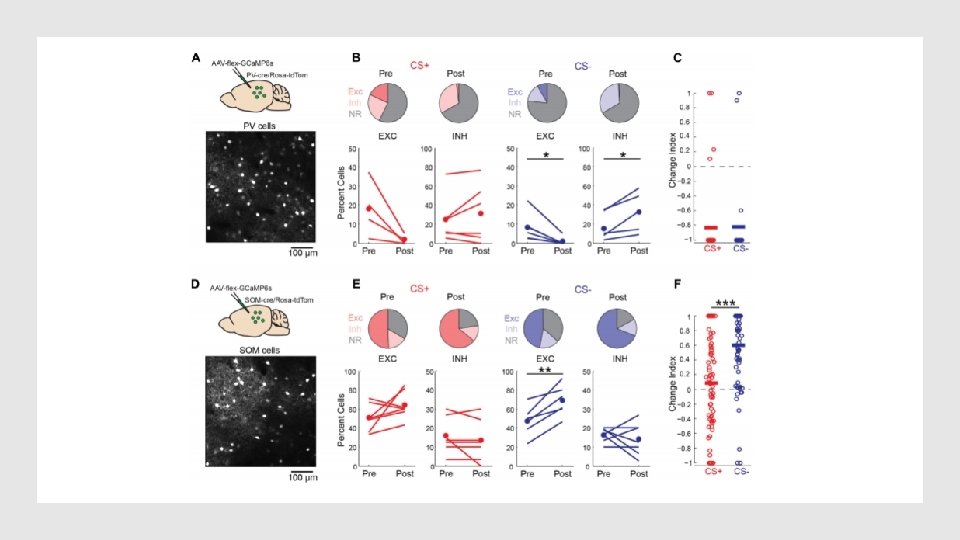
• Considered that changes in interneuron activity can create/influence effects of fear conditioning on pyramidal cell activity • Considered 2 main cortical Gabaergic neurons: paravalbumin (pv) and somatostatin (Som) • used conditional expression of GCa. MP 6 s in PV-cre and SOM-cre mice
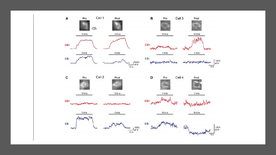
• Discriminative fear learning modulates cortical sensory representations via suppression of cortical habituation • Auditory Cortex has critical role in discriminative fear learning • CS- representations are reduced following fear conditioning • Decline due to habituation • Could be due to inhibitory circuits contributing to induction of auditory fear learning • CS+ representations are maintained Conclusions
• Amygdala: receives sensory information from both auditory thalamus and auditory cortex • Fear conditioning can be mediated by either pathway • Auditory Cortex involvement is likely dependent on intricacy of stimuli • Optogenetic cortical inactivation did not block fear expression when learning was based on a simple fear conditioning protocol. • Cortical silencing disrupted fear memory retrieval during discriminative fear learning Conclusions
• Sensory representations are bidirectionally modified based on the behavioral relevance of sensory stimuli. • Discriminative fear conditioning leads to a selective increase in SOM cell activity to CS− but not CS+ tones • Results indicate that discriminative fear conditioning regulates cortical sensory processing by preventing habituation • Cortical representations of valued sensory stimuli are retained while representations of stimuli lacking relevance can be reduced Conclusions
Zhou Paper
Research into Cellular and Molecular Mechanisms of Memories • Encoding and recall do not occur in the same condition • CA 1 Regions have been shown to be important in memory and generalization • Overgeneralization occurs with PTSD • Means this research could potentially affect these neural fields
Questions • Are CA 1 regions, and which, local, separate mechanisms are involved following memories? • How does generalization occur from original memories? • Does the hippocampus learn quickly enough to interact with cortical system, that then creates the elements of memory for generalization? • Can and how do Hippocampal CA 1 regions process fear memory and generalization through recruitment of other regions?
Memories include activitydependent fast changes in CA 3 -CA 1 synapses as a key mechanism Background medial prefrontal cortex (m. PFC) controls generalization of contextual fear memory during acquisition (via the nucleus reuniens of the thalamus) • signals to the hippocampus and also back to the m. PFC
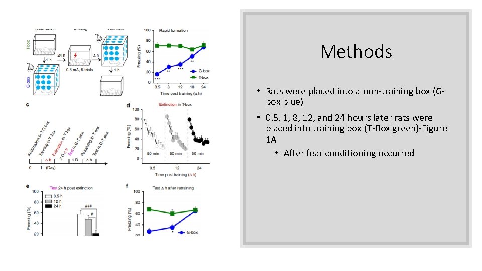
Methods • Rats were placed into a non-training box (Gbox blue) • 0. 5, 1, 8, 12, and 24 hours later rats were placed into training box (T-Box green)-Figure 1 A • After fear conditioning occurred
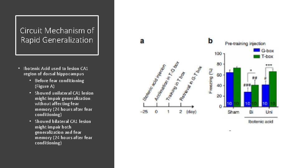
Circuit Mechanism of Rapid Generalization • Ibotenic Acid used to lesion CA 1 region of dorsal hippocampus • Before fear conditioning (Figure A) • Showed unilateral CA 1 lesion might impair generalization without affecting fear memory (24 hours after fear conditioning) • Showed bilateral CA 1 lesion might impair both generalization and fear memory (24 hours after fear conditioning)
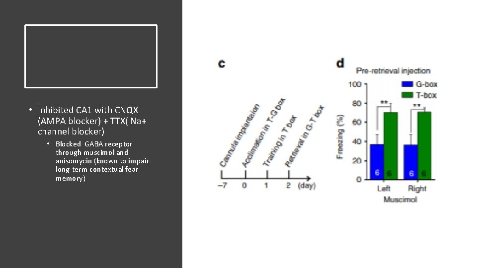
• Inhibited CA 1 with CNQX (AMPA blocker) + TTX( Na+ channel blocker) • Blocked GABA receptor through muscimol and anisomycin (known to impair long-term contextual fear memory)
Examined if left or right CA 1 regions were symmetrically required for generalization • Injection of muscimol to inhibit left or right before retrieval tests led to identical results (impairs generalization) • Unilateral lesion of dorsal hippocampus has no effect on contextual fear memory Generalization requires bilateral CA 1 activity • Means that dorsal hippocampus commissure is critical (DHC) • Highly preserved in mammals • Believed to be functional in humans as well
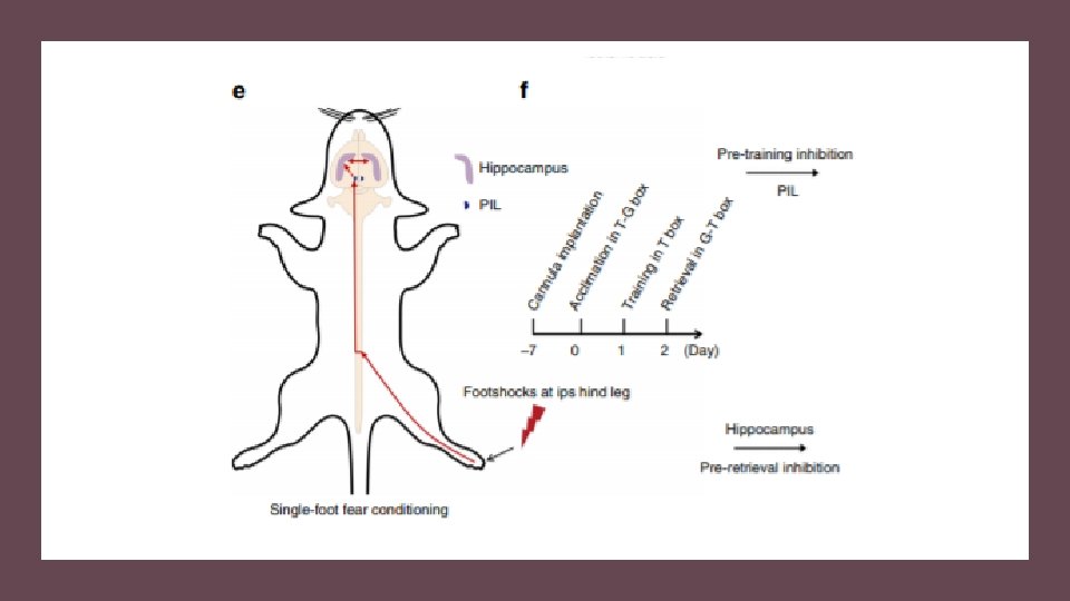
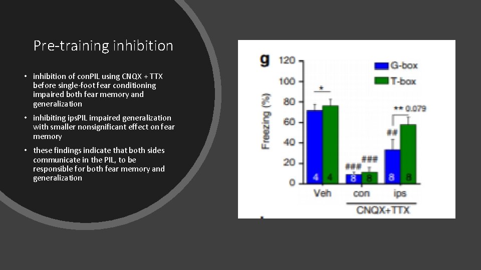
Pre-training inhibition • inhibition of con. PIL using CNQX + TTX before single-foot fear conditioning impaired both fear memory and generalization • inhibiting ips. PIL impaired generalization with smaller nonsignificant effect on fear memory • these findings indicate that both sides communicate in the PIL, to be responsible for both fear memory and generalization
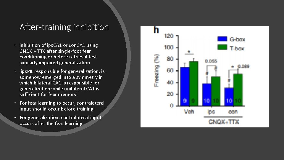
After-training inhibition • inhibition of ips. CA 1 or con. CA 1 using CNQX + TTX after single-foot fear conditioning or before retrieval test similarly impaired generalization • ips. PIL responsible for generalization, is somehow emerged into a symmetry in which bilateral CA 1 is responsible for generalization while unilateral CA 1 is sufficient for fear memory. • For fear learning to occur, contralateral input should occur before training • For generalization, contralateral input occurs after the fear learning
Generalization Recap: • Depends on • Bilataerl CA 1 activity • Requires symmetrical ipas. CA 1 activity and con. CA 1 activity (even in single foot response) • DHC could provide a route for interhemispheric exchanges between ips. CA 1 and con. CA 1 to lead to such symmetry in CA 1. • Pathway was reported to have only weak bilateral connections
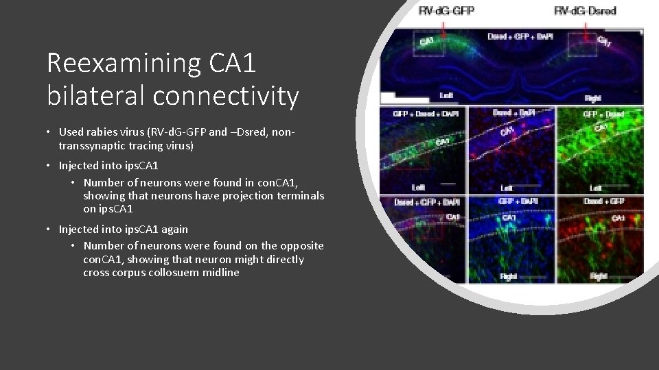
Reexamining CA 1 bilateral connectivity • Used rabies virus (RV-d. G-GFP and –Dsred, nontranssynaptic tracing virus) • Injected into ips. CA 1 • Number of neurons were found in con. CA 1, showing that neurons have projection terminals on ips. CA 1 • Injected into ips. CA 1 again • Number of neurons were found on the opposite con. CA 1, showing that neuron might directly cross corpus collosuem midline
RV-d. G-GFP injected directly into midline • Even conncetions between ips. CA 1 and con. CA 1 • Covered bilateral CA 1 region as a whole RV-d. G-GFP injected into ips. CA 1 stratum • Neurons found in ips. CA 3 and con. CA 3, but not CA 1 ipas. CA 1 • Receives bilateral input of CA 3 at stratum • Can only sense con. CA 1 at stratum
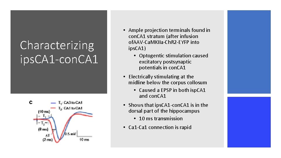
Characterizing ips. CA 1 -con. CA 1 • Ample projection terminals found in con. CA 1 stratum (after infusion of. AAV-Ca. MKIIa-Ch. R 2 -EYFP into ips. CA 1) • Optogentic stimulation caused excitatory postsynaptic potentials in con. CA 1 • Electrically stimulating at the midline below the corpus collosum • Caused a EPSP in both isp. CA 1 and con. CA 1 • Shows that ips. CA 1 -con. CA 1 is in the dorsal part of the hippocampus • 10 ms transmission • Ca 1 -Ca 1 connection is rapid
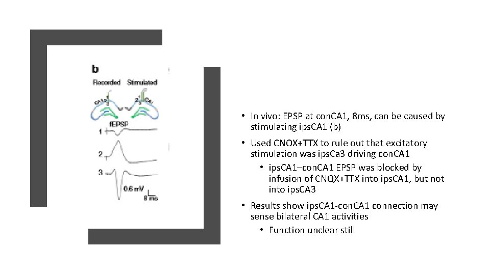
• In vivo: EPSP at con. CA 1, 8 ms, can be caused by stimulating ips. CA 1 (b) • Used CNOX+TTX to rule out that excitatory stimulation was ips. Ca 3 driving con. CA 1 • ips. CA 1–con. CA 1 EPSP was blocked by infusion of CNQX+TTX into ips. CA 1, but not into ips. CA 3 • Results show ips. CA 1 -con. CA 1 connection may sense bilateral CA 1 activities • Function unclear still
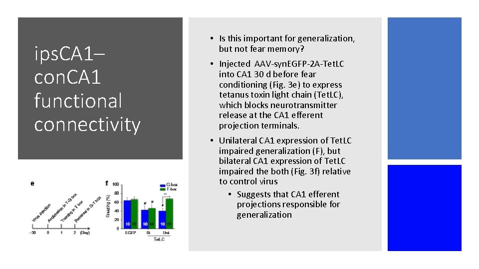
ips. CA 1– con. CA 1 functional connectivity • Is this important for generalization, but not fear memory? • Injected AAV-syn. EGFP-2 A-Tet. LC into CA 1 30 d before fear conditioning (Fig. 3 e) to express tetanus toxin light chain (Tet. LC), which blocks neurotransmitter release at the CA 1 efferent projection terminals. • Unilateral CA 1 expression of Tet. LC impaired generalization (F), but bilateral CA 1 expression of Tet. LC impaired the both (Fig. 3 f) relative to control virus • Suggests that CA 1 efferent projections responsible for generalization
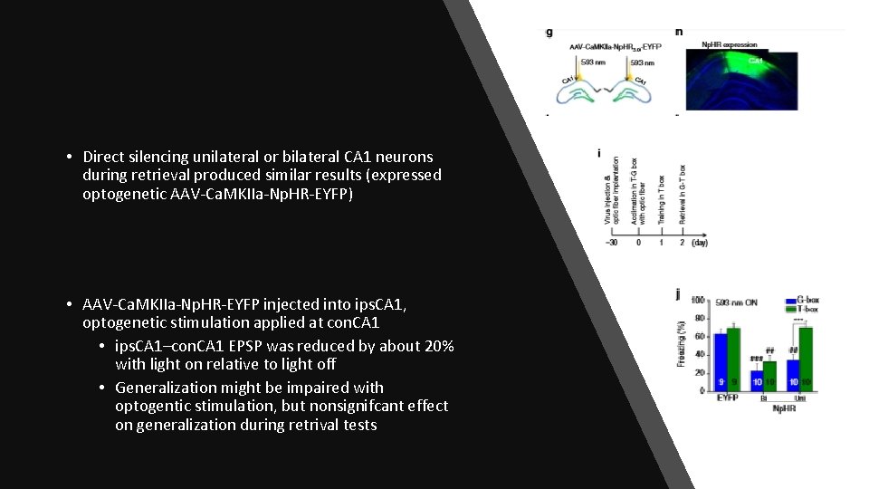
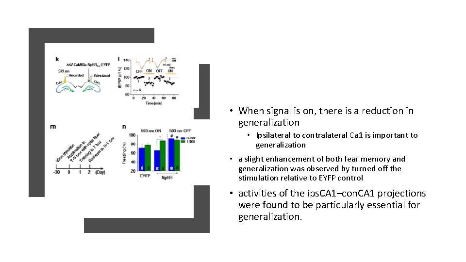
• Direct silencing unilateral or bilateral CA 1 neurons during retrieval produced similar results (expressed optogenetic AAV-Ca. MKIIa-Np. HR-EYFP) • AAV-Ca. MKIIa-Np. HR-EYFP injected into ips. CA 1, optogenetic stimulation applied at con. CA 1 • ips. CA 1–con. CA 1 EPSP was reduced by about 20% with light on relative to light off • Generalization might be impaired with optogentic stimulation, but nonsignifcant effect on generalization during retrival tests
• When signal is on, there is a reduction in generalization • Ipsilateral to contralateral Ca 1 is important to generalization • a slight enhancement of both fear memory and generalization was observed by turned off the stimulation relative to EYFP control • activities of the ips. CA 1–con. CA 1 projections were found to be particularly essential for generalization.
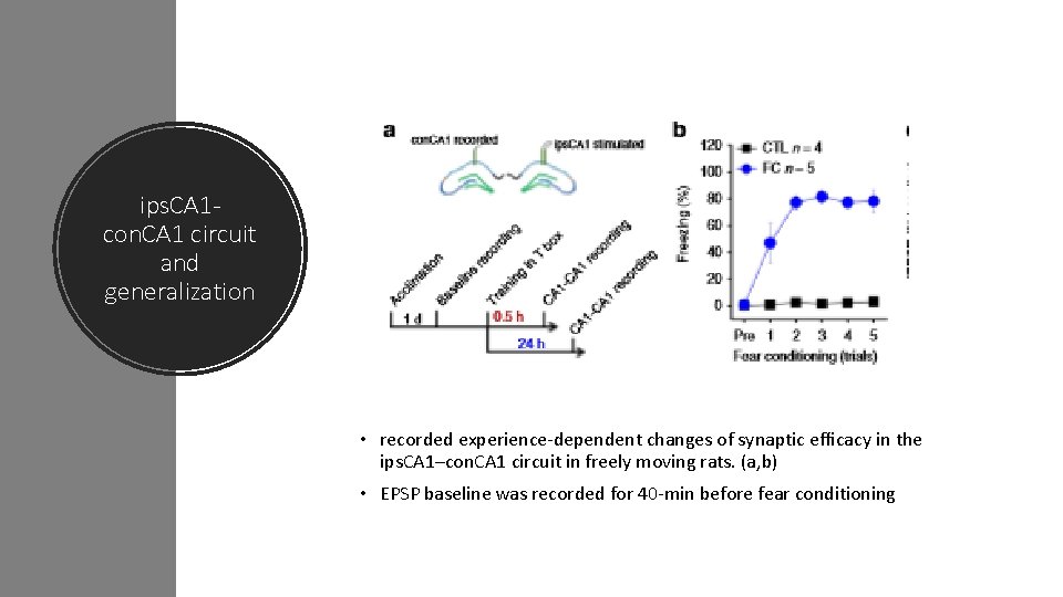
ips. CA 1 con. CA 1 circuit and generalization • recorded experience-dependent changes of synaptic efficacy in the ips. CA 1–con. CA 1 circuit in freely moving rats. (a, b) • EPSP baseline was recorded for 40 -min before fear conditioning
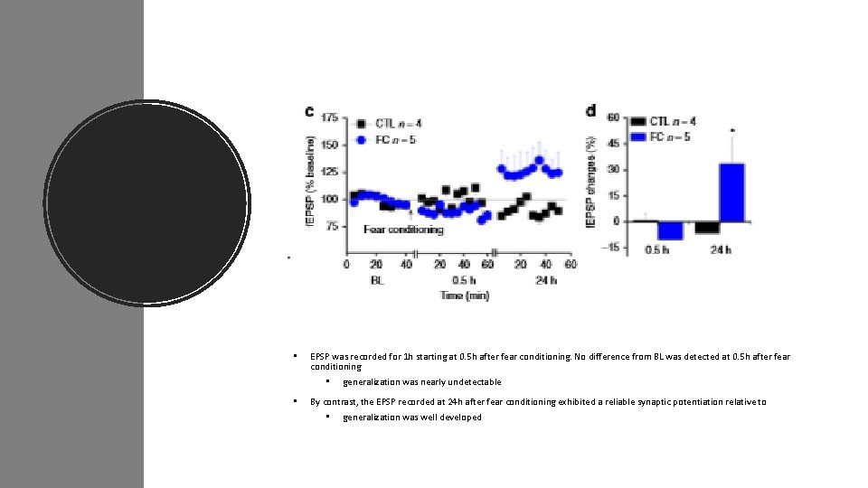
• EPSP was recorded for 1 h starting at 0. 5 h after fear conditioning. No difference from BL was detected at 0. 5 h after fear conditioning • • generalization was nearly undetectable By contrast, the EPSP recorded at 24 h after fear conditioning exhibited a reliable synaptic potentiation relative to • generalization was well developed
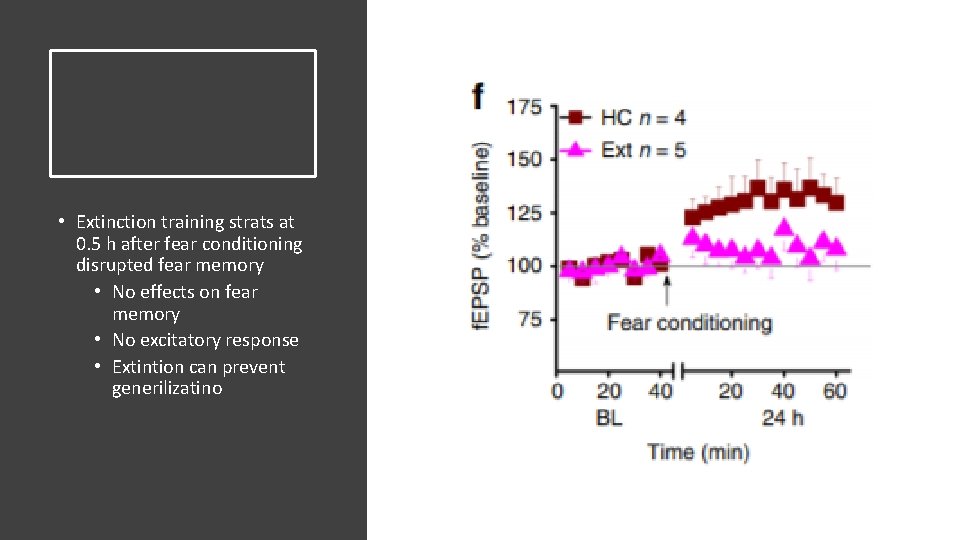
• Extinction training strats at 0. 5 h after fear conditioning disrupted fear memory • No effects on fear memory • No excitatory response • Extintion can prevent generilizatino
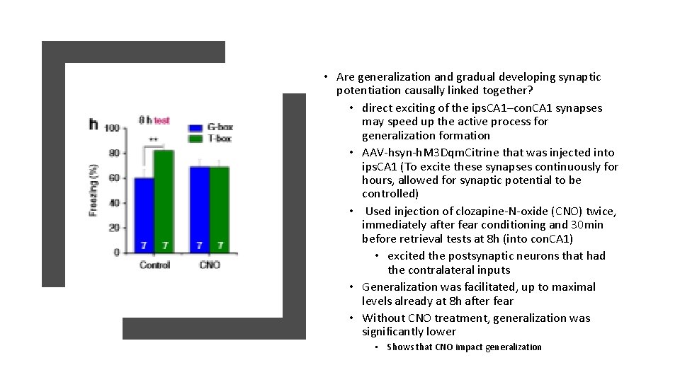
• Are generalization and gradual developing synaptic potentiation causally linked together? • direct exciting of the ips. CA 1–con. CA 1 synapses may speed up the active process for generalization formation • AAV-hsyn-h. M 3 Dqm. Citrine that was injected into ips. CA 1 (To excite these synapses continuously for hours, allowed for synaptic potential to be controlled) • Used injection of clozapine-N-oxide (CNO) twice, immediately after fear conditioning and 30 min before retrieval tests at 8 h (into con. CA 1) • excited the postsynaptic neurons that had the contralateral inputs • Generalization was facilitated, up to maximal levels already at 8 h after fear • Without CNO treatment, generalization was significantly lower • Shows that CNO impact generalization
• Hippocampal CA 1 regions are used in both fear memory and generalization • How do they function without interference? • spatiotemporally separated mechanisms within CA 1 (at least in the early stage of memory processing) • CA 3 neurons send commissural projections mainly onto synapses in stratum of ips. CA 1 and con. CA 1, Conclusions • Generalization is dependent on symmetrical ips. CA 1– con. CA 1 activity, • maintained by gradual developing potentiation of synaptic efficacy in the ips. CA 1–con. CA 1 circuit. • fear memory is widely believed to involve fast potentiation of synaptic efficacy in the synapses from CA 3 to CA 1 • Generalization is gradual developing • Sensitive to extinction training
Conclusions • “hypothesis for which the formation of generalization is gradual but have actively rapid and passively slow phases, likely corresponding to cellular consolidation and systems consolidation of memory when the contextual components are more or less specific” • “This explanation seems consistent with clinical recommendation that exposurebased therapy early after trauma in susceptible individuals may produce better efficacy in reducing overgeneralization of conditioned fear and prevalence of PTSD”
• Fear memory is processed by contralateral sides of amygdala or thalamus (during fear conditioning) • Fear memory is likely duplicated in bilateral CA 1 regions so that either CA 1 is sufficient for specific recall Conclusions • Generalization requires symmetrical activity of bilateral CA 1 • Selection of which CA 1 could be due to hippocampal activity • Sycroizes ips. CA 1 and con. CA 1 at an early stage creates functional connectivity in bilateral CA 1 neurons • Enables effective generalization formation at rapid phase
• Gradual internal learning is continuously readjusting the functional connectivity of the neural circuits not only for minimizing interference but also for reducing unpredicted errors under varying circumstances. Conclusions • Suggest that rapid generalization requires “gradual internal learning” over 24 h (dependent on bilateral CA 1 activities to readjust the interhemispheric CA 1–CA 1 synaptic efficacy for the selective formation and also extinction of generalization)
• Fear memory is quickly learned Conclusions • Generalization develops over a 24 hour period • Impaired by inhibiting ipsilateral (ips) or contralateral (con) CA 1, and by optogenetic silencing of the ips. CA 1 projections onto con. CA 1 • in vivo f. EPSP recordings reveal that ips. CA 1–con. CA 1 synaptic efficacy is increased with delay over 24 h when generalization is formed but it is unchanged if generalization is disrupted • Direct excitation of ips. CA 1–con. CA 1 synapses using chemogenetic h. M 3 Dq facilitates generalization formation • Rapid generalization is an active process dependent on bilateral CA 1 regions, and encoded by gradual synaptic learning in ips. CA 1–con. CA 1 circuit
Abs paper
Questions 1 How are the process of sensory input in the neoctorex encoded, based on behavioral relevance? 2 How is behavioral relevance encoded, integrated, and understood within the L 1 circuity? 3 L 1 circuity has not been able to be understood because of lack of selective genetic access • Cannot understand circuit or behavioral function of these specific INs
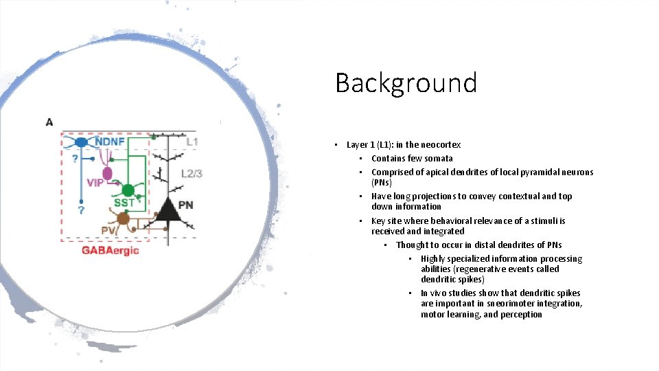
Background • Layer 1 (L 1): in the neocortex • Contains few somata • Comprised of apical dendrites of local pyramidal neurons (PNs) • Have long projections to convey contextual and top down information • Key site where behavioral relevance of a stimuli is received and integrated • Thought to occur in distal dendrites of PNs • Highly specialized information processing abilities (regenerative events called dendritic spikes) • In vivo studies show that dendritic spikes are important in sneorimoter integration, motor learning, and perception
Backgroundinhibition of L 1 • Dendritic Computations are controlled by inhibition • Distal dendritic inhibition in L 1 occurs from projections (SST) of positive Marinotti cells locate din deeper layers • SST (somatostatin) interneurons are dirven by local PNS • Have little input from thalamus • Thought to provide the dendritic inhibition • Based on proportion and activity similar to the PN levels
Background-inhibition of L 1 • Second source of inhibition, not as well understood • Comes from L 1 -IN • Receive input from top down projections • Includes cholinergic system, higher order thalamus, and cortico-cortical feedback (in rodents) • Recent studies point to strong cholinergic in L 1 IN in human neocortex • Suggests that inhibition from L 1 Ins may be internally generated activity • Represents the behavioral relevance of sensory information • Similar to distal dendritic excitation
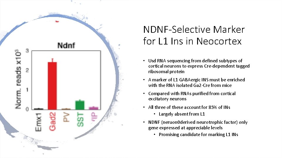
NDNF-Selective Marker for L 1 Ins in Neocortex • Usd RNA sequencing from defined subtypes of cortical neurons to express Cre-dependent tagged ribosomal protein • A marker of L 1 GABAergic INS must be enriched with the RNA isolated Ga 2 -Cre from mice • Compared with RNAs purified from cortical excitatory neurons • All three of these account for 85% of INs • Largely absent from L 1 • NDNF (neruon 0 derived neurotrophic factor) only gene expressed at appreciable levels • Promising candidate for marking L 1 INs
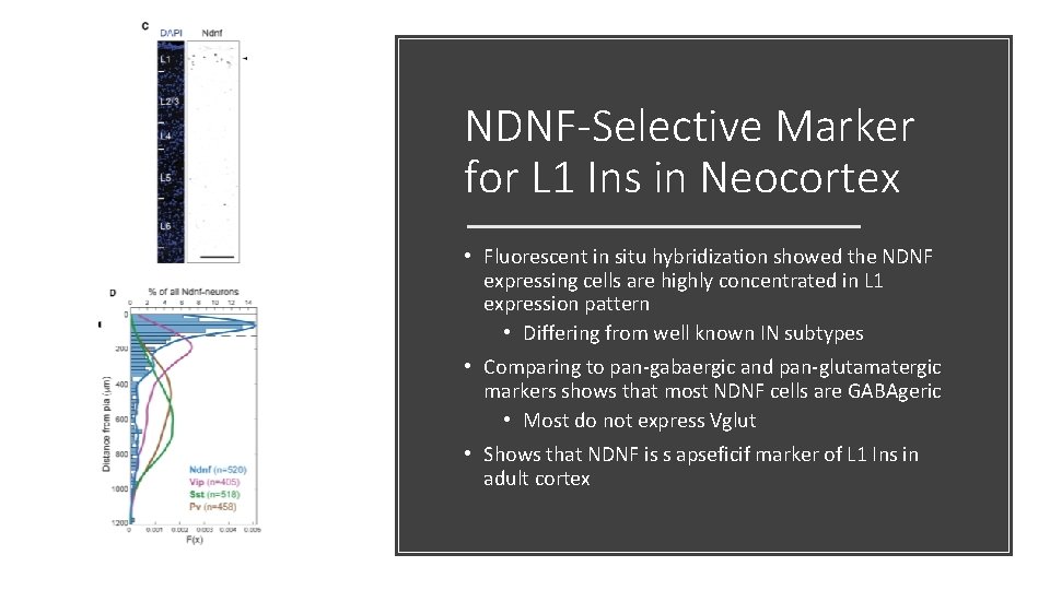
NDNF-Selective Marker for L 1 Ins in Neocortex • Fluorescent in situ hybridization showed the NDNF expressing cells are highly concentrated in L 1 expression pattern • Differing from well known IN subtypes • Comparing to pan-gabaergic and pan-glutamatergic markers shows that most NDNF cells are GABAgeric • Most do not express Vglut • Shows that NDNF is s apseficif marker of L 1 Ins in adult cortex
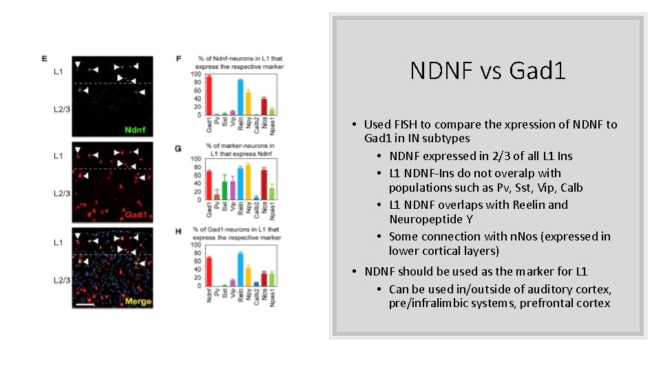
NDNF vs Gad 1 • Used FISH to compare the xpression of NDNF to Gad 1 in IN subtypes • NDNF expressed in 2/3 of all L 1 Ins • L 1 NDNF-Ins do not overalp with populations such as Pv, Sst, Vip, Calb • L 1 NDNF overlaps with Reelin and Neuropeptide Y • Some connection with n. Nos (expressed in lower cortical layers) • NDNF should be used as the marker for L 1 • Can be used in/outside of auditory cortex, pre/infralimbic systems, prefrontal cortex
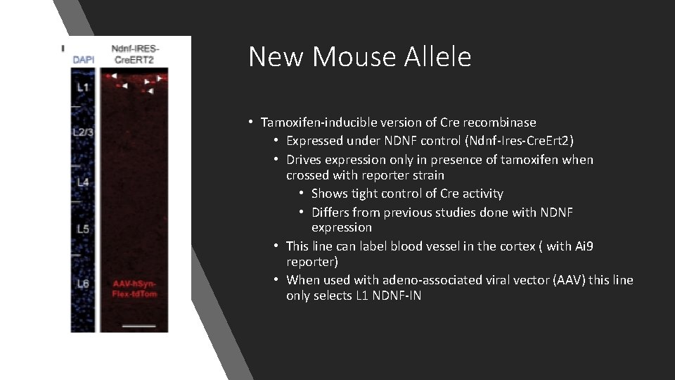
New Mouse Allele • Tamoxifen-inducible version of Cre recombinase • Expressed under NDNF control (Ndnf-Ires-Cre. Ert 2) • Drives expression only in presence of tamoxifen when crossed with reporter strain • Shows tight control of Cre activity • Differs from previous studies done with NDNF expression • This line can label blood vessel in the cortex ( with Ai 9 reporter) • When used with adeno-associated viral vector (AAV) this line only selects L 1 NDNF-IN
Second New Mouse Allele • Flp Recombinase expressed under NDNF locus (Ndnf-Ires-Flp. O) • Showed similar selectivity for L 1 • Similar for labeling NDNF neurons in the Cre line • Shows both lines can be important in circuit dissection • L 1 NDNf-Ins are faithfully used by both lines in prefrontal cortex • Allows for specific labeling and manipulation of L 1 NDNF-IN in adult cortex
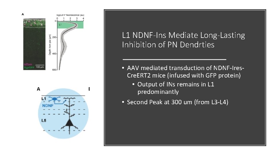
L 1 NDNF-Ins Mediate Long-Lasting Inhibition of PN Dendrties • AAV mediated transduction of NDNF-Ires. Cre. ERT 2 mice (infused with GFP protein) • Output of INs remains in L 1 predominantly • Second Peak at 300 um (from L 3 -L 4)
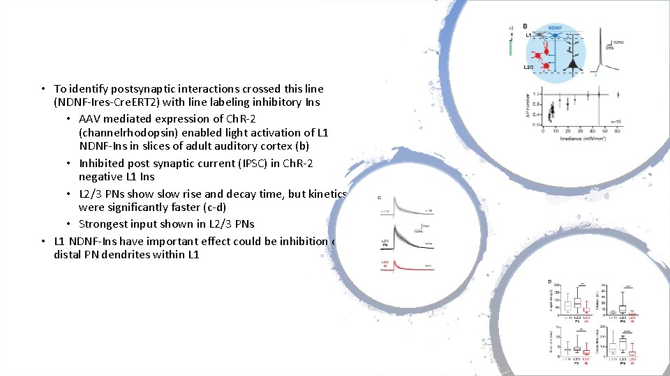
• To identify postsynaptic interactions crossed this line (NDNF-Ires-Cre. ERT 2) with line labeling inhibitory Ins • AAV mediated expression of Ch. R-2 (channelrhodopsin) enabled light activation of L 1 NDNF-Ins in slices of adult auditory cortex (b) • Inhibited post synaptic current (IPSC) in Ch. R-2 negative L 1 Ins • L 2/3 PNs show slow rise and decay time, but kinetics were significantly faster (c-d) • Strongest input shown in L 2/3 PNs • L 1 NDNF-Ins have important effect could be inhibition of distal PN dendrites within L 1
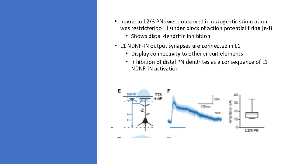
• Inputs to L 2/3 PNs were observed in optogentic stimulation was restricted to L 1 under block of action potential firing (e-f) • Shows distal dendritic inhibition • L 1 NDNF-IN output synapses are connected in L 1 • Display connectivity to other circuit elements • Inhibition of distal PN dendrites as a consequence of L 1 NDNF-IN activation
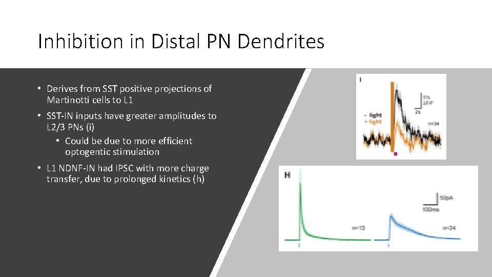
Inhibition in Distal PN Dendrites • Derives from SST positive projections of Martinotti cells to L 1 • SST-IN inputs have greater amplitudes to L 2/3 PNs (i) • Could be due to more efficient optogentic stimulation • L 1 NDNF-IN had IPSC with more charge transfer, due to prolonged kinetics (h)
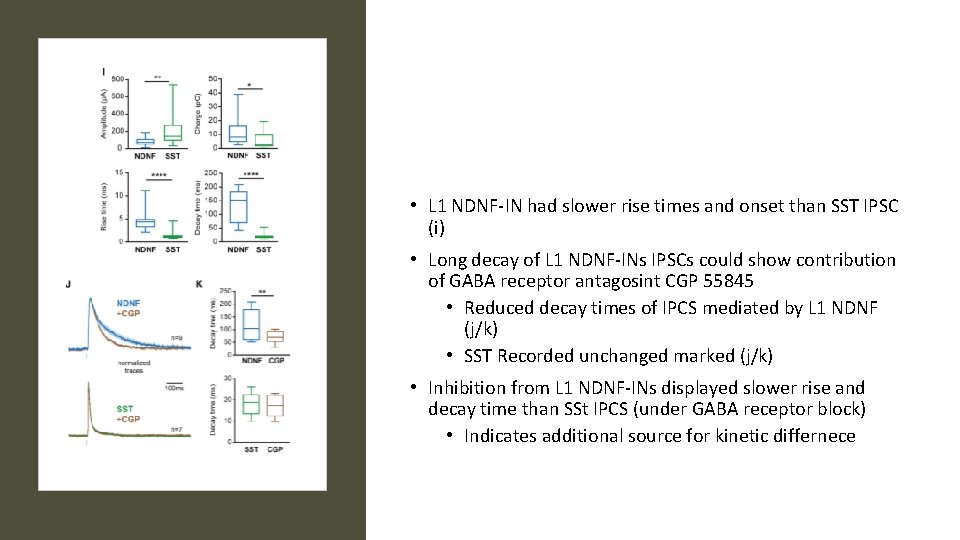
• L 1 NDNF-IN had slower rise times and onset than SST IPSC (i) • Long decay of L 1 NDNF-INs IPSCs could show contribution of GABA receptor antagosint CGP 55845 • Reduced decay times of IPCS mediated by L 1 NDNF (j/k) • SST Recorded unchanged marked (j/k) • Inhibition from L 1 NDNF-INs displayed slower rise and decay time than SSt IPCS (under GABA receptor block) • Indicates additional source for kinetic differnece
• Inhibition from two sources controls activity of PN desntrites • Identify L 1 NDNF-INs as genetic source of inhibition in PN dendrites • In comparison with SST Martinotti cells (based on kinetics and receptors) • Inhibition from L 1 NDNF-INs and SST-Ins could contribute to oscillations in differing frequency bands
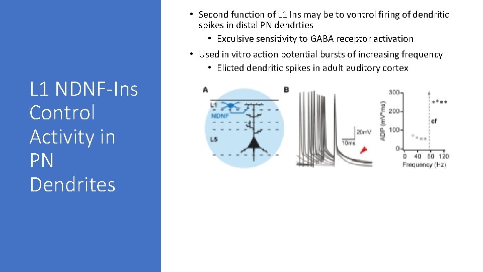
• Second function of L 1 Ins may be to vontrol firing of dendritic spikes in distal PN dendrties • Exculsive sensitivity to GABA receptor activation • Used in vitro action potential bursts of increasing frequency • Elicted dendritic spikes in adult auditory cortex L 1 NDNF-Ins Control Activity in PN Dendrites
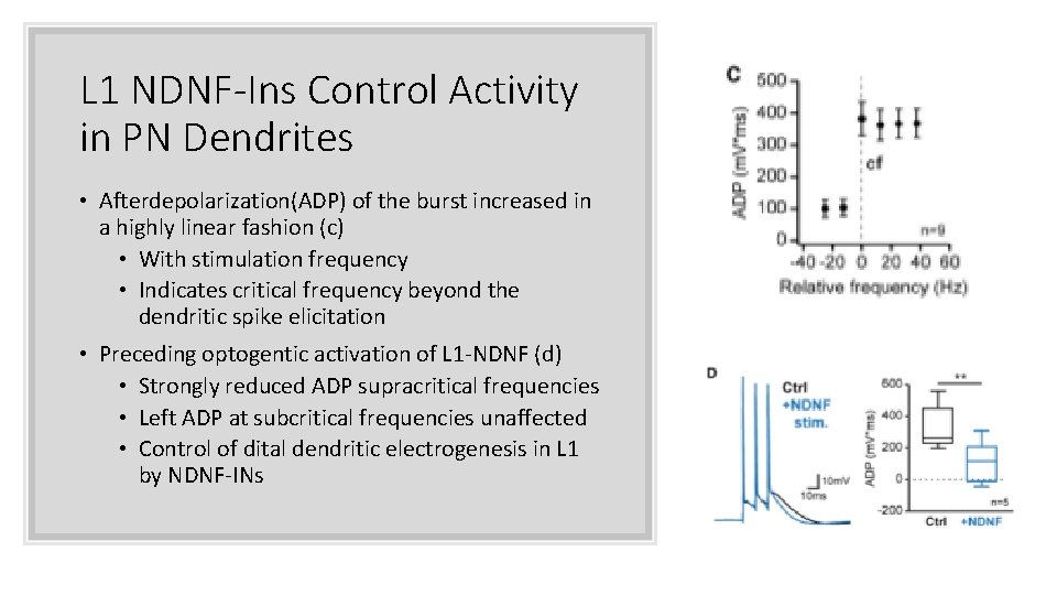
L 1 NDNF-Ins Control Activity in PN Dendrites • Afterdepolarization(ADP) of the burst increased in a highly linear fashion (c) • With stimulation frequency • Indicates critical frequency beyond the dendritic spike elicitation • Preceding optogentic activation of L 1 -NDNF (d) • Strongly reduced ADP supracritical frequencies • Left ADP at subcritical frequencies unaffected • Control of dital dendritic electrogenesis in L 1 by NDNF-INs
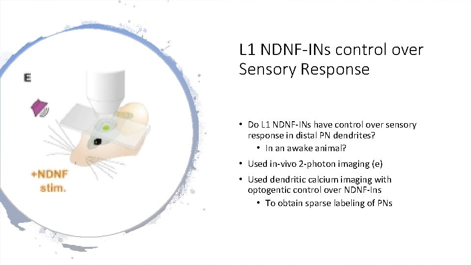
L 1 NDNF-INs control over Sensory Response • Do L 1 NDNF-INs have control over sensory response in distal PN dendrites? • In an awake animal? • Used in-vivo 2 -photon imaging (e) • Used dendritic calcium imaging with optogentic control over NDNF-Ins • To obtain sparse labeling of PNs
• Injected retrograde vector carrying Cre recombinase into subcortical targets of auditory cortex in Ndnf-Ires-Flp. O mice • Allow selective expression of GCa. MP 6 (calcium indicator) and td. Tomato (motion detection) • Simultaneously, a Flp-dependent AAV was injected in the auditory cortex • Enable expression at optogenetic activator (Chrimson) • Ideal due to use in combination with 2 -photon imaging and red shifted excitation spectrum
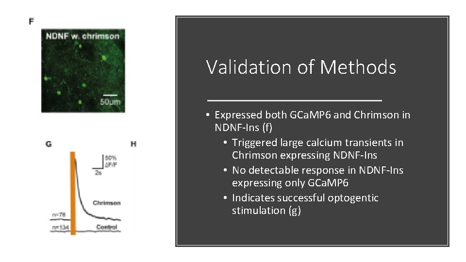
Validation of Methods • Expressed both GCa. MP 6 and Chrimson in NDNF-Ins (f) • Triggered large calcium transients in Chrimson expressing NDNF-Ins • No detectable response in NDNF-Ins expressing only GCa. MP 6 • Indicates successful optogentic stimulation (g)
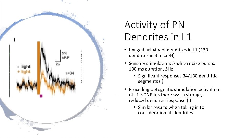
Activity of PN Dendrites in L 1 • Imaged activity of dendrites in L 1 (130 dendrites in 3 mice-H) • Sensory stimulation: 5 white noise bursts, 100 ms duration, 5 Hz • Significant responses 34/130 dendritic segments (I) • Preceding optogentic stimulation activation of L 1 NDNF-Ins there was a strongly reduced dendritic response (I) • Similar results when taking in to consideration all dendrites
Quick Review: Conclusions • Considering the observation that L 1 NDNF-IN output synapses are enriched in L 1, where there is direct contact with PN distal dendrites • In combination with previous slide data • Provides thought that there is direct control over dendritic activity by L 1 NDNF-Ins in the intact animal • Cannot rule out contribution of somatic inhibition to account for this reduced dendritic activity • Suggests that inhibition from NDNF-Ins control firing of spikes in distal PN dendrites in L 1 • Inhibition lasted several seconds, consistent with L 1 NDNF-IN inhibition in slice recordings • Larger responses were more strongly suppressed by NDNF-IN input
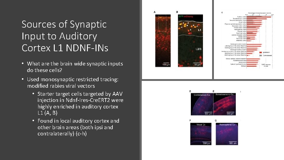
Sources of Synaptic Input to Auditory Cortex L 1 NDNF-INs • What are the brain wide synaptic inputs do these cells? • Used monosynaptic restricted tracing: modified rabies viral vectors • Starter target cells targeted by AAV injection in Ndnf-Ires-Cre. ERT 2 were highly enriched in auditory cortex L 1 (A, B) • Found in local auditory cortex and other brain areas (both ipsi and contralaterally) (c-h)
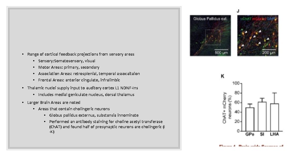
• Range of cortical feedback projections from sensory areas • Sensory: Somatosensory, visual • Motor Areas: primary, secondary • Association Areas: retrosplenial, temporal assocaitaion • Frontal Areas: anterior cingulate, infralimbic • Thalamic nuclei supply input to auditory cortex L 1 NDNF-Ins • Includes medial geniculate nucleus, dorsal thalamus • Larger Brain Areas are noted • Areas that contain cholingeric neurons • Globus pallidus externus, substansia innominate • Performed an antibody staining for choline acetyl transferase (Ch. AT) and found half of presynaptic neurons are cholingeric (I -K)
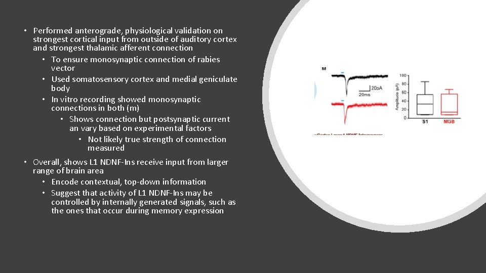
• Performed anterograde, physiological validation on strongest cortical input from outside of auditory cortex and strongest thalamic afferent connection • To ensure monosynaptic connection of rabies vector • Used somatosensory cortex and medial geniculate body • In vitro recording showed monosynaptic connections in both (m) • Shows connection but postsynaptic current an vary based on experimental factors • Not likely true strength of connection measured • Overall, shows L 1 NDNF-Ins receive input from larger range of brain area • Encode contextual, top-down information • Suggest that activity of L 1 NDNF-Ins may be controlled by internally generated signals, such as the ones that occur during memory expression
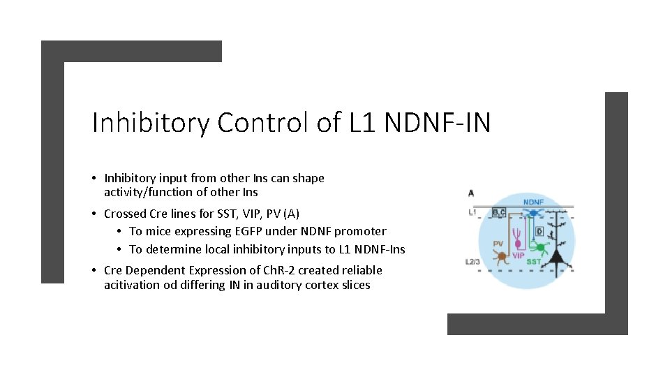
Inhibitory Control of L 1 NDNF-IN • Inhibitory input from other Ins can shape activity/function of other Ins • Crossed Cre lines for SST, VIP, PV (A) • To mice expressing EGFP under NDNF promoter • To determine local inhibitory inputs to L 1 NDNF-Ins • Cre Dependent Expression of Ch. R-2 created reliable acitivation od differing IN in auditory cortex slices
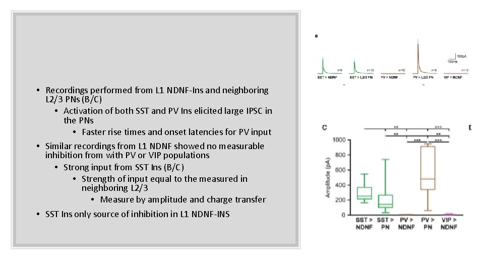
• Recordings performed from L 1 NDNF-Ins and neighboring L 2/3 PNs (B/C) • Activation of both SST and PV Ins elicited large IPSC in the PNs • Faster rise times and onset latencies for PV input • Similar recordings from L 1 NDNF showed no measurable inhibition from with PV or VIP populations • Strong input from SST Ins (B/C) • Strength of input equal to the measured in neighboring L 2/3 • Measure by amplitude and charge transfer • SST Ins only source of inhibition in L 1 NDNF-INS
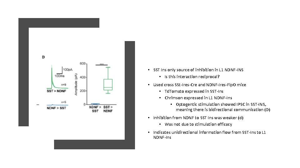
• SST Ins only source of inhibition in L 1 NDNF-INS • Is this interaction reciprocal? • Used cross SSt-Ires-Cre and NDNF-Ires-Flp. O mice • Td. Tomato expressed in SST-Ins • Chrimson expressed in L 1 NDNF-Ins • Optogentic stimulation showed IPSC in SST-INS, meaning there is bidirectional communication (D) • Inhibition from NDNF to SST Ins was weaker (d) • Was not due to stimulation efficacy • Indicates unidirectional information flow from SST-Ins to L 1 NDNF-Ins
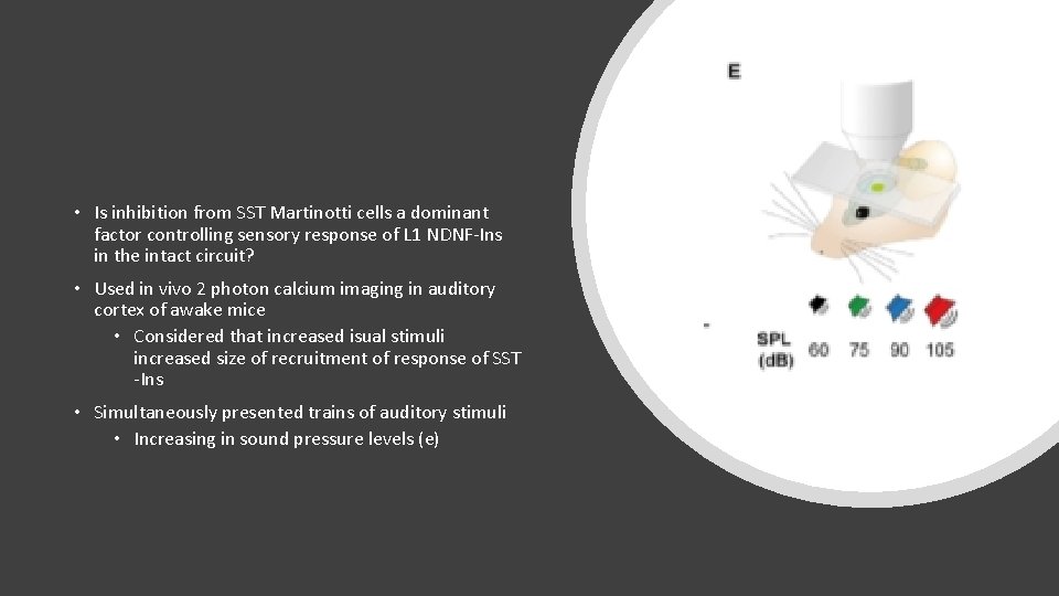
• Is inhibition from SST Martinotti cells a dominant factor controlling sensory response of L 1 NDNF-Ins in the intact circuit? • Used in vivo 2 photon calcium imaging in auditory cortex of awake mice • Considered that increased isual stimuli increased size of recruitment of response of SST -Ins • Simultaneously presented trains of auditory stimuli • Increasing in sound pressure levels (e)
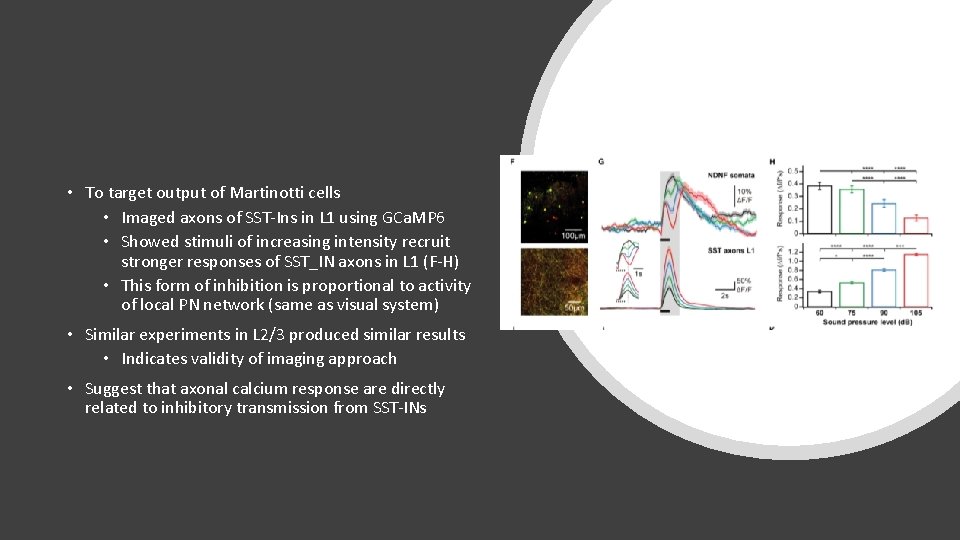
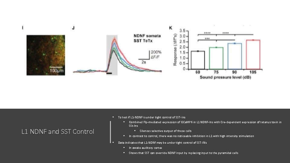
• To target output of Martinotti cells • Imaged axons of SST-Ins in L 1 using GCa. MP 6 • Showed stimuli of increasing intensity recruit stronger responses of SST_IN axons in L 1 (F-H) • This form of inhibition is proportional to activity of local PN network (same as visual system) • Similar experiments in L 2/3 produced similar results • Indicates validity of imaging approach • Suggest that axonal calcium response are directly related to inhibitory transmission from SST-INs
• To test if L 1 -NDNF is under tight control of SST-Ins • L 1 NDNF and SST Control • • Combined Flp-mediated expression of GCa. MP 6 in L 1 NDNF-Ins with Cre-dependent expression of tetanus toxin in SSt-Ins • Silences selective output of these cells In contrast to control, there was no noticeable inhibition in L 1 with high intensity stimulation Data indicates that L 1 -NDNf may be under tight control of SST-INs • • In awake auditory cortex Shows that SST can override NDNF input by replacing input to the pyramidal cells
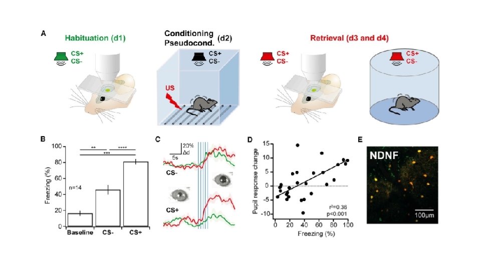
Learning related Plasticity of Sensory Responses in L 1 NDNF-Ins • Used associative auditory fear conditioning • Allowed for retrieval of the same L 1 NDNFIns during CS is neutral and during memory retrieval • Used freezing and pupil dilation (positive correlation) to show successful memory acquisition and to examine fear • Greater pupil response after fear learning • Shows successful fear memory retrieval in the mouse
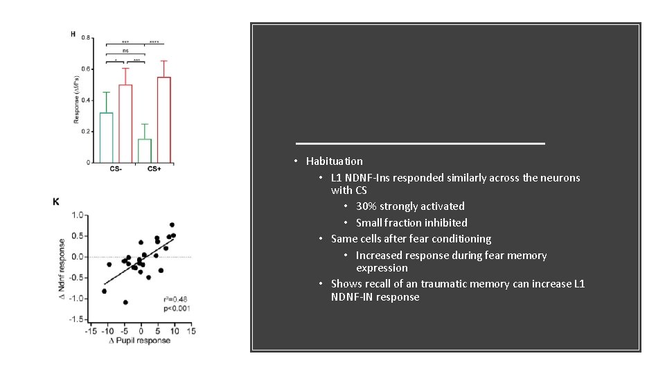
• Habituation • L 1 NDNF-Ins responded similarly across the neurons with CS • 30% strongly activated • Small fraction inhibited • Same cells after fear conditioning • Increased response during fear memory expression • Shows recall of an traumatic memory can increase L 1 NDNF-IN response
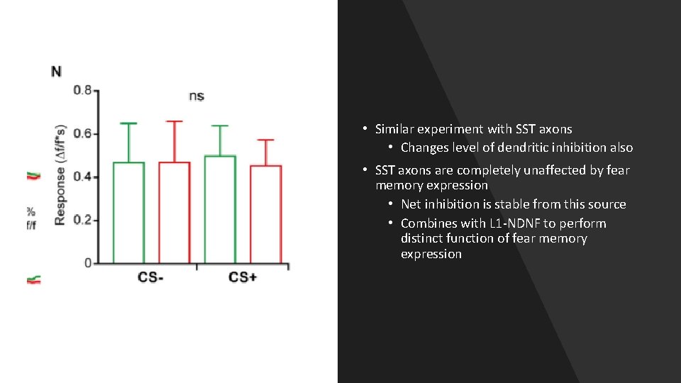
• Similar experiment with SST axons • Changes level of dendritic inhibition also • SST axons are completely unaffected by fear memory expression • Net inhibition is stable from this source • Combines with L 1 -NDNF to perform distinct function of fear memory expression
Inhibition of Martinotti cells remains unchanged after conditioning • Tightly control sensory response in NDNF-Ins • Shows genetically dependent form of dendritic inhibition • Highly experience dependent • Salient stimuli are encoded at elevated levels of distal dendritic inhibition • Along with disinhibition Conclusions • L 1 NDNF-Ins strong source of inhibition of distal PN dendrites • Differs from SST, but is complementary • Have slow time course and strong GABA receptor contribution • Probably located in neocortex • Recruited by cholinergic input and top down afferent inputs
• NDNF is a highly selective marker of INs in adult auditory and prefrontal cortex • Is the L 1 NDNF-Ins in the prefrontal cortex also? • Generate genetic responses to target these nerons (tamoxifen-inducible Cre allele, Flp allele) Conclusions • Auditory Cortex has a prominent interaction with L 1 NDNF Ins • Strong interaction with SST-Ins (in vivo and in vitro) • Induce Auditory Cortex plasticity by associate fear condition • Based on L 1 NDNF and SST-In responses of expression during behavioral memory • L 1 NDNF response is related to fear memory
Conclusions • SST Inhibition dominates in conditions of weak and imprecise stimulus encoding in PNs • L 1 NDNF-INs occurs when sensory input is relevant to the animal
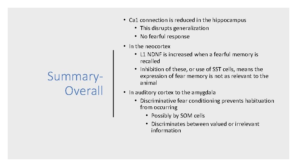
• Ca 1 connection is reduced in the hippocampus • This disrupts generalization • No fearful response Summary. Overall • In the neocortex • L 1 NDNF is increased when a fearful memory is recalled • Inhibition of these, or use of SST cells, means the expression of fear memory is not as relevant to the animal • In auditory cortex to the amygdala • Discriminative fear conditioning prevents habituation from occurring • Possibly by SOM cells • Discriminates between valued or irrelevant information
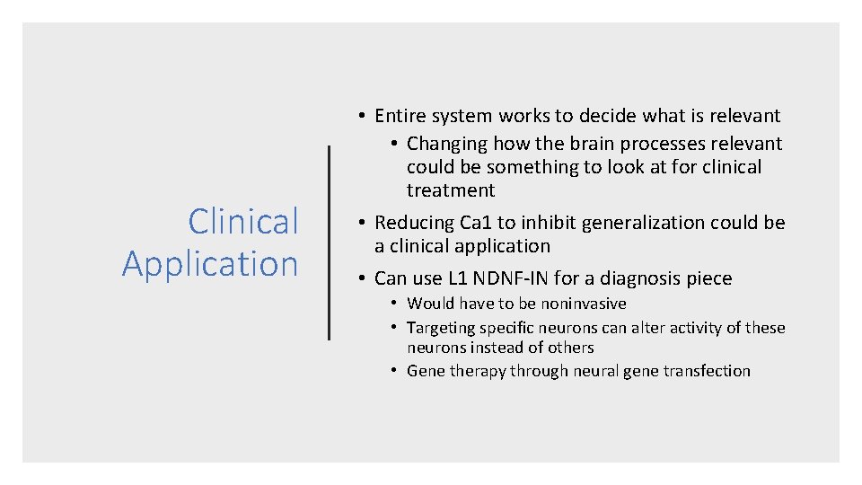
Clinical Application • Entire system works to decide what is relevant • Changing how the brain processes relevant could be something to look at for clinical treatment • Reducing Ca 1 to inhibit generalization could be a clinical application • Can use L 1 NDNF-IN for a diagnosis piece • Would have to be noninvasive • Targeting specific neurons can alter activity of these neurons instead of others • Gene therapy through neural gene transfection