34 1 Reproductive Anatomy KEY CONCEPT Female and


- Slides: 12

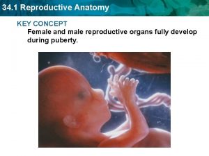
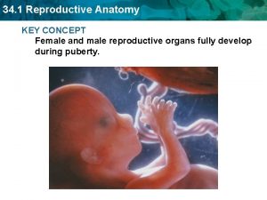
34. 1 Reproductive Anatomy KEY CONCEPT Female and male reproductive organs fully develop during puberty.

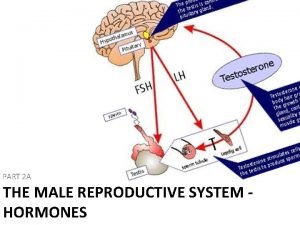
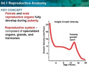
34. 1 Reproductive Anatomy The female reproductive system produces ova. • The reproductive system is a collection of specialized organs, glands, and hormones that help produce a new human being. • Females and males reach sexuality maturity, or the ability to produce offspring, only after puberty. • Puberty marks the time in your life when your hypothalamus and your pituitary gland release hormones. – Follicle stimulating hormones (FSH) and luteinizing hormones(LH)

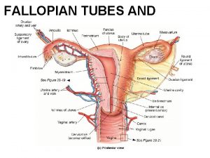
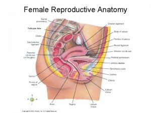
34. 1 Reproductive Anatomy The female reproductive system produces ova. • There are two main functions of the female reproductive system. – produce ova, or egg cells – provide a place where a zygote develops fallopian tube ovary uterus cervix pubic bone urinary bladder urethra rectum vagina

34. 1 Reproductive Anatomy The female reproductive system produces ova. • The egg cells are produced in the ovaries. • The ovaries are paired organs located on either side of the uterus, or womb. • When a female baby is born, she has all of her eggs.

34. 1 Reproductive Anatomy • Follicle-stimulating hormones and Luteinizing hormones stimulate the release of Estrogen has three main functions: – develop female sexual characteristics Ex: widening the pelvis, increasing fat deposits and bone mass, and enlarging the breasts. – develop eggs – prepare uterus for pregnancy and maintain a pregnancy when it occurs

34. 1 Reproductive Anatomy • When the egg cell matures each month, it is released from an ovary and enters the fallopian tube. • An egg takes several days to travel through the fallopian tube, during that time it can be fertilized by sperm that enters the tube. • When the egg is fertilized it will attach to the wall of the uterus, if it is unfertilized it will eventually be broken down and discarded.

34. 1 Reproductive Anatomy • The uterus is made up of 3 layers – A thin inner layer of epithelial cells – A thick middle layer of muscle – An outer layer of connective tissue The lower end is called the cervix, which opens to the vagina. During a normal birth, a baby is pushed down the canal of the vagina to exit the mothers body.

34. 1 Reproductive Anatomy The male reproductive system produces sperm. • There are two main functions of the male reproductive system. – produce sperm cells – deliver sperm to the female reproductive system urinary bladder seminal vesicle vas deferens pubic bone prostate gland rectum penis urethra epididymis scrotum testis bulbourethral gland

34. 1 Reproductive Anatomy The male reproductive system produces sperm. • Males do not produce sperm until after they reach puberty • Sperm production takes place in the testicles, or testes • Each testis contains hundreds of tiny tubules where millions of sperm cells are produced.

34. 1 Reproductive Anatomy • LH stimulates the release of testosterone • Testosterone has two main functions. – Controls developing male sexual characteristics Ex: deeper voice, more body hair, greater bone density, increased muscle mass – producing sperm

34. 1 Reproductive Anatomy • The testes are enclosed in a pouch called the scrotum which hangs below the pelvis outside the body. – It is 2 -3 degrees cooler then the core body temperature – Sperm cannot develop if the temperature is too high • When the sperm leaves the testes, they travel through a duct to a long coiled tube known as the epididymus. – Sperm matures there and remains until expelled • During sexual stimulation, the sperm travel into another long duct called the vas deferens

34. 1 Reproductive Anatomy • Secondary sex glands secrete fluids into the vas deferens to nourish and protect the sperm. • The prostate gland produces a fluid that helps sperm move more easily. • The bulbourethral gland the seminal vesicle secrete basic fluids that help neutralize the acidity in the urethra and in the females vagina. • The fluids from all 3 glands plus sperm form semen. • Semen moves from the vas deferens into the urethra and ejects it from the penis.
 Cow reproductive anatomy
 Female reproductive system anatomy
 Uterus diagram
 Female reproductive anatomy
 Frog testis labeled
 Female and male reproductive system
 Similarities between male and female reproductive system
 Parts of and functions of female reproductive system
 Function of reproductive organs
 Male reproductive system table
 90/2
90/2 Circumcised dick
 Male fish reproductive system
