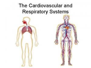
The Respiratory System Organs of the Respiratory system


- Slides: 34

The Respiratory System

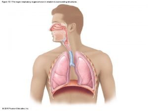
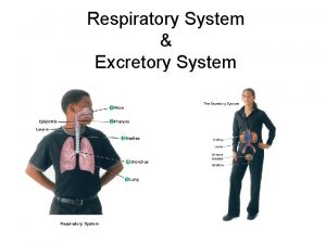
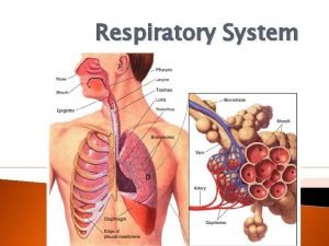
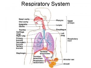
Organs of the Respiratory system • • • Nose Pharynx Larynx Trachea Bronchi Lungs – alveoli Figure 13. 1

Function of the Respiratory System • Oversees gas exchanges between the blood and external environment • Exchange of gasses takes place within the alveoli • Passageways to the lungs purify, warm, and humidify the incoming air

The Nose • The only externally visible part of the respiratory system • Air enters the nose through the external nares (nostrils) • The interior of the nose consists of a nasal cavity divided by a nasal septum

Anatomy of the Nasal Cavity • Olfactory receptors are located in the mucosa on the superior surface • The rest of the cavity is lined with respiratory mucosa – Moistens air – Traps incoming foreign particles

Sinuses Cavities within bones surrounding the nasal cavity • Function of the sinuses – Lighten the skull – Act as resonance chambers for speech – Produce mucus that drains into the nasal cavity

Pharynx (Throat) • Muscular passage from nasal cavity to larynx • The oropharynx and laryngopharynx are common passageways for air and food • Auditory tubes enter the nasopharynx • Tonsils of the pharynx

Larynx (Voice Box) • Routes air and food into proper channels • Plays a role in speech • Made of eight rigid hyaline cartilages and a spoon-shaped flap of elastic cartilage (epiglottis) • Vocal cords - vibrate with expelled air to create sound (speech)

Structures of the Larynx • Thyroid cartilage – Largest hyaline cartilage – Protrudes anteriorly (Adam’s apple) • Epiglottis – Superior opening of the larynx – Routes food to the larynx and air toward the trachea • Glottis – opening between vocal cords

Trachea (Windpipe) • Connects larynx with bronchi • Lined with ciliated mucosa • Walls are reinforced with C-shaped hyaline cartilage

Lungs • Ocupy most of the thoracic cavity – Apex is near the clavicle (superior portion) – Each lung is divided into lobes by fissures • Left lung – two lobes • Right lung – three lobes

Lungs Figure 13. 4 b

Respiratory Tree Divisions • • • Primary bronchi Secondary bronchi Tertiary bronchi Bronchioli Terminal bronchioli

Bronchioles • Smallest branches of the bronchi • All but the smallest branches have reinforcing cartilage • Terminal bronchioles end in alveoli Figure 13. 5 a

Respiratory Zone • Structures – Respiratory bronchioli – Alveolar duct – Alveoli • Site of gas exchange

Alveoli • Structure of alveoli – Alveolar duct – Alveolar sac – Alveolus • Gas exchange takes place within the alveoli in the respiratory membrane

Gas Exchange • Gas crosses the respiratory membrane by diffusion – Oxygen enters the blood – Carbon dioxide enters the alveoli • Macrophages add protection • Surfactant coats gas-exposed alveolar surfaces

Respiratory Membrane (Air. Blood Barrier) Figure 13. 6

Events of Respiration • Pulmonary ventilation – moving air in & out of the lungs • External respiration – gas exchange between pulmonary blood and alveoli • Respiratory gas transport – transport of oxygen & carbon dioxide via the bloodstream • Internal respiration – gas exchange between blood and tissue cells in systemic capillaries

Mechanics of Breathing (Pulmonary Ventilation) • Mechanical process • 2 phases – Inspiration – flow of air into lung – Expiration – air leaving lung

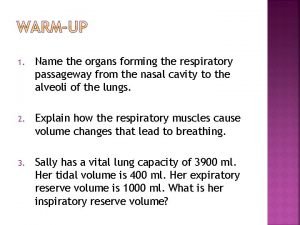
Inspiration • Diaphragm and intercostal muscles contract • The size of the thoracic cavity increases • External air is pulled into the lungs due to an increase in intrapulmonary volume

Expiration Figure 13. 7 b

Pressure Differences in the Thoracic Cavity • Normal pressure within the pleural space is always negative (intrapleural pressure) • Differences in lung and pleural space pressures keep lungs from collapsing

Respiratory Volumes and Capacities • Normal breathing moves about 500 ml of air with each breath - tidal volume (TV) • Many factors that affect respiratory capacity – – A person’s size Sex Age Physical condition • Residual volume of air – after exhalation, about 1200 ml of air remains in the lungs

Respiratory Sounds • Sounds are monitored with a stethoscope • Bronchial sounds – produced by air rushing through trachea and bronchi • Vesicular breathing sounds – soft sounds of air filling alveoli

Internal Respiration • Exchange of gases between blood and body cells • An opposite reaction to what occurs in the lungs – Carbon dioxide diffuses out of tissue to blood – Oxygen diffuses from blood into tissue

Internal Respiration Figure 13. 11

Emphysema • Alveoli enlarge as adjacent chambers break through • Chronic inflammation promotes lung fibrosis • Airways collapse during expiration • Patients use a large amount of energy to exhale • Over-inflation of the lungs leads to a barrel chest • Cyanosis appears late in the disease

Chronic Bronchitis • Inflammation of the mucosa of the lower respiratory passages • Mucus production increases • Pooled mucus impairs ventilation & gas exchange • Increased lung infection • Pneumonia is common • Hypoxia & cyanosis

Asthma • Chronic inflammation if the bronchiole passages • Response to irritants with dyspnea, coughing, and wheezing

Pneumonia • An infection – inflames the air sacs – The air sacs may fill with fluid or pus – Symptoms: • cough with phlegm or pus • Fever & chills • difficulty breathing

Developmental Aspects of the Respiratory System • Lungs are filled with fluid in the fetus • Lungs are not fully inflated with air until two weeks after birth • Surfactant that lowers alveolar surface tension is not present until late in fetal development and may not be present in premature babies

Aging Effects • • Elasticity of lungs decreases Vital capacity decreases Blood oxygen levels decrease Stimulating effects of carbon dioxide decreases • More risks of respiratory tract infection

Respiratory Rate Changes Throughout Life Respiration rate: • Newborns – 40 to 80 min. • Infants – 30 min. • Age 5 – 25 min. • Adults – 12 to 18 min • Rate often increases with old age
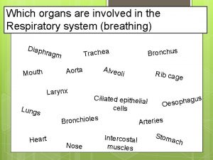
 Which organs are involved in respiratory system
 Chapter 13 respiratory system
 Invertebrate digestive system
 Germ layers
 Organs forming the respiratory passageway
 Upper respiratory system labeled
 Figure 13-1 respiratory system
 Conducting zone of the respiratory system function
 Respiratory digestive and circulatory system
 Sensory system organs
 Excretory system function
 Major and accessory organs of the digestive system
 Accessory organs of the digestive system
 Digestive accessory organs
 Red and white blood cells difference
 Formation of lymph
 Lymphatic system organs
 Function of lymphatic system
 Organs of the sensory system
 Chapter 16
 How respiratory system work with circulatory system
 Circulatory system and respiratory system work together
 Organs of wto
 Homologous structures
 Vowel sounds with examples
 Primary and secondary retroperitoneal organs
 Is the uterus intraperitoneal
 Ligaments of stomach
 Epiploic foramen
 Kidney is a retroperitoneal organ
 Pollen basket
 Urinary structure
 5 functions of the stomach
 Sense organs
 Proves anatòmiques
