The Microscope and Forensic Identification Magnification of Images


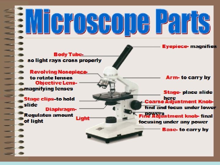
 • Body tube or head:")
")
")
 or via")


- Slides: 26
The Microscope and Forensic Identification
Magnification of Images • A microscope is an optical instrument that uses a lens or a combination of lenses to magnify and resolve the fine details of an object. • The magnified image seen by looking through a lens is known as a virtual image, whereas an image viewed directly is known as a real image. – Virtual image- an image that cannot be seen directly. It can only be seen by a viewer by looking through a lens. Ex. –Looking through a microscope. – Real image- an image formed by the actual convergence of light rays on a screen. Ex. Movie projected on a screen. • The magnification provided by a lens comes from the process of refraction: light rays bend as they pass through the lens.
Refraction!
Focal Point & Focal Length • The simplest microscope is a magnifying glass • The point at which parallel rays are converged to an image is the focal point of the lens. • The distance of this point from the lens is the focal length.
A Simple Magnifier Virtual image I • Object O is placed close to the lens, the rays from it bend but do not intersect. • The observer’s eye follows rays back to the point of apparent origin (I). • The virtual image (I) appears bigger than the object (O) • Moving the lens closer to the eye increases magnification.
The Compound Light Microscope • Robert Hooke- 1665 first to use light and a lens system to magnify. • Only used on objects light can pass through • Usually specimen needs to be stained
Terms in Microscopy • Magnification: – How much the image is being increased – Depends on the refractive index, curvature and thickness of the lens • Field of View: – Everything that is visible through the eyepiece – When you increase magnification, you decrease field of view 10 x 20 x 40 x
Terms in Microscopy • Depth of Focus: – The thickness of the specimen that can be seen clearly – Can be used to determine layering of objects – Decreases at higher magnification • Resolution: – Ability to distinguish objects close together – Increases with numerical aperture; wavelength of radiation and refractive index
Types of Microscopes • Compound Microscope • Comparison Microscope • Stereoscopic Microscope • Polarizing Microscope • Electron Microscopes
Compound Microscope • Lenses: Ocular and objective (with mirror) • Body tube or head: hollow tube that holds the objective and eyepiece lenses • Stage: platform that supports the specimen • Lenses (Eyepiece and objectives)- magnify • Diaphragm- regulates amount of light. • Coarse Adjustment knob- to find and focus under lower powers • Fine adjustment knob- to final focus under any power
The Compound Microscope See also Figure 4 -4 • Rays from the object (O) pass first through the objective lens forming a real, slightly enlarged, inverted image (I 1). • The second lens (eyepiece) acts as a simple magnifier to create an even bigger image (I 2).
Compound Microscope • A compound microscope has at least two lenses: – Objective (lower) lens: produces a magnified and inverted version of the object (4 x, 10 x, 40 x) – Ocular (smaller) lens: produces a virtual image in the viewer’s brain (10 x) • Magnifying power = power of the objective lens x power of the ocular lens 4 x X 10 x= 40 x • Working distance = distance between the objective lens and the stage
Comparison Microscopes • Can be lighted from below the stage (transmission illumination) or via a vertical or reflected illumination system from the top.
Comparison Microscope • Provides a side by side comparison of specimens • Structure consists of 2 compound microscopes combined into one unit. • Designed to use a bridge incorporating a series of mirrors and lenses to join 2 independent objective lenses into a singular binocular unit. • Viewer will see a circular field divided into 2 equal parts by a thin line. • The specimen on the left will be seen in the left half of the field of view and the specimen on the right will be seen in the right half of the field of view.
Stereoscopic Microscope The most commonly used microscope in crime labs Stereo Microscope Compound Microscope
Stereoscopic Microscope • Two separate monocular microscopes each with its own set of lenses – Produce a three-dimensional image with a rightside-up, frontward orientation – Offers a large working distance for bulky items – Relatively low magnification (10 x-125 x) – Can be lighted from below or vertically from above – This microscope is actually two separate monocular microscopes except for the lowest objective lens, which is common to both microscopes – Wide field of view and great depth of focus
Polarizing Microscopes • Polarizing microscopes – Include two polarizing filters, a polarizer lens (fixed below the specimen), and an analyzer lens (fixed above the specimen) – The stage with the sample is rotated to determine how the polarized light interacts with the sample
Polarized Light
Polarizing Microscopes This can provide information about the shape, color, and size of minerals and it is used to identify hair, human-made fibers and paint. Hair Sample: Natural Light Hair Sample: Polarized Light
Scanning Electron Microscope • Can magnify 100, 000 X • Has a depth of focus more than 300 X that of an optical microscope • Uses electrons rather than light • Offer much greater resolution than with a light microscope • Scans the surface of a specimen usually coated with gold, holes are dark and knobs are light.
Scanning Electron Microscope • Beam of electrons is emitted from a tungsten film and focused by means of electromagnets onto the surface of the specimen. • This primary beam causes electrons to leave the surface of the specimen. All back scattered electrons (those not absorbed by the specimen but reflected back) are collected and the amplified signal is displayed in a 3 -D image focused on the viewing screen. • High magnification and resolving power results in a superb picture!
Scanning Electron Microscope
Scanning Electron Microscope Putting it all together—making the image:
Scanning Electron Microscope The SEM shows very detailed 3 -dimensional images created without light waves.
Transmission Electron Microscopes • Invented in the 1950’s • Magnifies up to 350, 000 x • Allows scientists to observe viruses and substructures – Aims a beam at a thin section of specimen, beam is transmitted through the specimen. – Beam is directed by electromagnets – Specimen has two types of areas • Electron transparent- e- will pass through and appear light • Electron opaque- e- will scatter and will appear dark Specimen needs to be dried, coated in heavy metal to enhance contrast, embedded in plastic and sliced into thin sections