
Title Magnification Lesson Objectives 1 Can I calculate
Title: Magnification Lesson Objectives: 1. Can I calculate magnification, image size and actual size of a cell? Grade C-A Starter: Recap quiz….
Recap quiz… What is the difference between a micrometer and micrometre? b. How many micrometres in a millimetre? c. What is the unit after micrometre? d. How many micrometres in a nanometre? e. Calculate the calibration: X 4 objective lens and a x 10 eyepiece (=) Micrometer is 1 mm long with 100 divisions (what is each division worth? ) a.
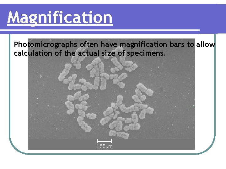
Magnification Photomicrographs often have magnification bars to allow calculation of the actual size of specimens. 4. 55μm
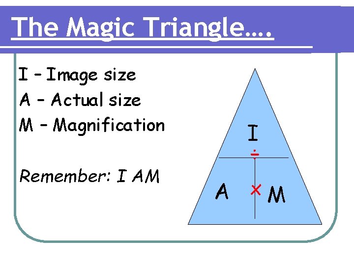
The Magic Triangle…. I – Image size A – Actual size M – Magnification Remember: I AM I ÷ A x. M
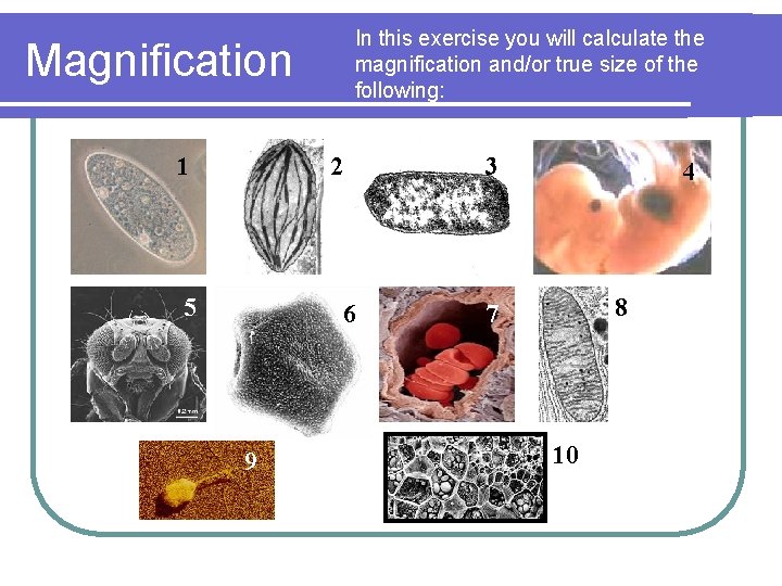
In this exercise you will calculate the magnification and/or true size of the following: Magnification 1 2 5 6 9 3 4 8 7 10
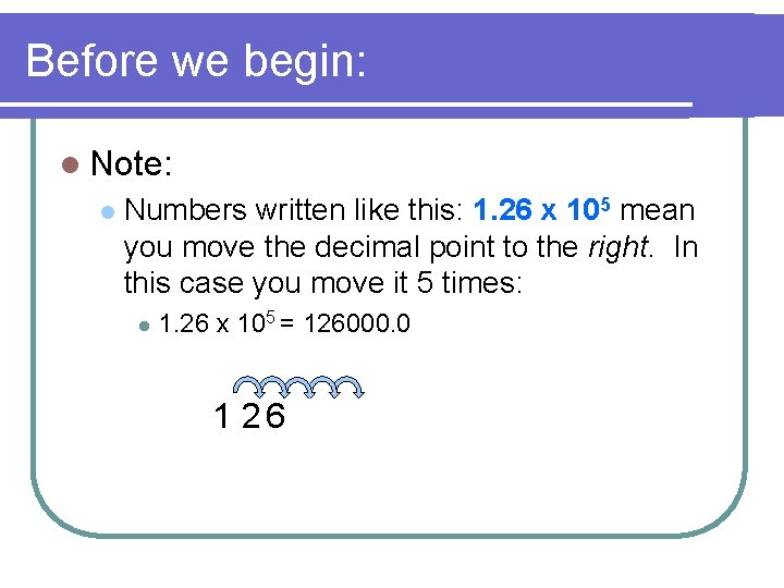
Before we begin: l Note: l Numbers written like this: 1. 26 x 105 mean you move the decimal point to the right. In this case you move it 5 times: l 1. 26 x 105 = 126000. 0 1. 2 6 0 0 0. 0
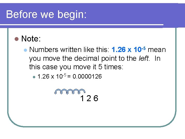
Before we begin: l Note: l Numbers written like this: 1. 26 x 10 -5 mean you move the decimal point to the left. In this case you move it 5 times: l 1. 26 x 10 -5 = 0. 0000126 0. 0 0 1. 2 6
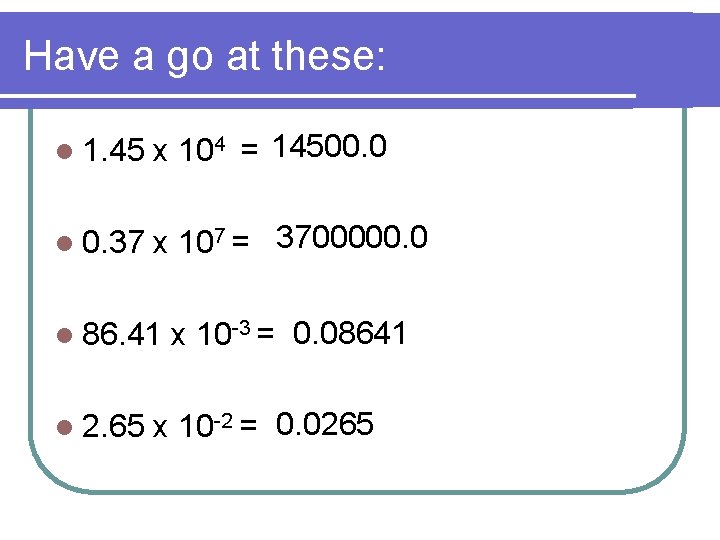
Have a go at these: l 1. 45 x 104 = 14500. 0 l 0. 37 x 107 = 3700000. 0 l 86. 41 l 2. 65 x 10 -3 = 0. 08641 x 10 -2 = 0. 0265
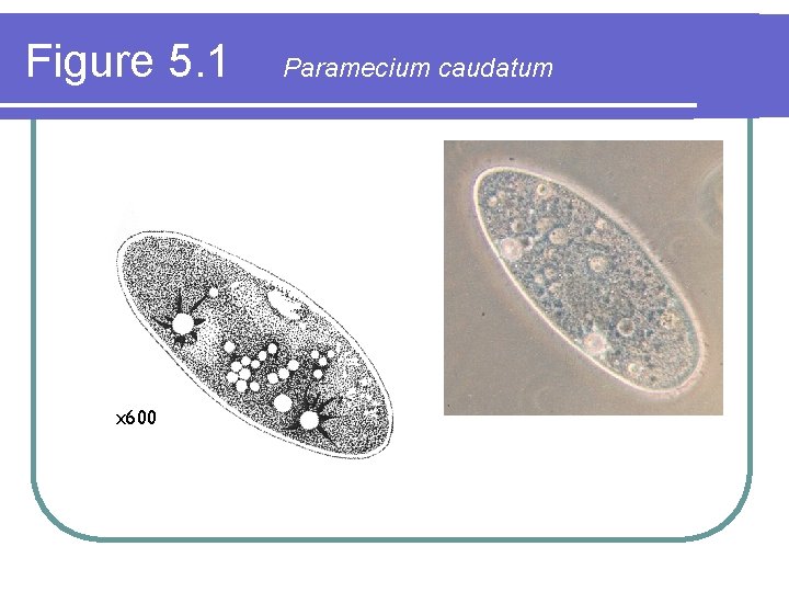
Figure 5. 1 x 600 Paramecium caudatum
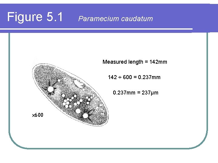
Figure 5. 1 Paramecium caudatum Measured length = 142 mm 142 ÷ 600 = 0. 237 mm = 237μm x 600
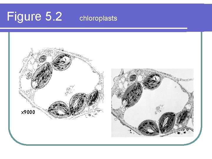
Figure 5. 2 x 9000 chloroplasts
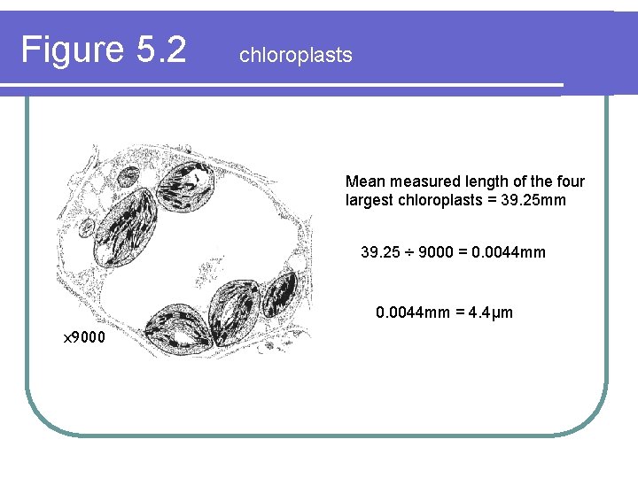
Figure 5. 2 chloroplasts Mean measured length of the four largest chloroplasts = 39. 25 mm 39. 25 ÷ 9000 = 0. 0044 mm = 4. 4μm x 9000
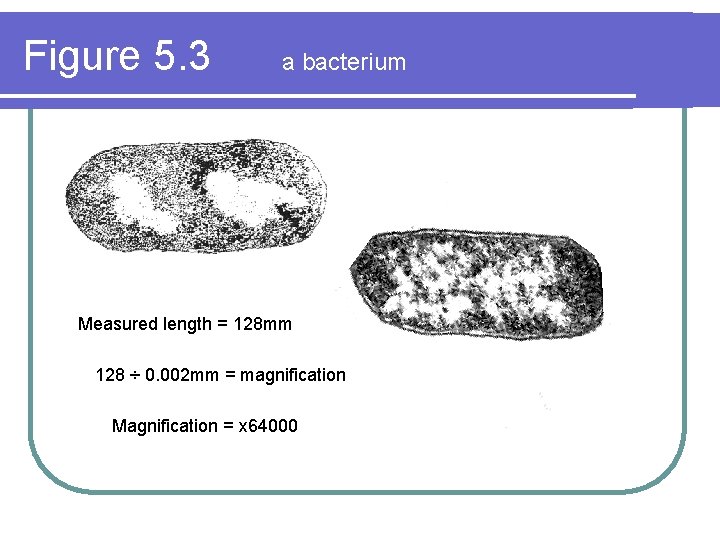
Figure 5. 3 a bacterium Measured length = 128 mm 128 ÷ 0. 002 mm = magnification Magnification = x 64000
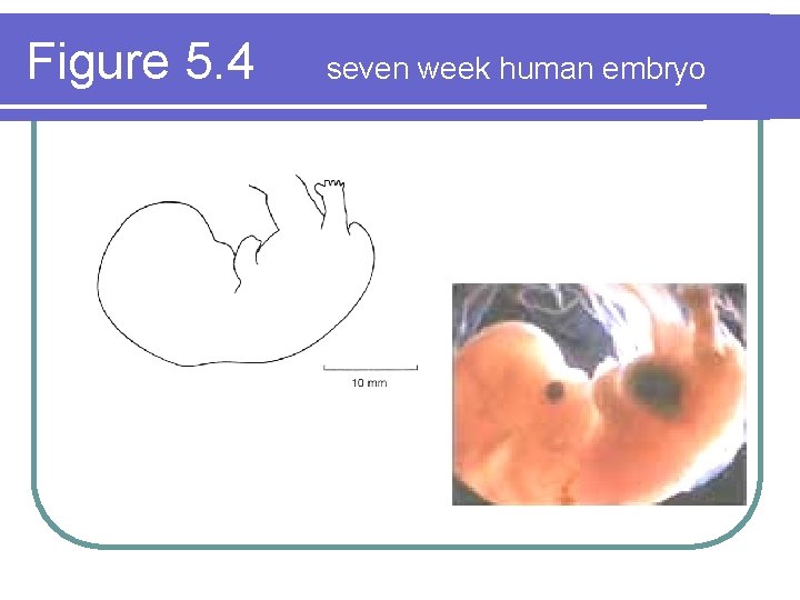
Figure 5. 4 seven week human embryo
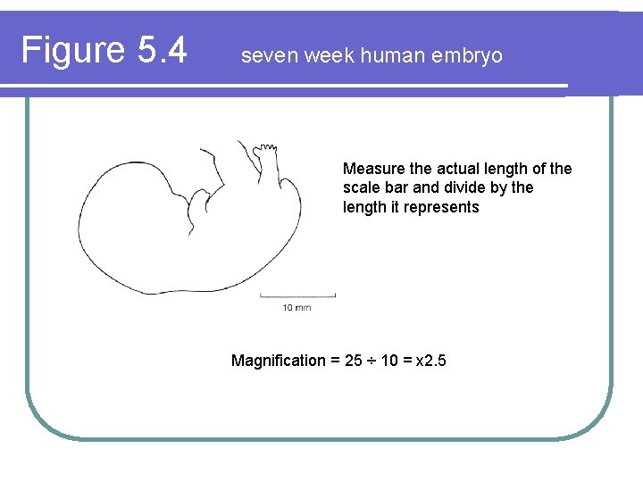
Figure 5. 4 seven week human embryo Measure the actual length of the scale bar and divide by the length it represents Magnification = 25 ÷ 10 = x 2. 5
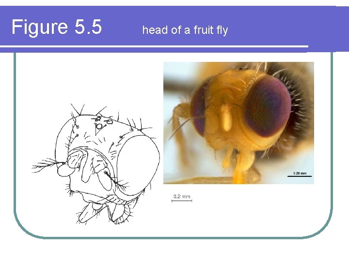
Figure 5. 5 head of a fruit fly
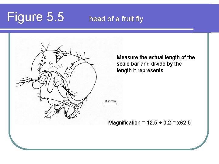
Figure 5. 5 head of a fruit fly Measure the actual length of the scale bar and divide by the length it represents Magnification = 12. 5 ÷ 0. 2 = x 62. 5
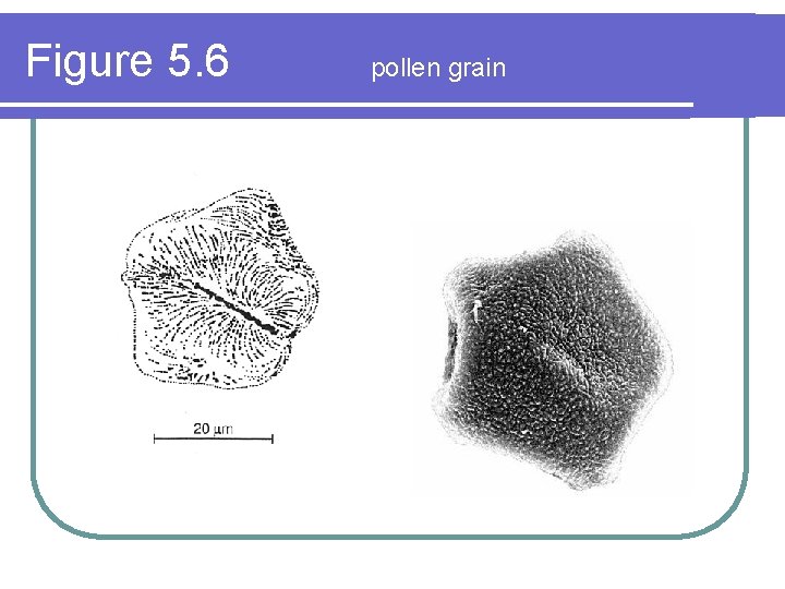
Figure 5. 6 pollen grain
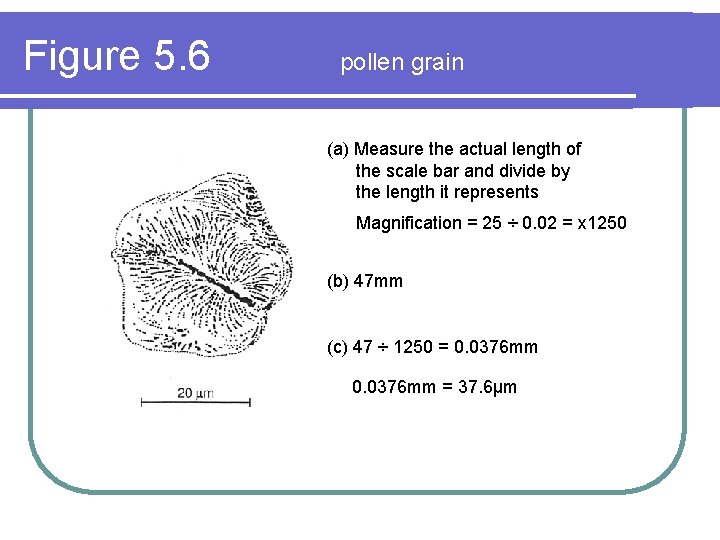
Figure 5. 6 pollen grain (a) Measure the actual length of the scale bar and divide by the length it represents Magnification = 25 ÷ 0. 02 = x 1250 (b) 47 mm (c) 47 ÷ 1250 = 0. 0376 mm = 37. 6μm
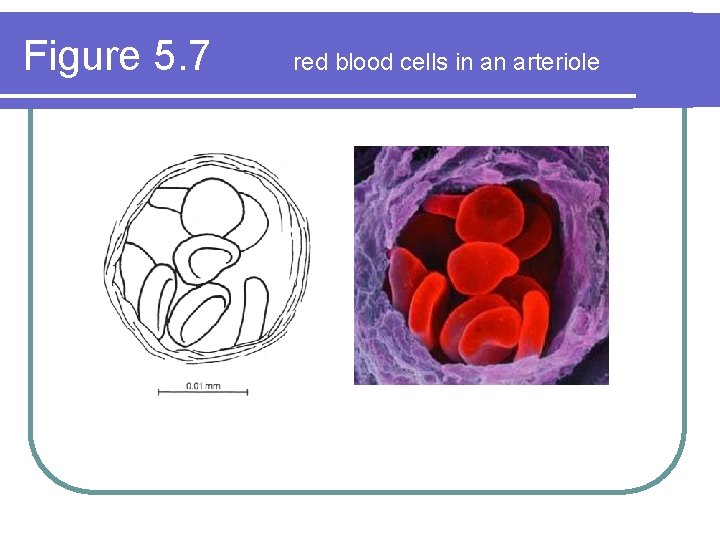
Figure 5. 7 red blood cells in an arteriole
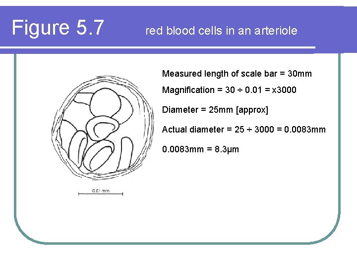
Figure 5. 7 red blood cells in an arteriole Measured length of scale bar = 30 mm Magnification = 30 ÷ 0. 01 = x 3000 Diameter = 25 mm [approx] Actual diameter = 25 ÷ 3000 = 0. 0083 mm = 8. 3μm
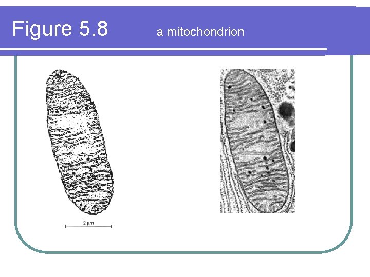
Figure 5. 8 a mitochondrion
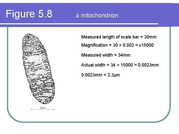
Figure 5. 8 a mitochondrion Measured length of scale bar = 30 mm Magnification = 30 ÷ 0. 002 = x 15000 Measured width = 34 mm Actual width = 34 ÷ 15000 = 0. 0023 mm = 2. 3μm
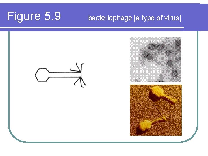
Figure 5. 9 bacteriophage [a type of virus]
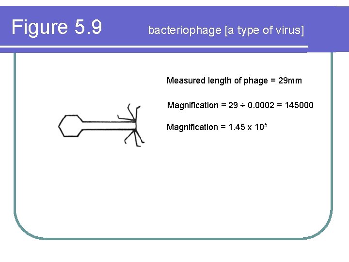
Figure 5. 9 bacteriophage [a type of virus] Measured length of phage = 29 mm Magnification = 29 ÷ 0. 0002 = 145000 Magnification = 1. 45 x 105
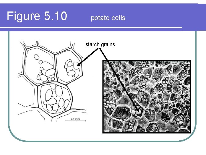
Figure 5. 10 potato cells starch grains
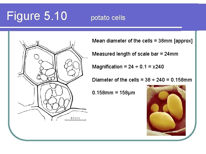
Figure 5. 10 potato cells Mean diameter of the cells = 38 mm [approx] Measured length of scale bar = 24 mm Magnification = 24 ÷ 0. 1 = x 240 Diameter of the cells = 38 ÷ 240 = 0. 158 mm = 158μm
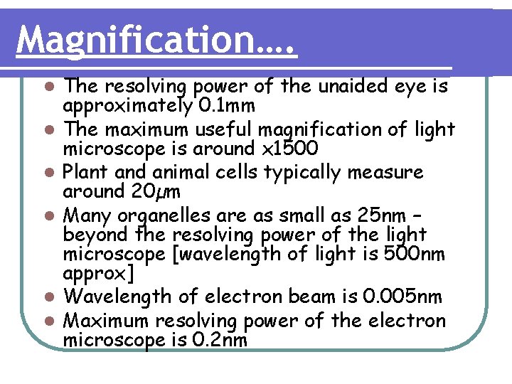
Magnification…. l l l The resolving power of the unaided eye is approximately 0. 1 mm The maximum useful magnification of light microscope is around x 1500 Plant and animal cells typically measure around 20µm Many organelles are as small as 25 nm – beyond the resolving power of the light microscope [wavelength of light is 500 nm approx] Wavelength of electron beam is 0. 005 nm Maximum resolving power of the electron microscope is 0. 2 nm
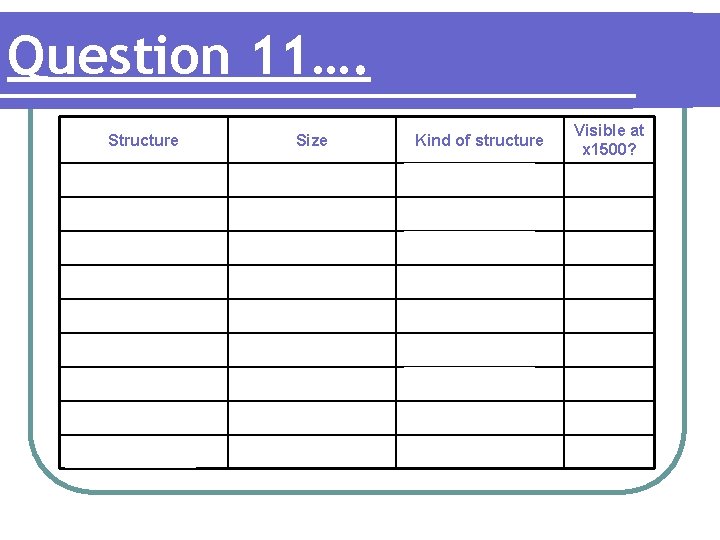
Question 11…. Structure Size Kind of structure Visible at x 1500? bacteriophage 0. 2μm virus bacterium 2. 0μm prokaryotic cell mitochondrion 2. 3μm eukaryotic organelle chloroplast 4. 4μm eukaryotic organelle red blood cell 8. 3μm eukaryotic cell pollen grain 38μm eukaryotic cell potato cell 158μm eukaryotic cell paramecium 237μm eukaryotic organism embryo 30 mm eukaryotic organism (just)
- Slides: 29