THE EYES AND VISION Vision is the dominant


- Slides: 21


THE EYES AND VISION • Vision is the dominant sense in humans • 70% of sensory receptors in humans are in the eyes • 40% of the cerebral cortex is involved in processing visual information • The eye (or eyeball) is the visual organ – It is globe shaped with diameter 2. 5 cm (1 inch) – It is slightly flattened from above downwards – Its anterior part is small and forms 1/6 of the eye ball while it posterior part is large and forms 5/6 of the eye ball. – Lies in bony orbit

Accessory structures of the eye The followings are the accessory parts of the eye: • Eyebrows • Eyelids or palpebrae – Upper & lower separated by palpebral fissure – Corners: medial & lateral canthi – Eyelashes 3

EYELIDS - It protects the eye ball - It opens and closes voluntarily and reflexly - The margins of the eyelids have sensitive hairs called cilia. - Each of the cilia has a follicle surrounded by a nerve plexus - Blinking of eyelids occur when sensory nerve are activated. - Prevent dust particles from reaching eyeball. - The opening between the two eyelids is known as palpebral fissure. 4

• Eyelid tarsal plates give structure – Where orbicularis oculi muscles attach (close eyes) • Levator palpebrae superioris muscle – Lifts upper lid voluntarily (inserts on tarsal plate) 5

• Tarsal glands – modified sebaceous (oil) glands in tarsal plates • Conjunctiva - transparent mucus membrane of stratified columnar epithelium which covers the exposed part of the eye. – Palpebral conjunctiva – Bulbar conjunctiva • Covers white of eye but not the cornea (transparent tissue over the iris and pupil) 6

• Conjunctiva • It covers the anterior surface and also reflected into the inner surface of eyelids. – Palpebral conjunctiva: The part of conjunctiva covering the eyelids – Bulbar conjunctiva : The part of conjunctiva covering the eye ball. . The surface of conjunctiva is lubricated by a thin film of tears secreted by lacrimal glands 7

Lacrimal apparatus • Responsible for tears – The fluid has mucus, antibodies and lysozyme • Lacrimal gland in orbit superolateral to eye • Tears pass out through puncta into canaliculi into sac into nasolacrimal duct • Empty into nasal cavity (sniffles) 8

LACRIMAL GLAND • It is situated in the shelter of bone forming the upper and outer border of wall of the eye socket • Tears flow from lacrimal gland over the surface of conjunctiva and drains into nose through lacrimal duct, lacrimal sac and nasolacrimal duct. • Conjunctiva is kept moist and is protected from infection due to its continuous washing and lubrication by tears • Tears also contains lysozyme that kills bacteria. • Secretion of tears is controlled by the parasympathetic fiber of facial nerve.

LACRIMAL GLAND • At birth the nasolacrimal duct may not be fully developed causing a watery eye. • Acquired obstruction of the nasolacrimal duct is a common cause of water eye in adult which may lead to acute infection of the sac.

Extraocular (extrinsic) eye muscles: 6 in # “EOMs intact” means they all work right • Four are rectus muscles (straight) – Originate from common tendinous or anular ring, at posterior point of orbit • Two are oblique: superior and inferior 11

Extraocular (extrinsic) eye muscles Cranial nerve innervations: • Lateral rectus: VI (Abducens n. ) – abducts eye outward • Medial, superior, inferior rectus & inf oblique: III (Oculomotor n. ) – able to look up and in if all work • Superior oblique: IV (Trochlear n. ) – moves eye down and out 12

Innervation 13

• Double vision: diplopia (what the patient experiences) – Eyes do not look at the same point in the visual field • Misalignment: strabismus (what is observed when shine a light: not reflected in the same place on both eyes) – can be a cause of diplopia – – Cross eyed Gaze & movements not conjugate (together) Medial or lateral, fixed or not Many causes • Weakness or paralysis of extrinsic muscle of eye – Surgical correction necessary • Oculomotor nerve problem, other problems • Lazy eye: amblyopia – Cover/uncover test at 5 yo – If don’t patch good eye by 6, brain ignores lazy eye and visual pathway degenerates: eye functionally blind NOTE: some neurological development and connections have a window of time - need stimuli to develop, or ability lost 14

3 Layers form the external wall of the eye 1. (outer) Fibrous: dense connective tissue – – Sclera – white of the eye Cornea • • • 2. 100 s of sheets of collagen fibers between sheets of epithelium and endothelium Clear because regular alignment Role in light bending Avascular but does have pain receptors Regenerates (middle) Vascular: uvea – – – 3. Choroid – posterior, pigmented Ciliary body Iris (colored part: see next slide) (inner) Sensory – Retina and optic nerve 15

1. (outer layer) Fibrous: dense connective tissue – – 2. Sclera – white of the eye Cornea (middle) Vascular: uvea – – Choroid – posterior, pigmented Ciliary body • • • – 3. Muscles – control lens shape Processes – secrete aqueous humor Zonule (attaches lens) Iris (inner layer) Sensory – Retina and optic nerve 16

Layers of external wall of eye continued 1. (outer) Fibrous: dense connective tissue – – 2. Sclera – white of the eye Cornea (middle) Vascular: uvea – – Choroid – posterior, pigmented Ciliary body – Iris Pigmented put incomplete: pupil lets in light Sphincter of pupil: circularly arranged smooth muscle parasympathetic control for bright light and/or close vision Dilator of pupil: radiating smooth muscle – sympathetic control for dim light and/or distance vision (inner) Sensory 3. – Retina 17

Layers of external wall of eye continued 1. (outer) Fibrous: dense connective tissue – Sclera – white of the eye – Cornea 2. (middle) Vascular: uvea – Choroid – posterior, pigmented – Ciliary body – Iris 3. (inner) Sensory – Retina -------will cover after the chambers 18

some pictures… 19

Chambers and fluids (see previous pics) • Vitreous humor in posterior segment – Jellylike – Forms in embryo and lasts life-time • Anterior segment filled with aqueous humor – liquid, replaced continuously – Anterior chamber between cornea and iris – Posterior chamber between iris and lens – Glaucoma when problem with drainage resulting in increased intraocular pressure 20

THANK YOU
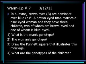
 What is the genotype of the man? ii i a i i b i i a i a
 Hazel and blue eye punnett square
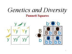
 Mendel squares genetics
 Are brown eyes a dominant gene
 Are almond eyes dominant
 Are brown eyes dominant
 Inherited traits of a lion
 Healthy eyes vs anemic eyes
 Green hazel eye color
 Outermost layer of eye
 Structured light
 Dominant and servient easements
 Phynelketonuria
 Autosomal dominant vs autosomal recessive
 Ab blood type punnett square
 Color blindness punnett square
 Dominant and servient easements
 Dominant and recessive genes
 Dominant on and off circuit
 Which is an electro-pneumatic device?
 Dominant and recessive genes
 Example of punnett square
