Testicular Torsion Faizah Shareef MS 4 Dr Christina
- Slides: 18
Testicular Torsion Faizah Shareef MS 4 Dr. Christina Le. Bedis Ultrasound
CASE HISTORY Patient is a 23 y/o Male with no significant past medical history who arrived to the ED due to increasing, constant and spontaneous groin pain for the last 4 days. He has abdominal pain, groin pain, nausea, scrotal swelling and vomiting. Urinalysis and STI testing were both negative. CBC and BMP were unremarkable.
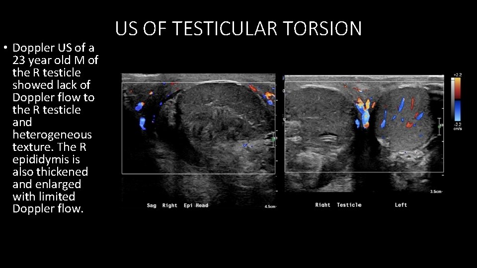
• Doppler US of a 23 year old M of the R testicle showed lack of Doppler flow to the R testicle and heterogeneous texture. The R epididymis is also thickened and enlarged with limited Doppler flow. US OF TESTICULAR TORSION
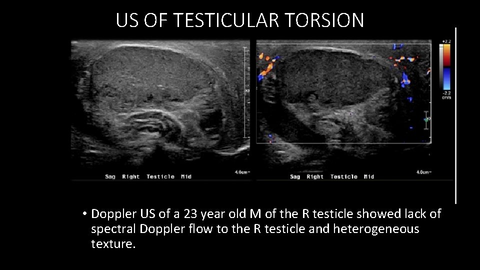
US OF TESTICULAR TORSION • Doppler US of a 23 year old M of the R testicle showed lack of spectral Doppler flow to the R testicle and heterogeneous texture.
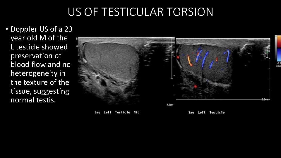
US OF TESTICULAR TORSION • Doppler US of a 23 year old M of the L testicle showed preservation of blood flow and no heterogeneity in the texture of the tissue, suggesting normal testis.
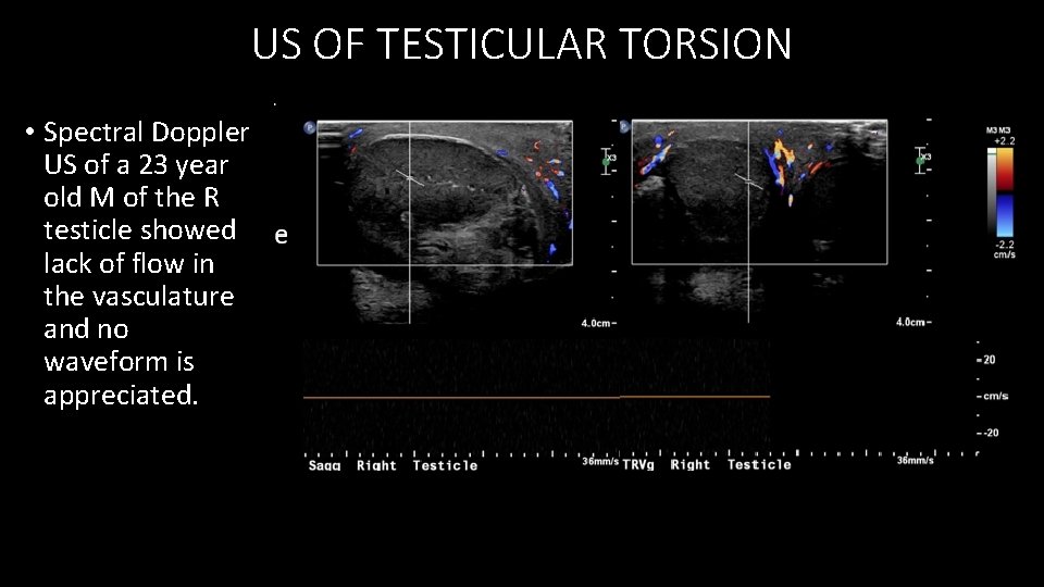
US OF TESTICULAR TORSION • Spectral Doppler US of a 23 year old M of the R testicle showed lack of flow in the vasculature and no waveform is appreciated.
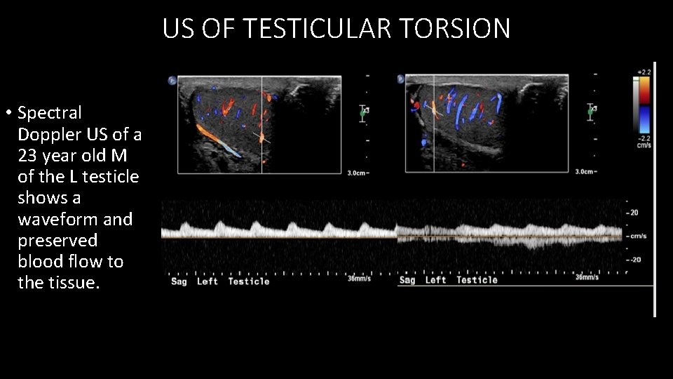
US OF TESTICULAR TORSION • Spectral Doppler US of a 23 year old M of the L testicle shows a waveform and preserved blood flow to the tissue.
CLINICAL FOLLOW UP Patient received emergency R orchiectomy and L Orchiopexy which showed a mottled black right testicle, no bleeding with incision into tunica albuginea. Pathology report indicated the testicular parenchyma, vas deferens and spermatic cord and epididymis with congestion, hemorrhage and coagulative necrosis, consistent with testicular torsion.
IN A NUTSHELL • Major Criteria (Ultrasound) • twisting of the spermatic cord is likely the most specific and sensitive finding in complete and incomplete torsion • Altered blood flow • Minor Criteria (Ultrasound) • Increase in size of the testis • Change in echotexture • Peripheral neovascularization • Complications • Ischemia and necrosis leading to loss of testicle • Recurrence • Infertility Remember • It can be confused with epididymo-orchitis (changes only confined to testis and spontaneously detorted testis (see differential diagnosis below).
VOICE RAD MODALITY AND DISEASE by Dr……. (year produced) Can follow IN A NUTSHELL CONTENT Template Just a guide: This is Dr…. and I wanted to discuss the diagnosis of (modality of. . disease). The most important findings of the (disease on modality) include (major criteria) Sometimes you may also find (minor criteria). You must always remember to look for (complications or other nuances of the disease) Other diseases to consider include (ddx) and you may want to recommend (other imaging or lab tests) (Duration 30 -45 seconds)
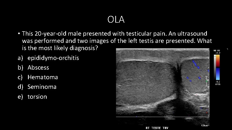
OLA • This 20 -year-old male presented with testicular pain. An ultrasound was performed and two images of the left testis are presented. What is the most likely diagnosis? a) epididymo-orchitis b) Abscess c) Hematoma d) Seminoma e) torsion
OLA • What is the initial imaging modality you would use in the case of acute scrotal pain? a) b) c) d) Pelvic CT with contrast MRI Nuclear Imaging Doppler US
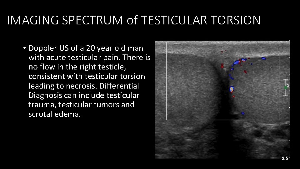
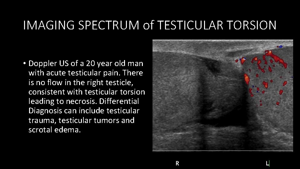
IMAGING SPECTRUM of TESTICULAR TORSION • Doppler US of a 20 year old man with acute testicular pain. There is no flow in the right testicle, consistent with testicular torsion leading to necrosis. Differential Diagnosis can include testicular trauma, testicular tumors and scrotal edema.
IMAGING SPECTRUM of TESTICULAR TORSION • Doppler US of a 20 year old man with acute testicular pain. There is no flow in the right testicle, consistent with testicular torsion leading to necrosis. Differential Diagnosis can include testicular trauma, testicular tumors and scrotal edema.
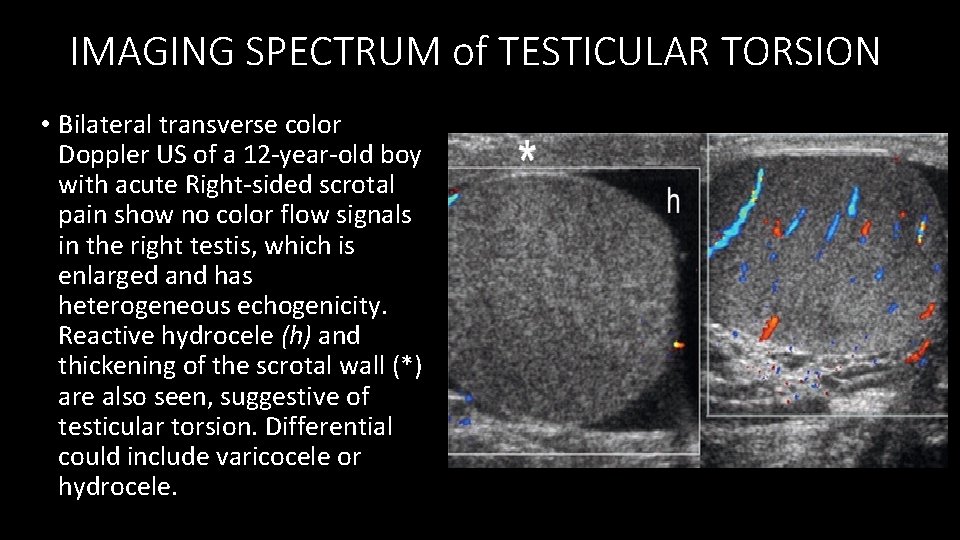
IMAGING SPECTRUM of TESTICULAR TORSION • Bilateral transverse color Doppler US of a 12 -year-old boy with acute Right-sided scrotal pain show no color flow signals in the right testis, which is enlarged and has heterogeneous echogenicity. Reactive hydrocele (h) and thickening of the scrotal wall (*) are also seen, suggestive of testicular torsion. Differential could include varicocele or hydrocele.
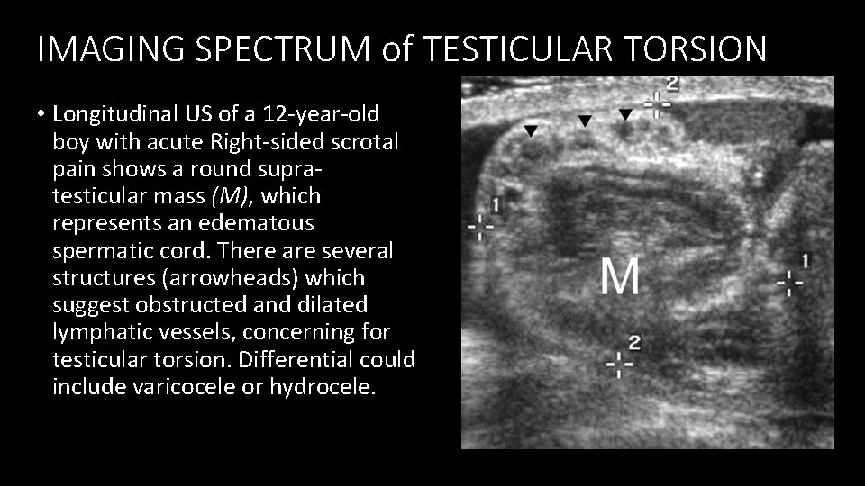
IMAGING SPECTRUM of TESTICULAR TORSION • Longitudinal US of a 12 -year-old boy with acute Right-sided scrotal pain shows a round supratesticular mass (M), which represents an edematous spermatic cord. There are several structures (arrowheads) which suggest obstructed and dilated lymphatic vessels, concerning for testicular torsion. Differential could include varicocele or hydrocele.
DISCUSSION • Testicular torsion is generally a clinical diagnosis that is also a surgical emergency, because prolonged lack of blood flow can lead to ischemia and necrosis of the tissue. If imaging is done, it is usually a Doppler US to prevent radiation to the area and the findings show altered blood flow and possible changes in echotexture in the tissue if the findings are early.
LINKS AND REFERENCES • References Radiopaedia Radiographics Up. To. Date