Testes By Dr Noori Mohammed Luaibi Testicle The

Testes By Dr. Noori Mohammed Luaibi

Testicle The testicle or testicle testis is the male reproductive gland in testis all animals, including humans. It is homologous to the female ovary. The functions of the testes are to produce both testes sperm androgens, primarily testosterone. sperm androgens testosterone Testosterone release is controlled by the anterior pituitary luteinizing hormone; whereas sperm production is controlled both by the anterior pituitary follicle-stimulating hormone and anterior pituitary follicle-stimulating hormone gonadal testosterone Appearance Males have two testicles of similar size contained within testicles the scrotum , which is an extension of the abdominal wall. scrotum Scrotal asymmetry is not unusual: one testicle extends testicle further down into the scrotum than the other due to scrotum differences in the anatomy of the vasculature.
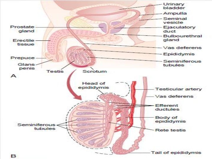

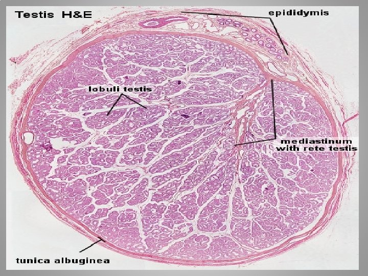
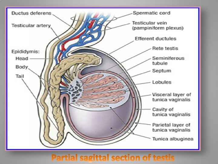
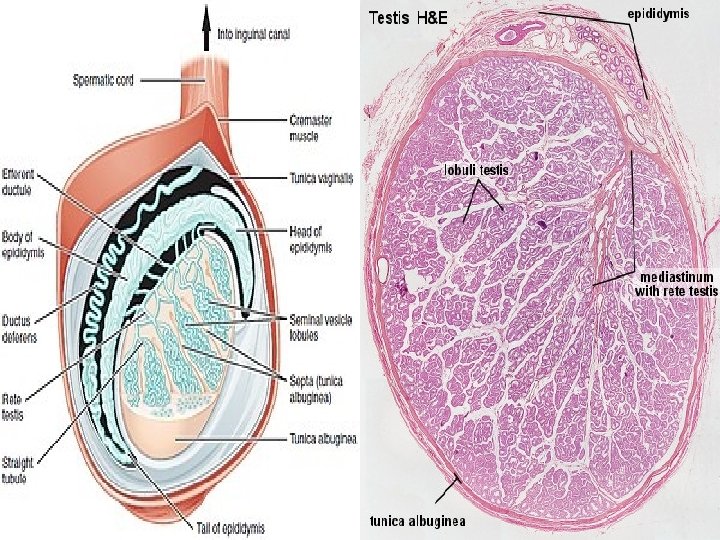
Internal structure Duct system The testes are covered by a tough membranous shell called testes the tunica albuginea. Within the testes are very fine coiled tunica albuginea testes tubes called seminiferous tubules. The tubules are lined with seminiferous tubules a layer of cells (germ cells) that develop from puberty germ cells through old age into sperm cells (also known as sperm cells spermatozoa or male gametes ). The developing spermatozoa male gametes travel through the seminiferous tubules to the rete seminiferous tubules testis located in the mediastinum testis , to the efferent testis mediastinum testis ducts , and then to the epididymis where newly created ducts epididymis sperm cells mature (spermatogenesis ). The sperm move into sperm cells the vas deferens, and are eventually expelled through the vas deferens urethra and out of the urethral orifice through muscular urethra contractions.

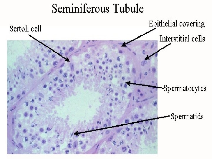
Primary cell types within the seminiferou • Germ cells develop into spermatogonia, Germ cells spermatogonia spermatocytes, spermatocytes spermatids and spermatozoon through the process of spermatids spermatozoon spermatogenesis. The gametes contain DNA for spermatogenesis fertilization of an ovum • Sertoli cells – the true epithelium of the seminiferous Sertoli cells epithelium, critical for the support of germ cell development into spermatozoa Sertoli cells secrete Sertoli cells inhibin • Peritubular myoid cells surround the seminiferous Peritubular myoid cells tubules
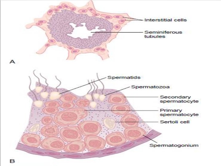

A germ cell is any biological cell that gives rise to the gametes of an organism that reproduces sexually. Germinal epithelium of the testicle. 1: basal lamina 2: spermatogonia 3: spermatocyte 1 st order 4: spermatocyte 2 nd order 5: spermatid 6: mature spermatid 7: Sertoli cell 8: tight junction (blood testis barrier)

Primary cell types between tubules (inter • Leydig cells – cells localized between seminiferous Leydig cells tubules that produce and secrete testosterone and other androgens important for sexual development and puberty, secondary sexual characteristics like facial hair, facial hair sexual behavior and sexual behavior libido, supporting spermatogenesis and erectile libido spermatogenesis erectile function. Testosterone also controls testicular function volume. • Also present are: Also present are v Immature Leydig cells v Interstitial macrophages and epithelial cells Interstitial macrophages
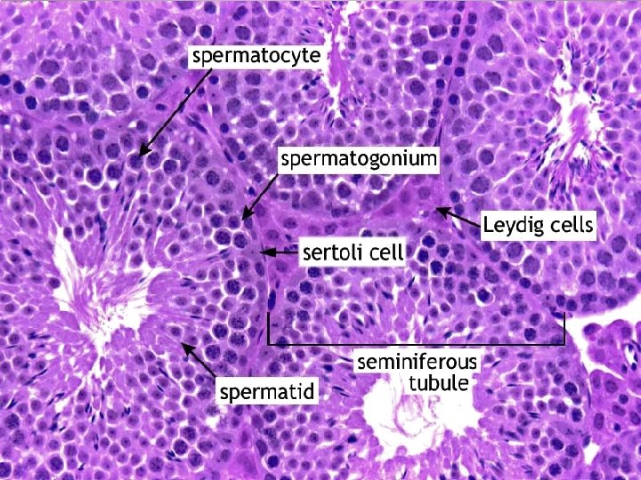

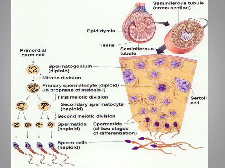
Cell division during spermatogenesis. During embryonic development the primordial germ cells migrate to the testis where they become spermatogonia. At puberty ( usually 12 to 14 years after birth), the spermatogonia proliferate rapidly by mitosis. Some begin meiosis to become primary spermatocytes and continue through meiotic division 1 to become secondary spermatocytes. After completion of meiotic division 2, the secondary spermatocytes produce spermatids, which differentiate to form spermatozoa.

Layers Many anatomical features of the adult testis reflect its testis developmental origin in the abdomen. The layers of tissue enclosing each testicle are derived from the layers of the testicle anterior abdominal wall. Notably, the cremasteric muscle arises from the internal oblique muscle. The blood–testis barrier Large molecules cannot pass from the blood into the lumen of a seminiferous tubule due to the presence of tight seminiferous tubule junctions between adjacent Sertoli cells. The spermatogonia are in the basal compartment (deep to the spermatogonia level of the tight junctions) and the more mature forms such as primary and secondary spermatocytes and spermatids are spermatocytes spermatids in the adluminal compartment

The function of the blood–testis barrier may be to prevent an auto-immune reaction. Mature sperm (and their antigens) arise long after immune tolerance is established in infancy. Therefore, since sperm are antigenically different from self tissue, a male animal can react immunologically to his own sperm. In fact, he is capable of making antibodies against them. Temperature regulation Spermatogenesis is enhanced at temperatures slightly less than core body temperature The spermatogenesis is less efficient at lower and higher temperatures than 33 °C. Because the testes are located outside the body, 33 °C the smooth tissue of the scrotum can move them closer or further away from the body.

The temperature of the testes is maintained at 35 testes 35 degrees Celsius (95 degrees Fahrenheit) two degrees 95 below the body temperature of 37 degrees Celsius 37 (98. 6 degrees Fahrenheit). Higher temperatures 98. 6 affect spermatogenesis. There a number of mechanisms to maintain the testes at the optimum temperature. The cremasteric muscle is part of the spermatic cord. cremasteric muscle When this muscle contracts, the cord is shortened and the testicle is moved closer up toward the body, testicle which provides slightly more warmth to maintain optimal testicular temperature. When cooling is required, the cremasteric muscle relaxes and the cremasteric muscle testicle is lowered away from the warm body and is testicle able to cool.

Development There are two phases in which the testes grow substantially; namely in embryonic and pubertal age. 1. Embryonic During mammalian development, the gonads are at first capable of becoming either ovaries or testes. In humans, starting at about week 4 the gonadal rudiments are present within the intermediate mesoderm adjacent to the mesoderm developing kidneys. At about week 6, sex cords develop kidneys within the forming testes. These are made up of early Sertoli cells that surround and nurture the germ cells that migrate cells germ cells into the gonads shortly before sex determination begins. In males, the sex-specific gene SRY that is found on the Ychromosome initiates sex determination by downstream regulation of sex-determining factors, which leads to

development of the male phenotype, including directing development of the early bipotential gonad down the male path of development. Testes follow the "path of descent" from high in the posterior feta path of descent abdomen to the inguinal ring and beyond to the inguinal canal and into the scrotum. In most cases both testes have descended by birth. In most other cases, only one testis fails to descend (cryptorchidism) and that will probably express itself within a year. cryptorchidism 2. Pubertal The testes grow in response to the start of spermatogenesis. Size depends on lytic function, sperm production, interstitial fluid, and Sertoli cell fluid production. After puberty, the volume of the testes can be increased by over 500% as compared to the prepubertal size. Testicles are fully descended before one reaches puberty.

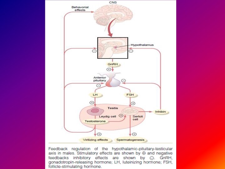
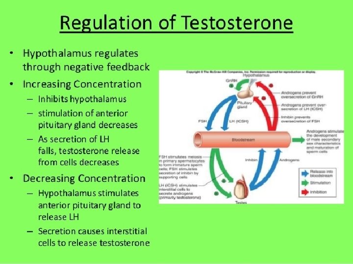
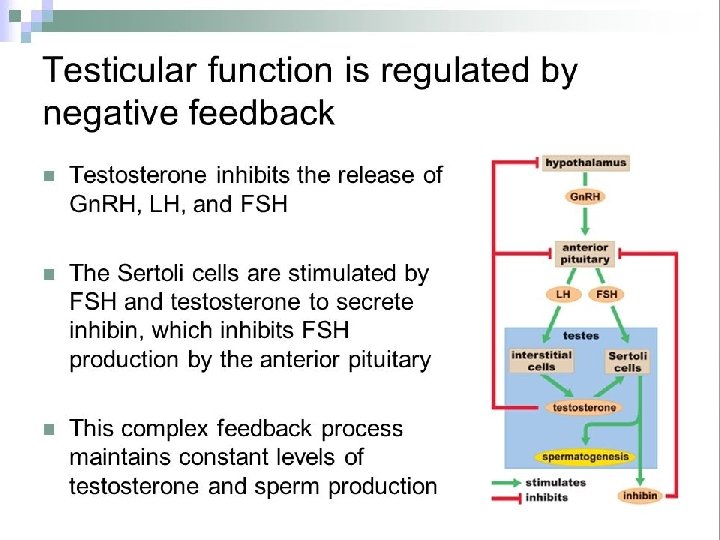
The testes of the male begin to produce testosterone at puberty in response to LH. LH Testosterone is the Testosterone primary male sex hormone and an anabolic steroid, it plays a key role in the development of male reproductive tissues such as testes and prostate, it promotes maturation of the male reproductive organs, development of secondary sex characteristics(facial and body hair, also deepens the voice of a male at a certain age and maintaining healthy levels of muscle and bone mass) and production of sperm by the testes Testosterone is biosynthesized in several steps from Testosterone cholesterol and is converted in the liver to inactive metabolites. It exerts its action through binding to and activation of the androgen receptor

. Hormone Produced by Regulation Action Testosterone Leydig Cells Gn. RH from the Hypothalamus causes LH secretion from the Pituitary Gland which stimulates the Leydig Cells. This hormone controls and maintains the growth and functions of the reproductive organs. It enhances libido and is essential for spermatogenesis. Inhibin Sertoli Cells Gn. RH from the Hypothalamus causes FSH secretion from the Pituitary Gland which stimulates the Sertoli Cells. Prevents secretion of further FSH from the Pituitary Gland. Oestradiol Sertoli Cells Gn. RH from the Hypothalamus causes FSH secretion from the Pituitary Gland which stimulates the Sertoli Cells Converted from testosterone, this hormone's function is complex. It may prevent apoptosis of male germ cells.
 axis")
The hypothalamic–pituitary–gonadal (HPG) axis


- Slides: 29