Swine diseases Coccidiosis confinement rearing and continuous farrowing
- Slides: 50
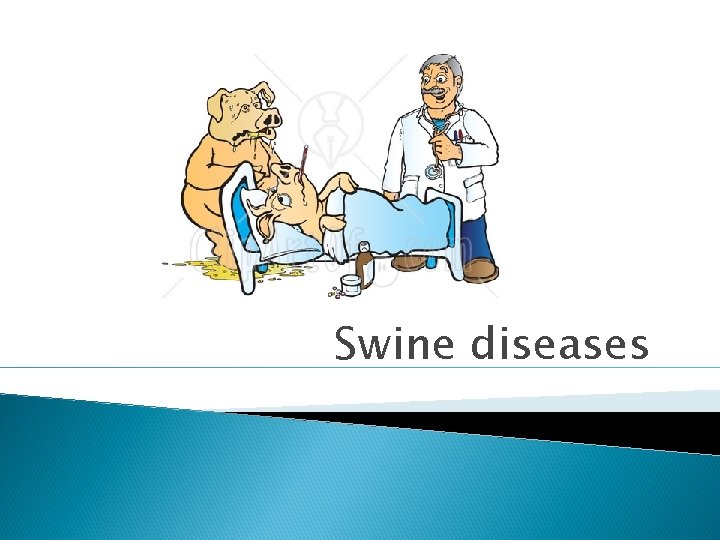
Swine diseases
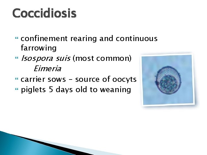
Coccidiosis confinement rearing and continuous farrowing Isospora suis (most common) Eimeria carrier sows – source of oocyts piglets 5 days old to weaning
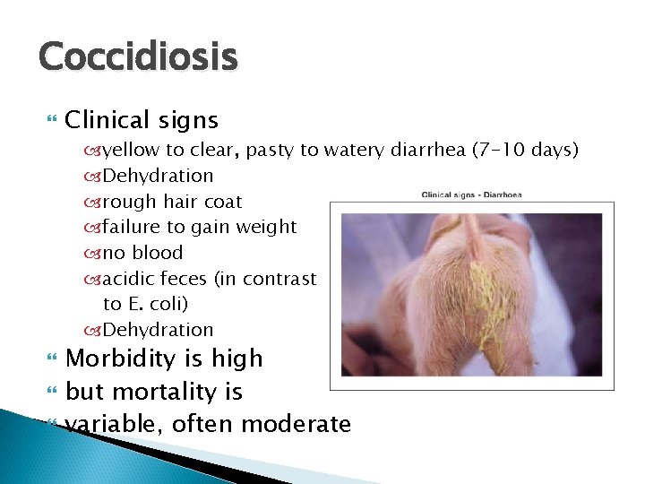
Coccidiosis Clinical signs yellow to clear, pasty to watery diarrhea (7 -10 days) Dehydration rough hair coat failure to gain weight no blood acidic feces (in contrast to E. coli) Dehydration Morbidity is high but mortality is variable, often moderate
Coccidiosis cont. . . Diagnosis Diarrheas in pigs <7 days old are not Isospora! Necropsy - fibrinonecrotic enteritis Histopathology - oocysts, merozoites Fecal flotation can be falsely negative Treatment Adding coccidiostats to feed is ILLEGAL amprolium to piglets Control - disinfection of farrowing area: ◦ Strong bleach or ammonium compounds ◦ Between farrowings, steam cleaning Installation of perforated metal or plastic flooring in the crates will be beneficial in the control of coccidiosis
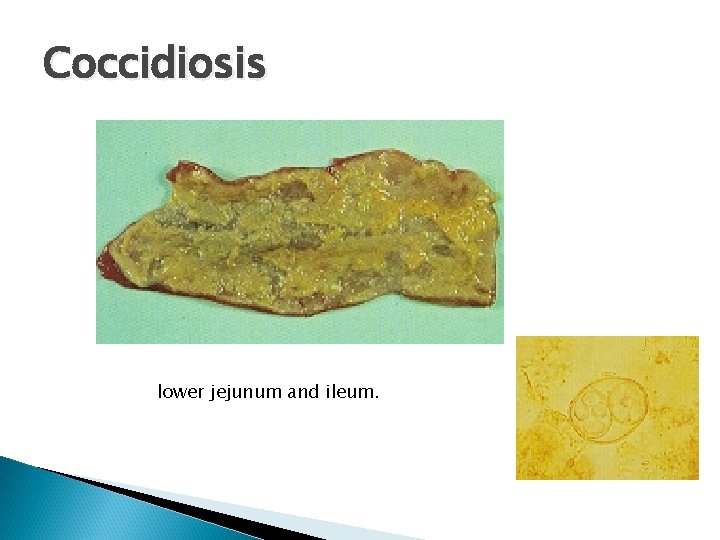
Coccidiosis lower jejunum and ileum.
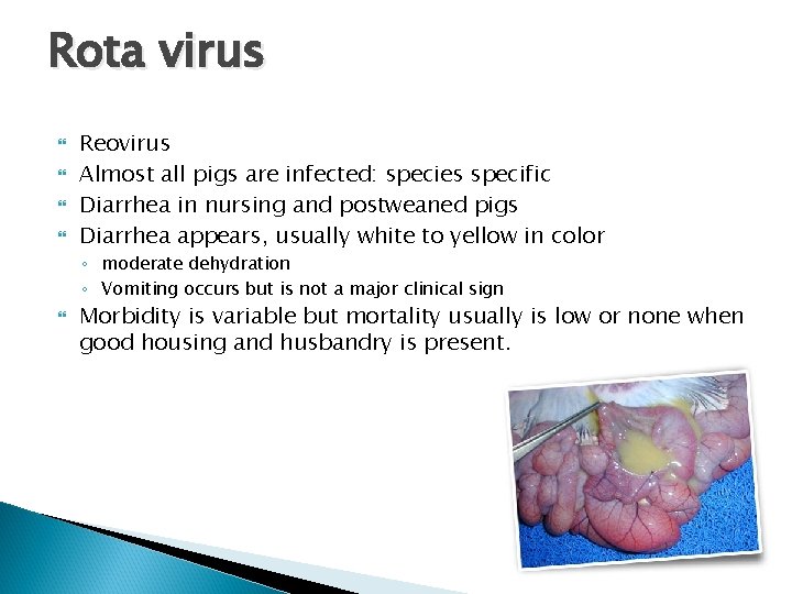
Rota virus Reovirus Almost all pigs are infected: species specific Diarrhea in nursing and postweaned pigs Diarrhea appears, usually white to yellow in color ◦ moderate dehydration ◦ Vomiting occurs but is not a major clinical sign Morbidity is variable but mortality usually is low or none when good housing and husbandry is present.
Rota virus Diagnosis - difficult Necropsy-thin walled small intestine Histopathology Flourescent antibody test Electron microscopy
Rota virus cont. . . Treatment Dextrose and fluids Antimicrobials for concurrent infections E. coli Isospora Control Rotaviruses are very stable in the environment: formaldehyde and chlorine-based disinfectants including chlorox Wean pigs on good nutritional diet MLV vaccine at 7 and 21 days (in water) and also for dams
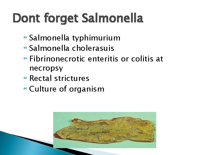
Dont forget Salmonella typhimurium Salmonella cholerasuis Fibrinonecrotic enteritis or colitis at necropsy Rectal strictures Culture of organism
Swine dysentery Brachyspira hyodysenteriae: Gram-negative, anaerobic spirochete ◦ Brachyspira (Serpulina) pilosicoli (similar but less severe c. s. ) Grower / finishers (not in < 3 weeks) Mortality can be up to 30% lagoon water: two months, moist feces: two months, soil: 18 days Transmission: fecal-oral and fomites, rats, birds
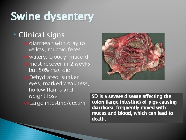
Swine dysentery Clinical signs diarrhea : with gray to yellow, mucoid feces watery, bloody, mucoid most recover in 2 weeks but 50% may die Dehydrated: sunken eyes, marked weakness, hollow flanks and weight loss SD is a severe disease affecting the Large intestine/cecum colon (large intestine) of pigs causing diarrhoea, frequently mixed with mucus and blood, which can lead to death.
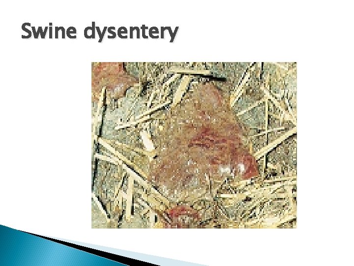
Swine dysentery
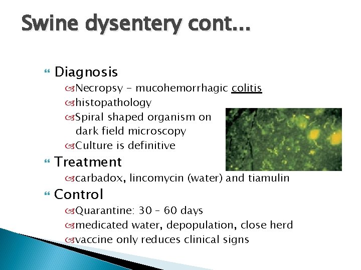
Swine dysentery cont. . . Diagnosis Necropsy - mucohemorrhagic colitis histopathology Spiral shaped organism on dark field microscopy Culture is definitive Treatment carbadox, lincomycin (water) and tiamulin Control Quarantine: 30 – 60 days medicated water, depopulation, close herd vaccine only reduces clinical signs
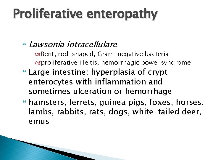
Proliferative enteropathy Lawsonia intracellulare Bent, rod-shaped, Gram-negative bacteria proliferative illeitis, hemorrhagic bowel syndrome Large intestine: hyperplasia of crypt enterocytes with inflammation and sometimes ulceration or hemorrhage hamsters, ferrets, guinea pigs, foxes, horses, lambs, rabbits, rats, dogs, white-tailed deer, emus
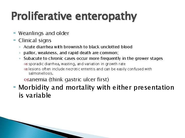
Proliferative enteropathy Weanlings and older Clinical signs ◦ Acute diarrhea with brownish to black unclotted blood ◦ pallor, weakness, and rapid death are common; ◦ Subacute to chronic cases occur more frequently in the grower stages sporadic diarrhea, wasting, and variation in growth rate lesions often include necrotic enteritis and can be easily confused with salmonellosis. anemia (think gastric ulcer first) Morbidity and mortality with either presentation is variable
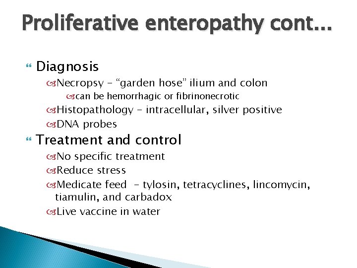
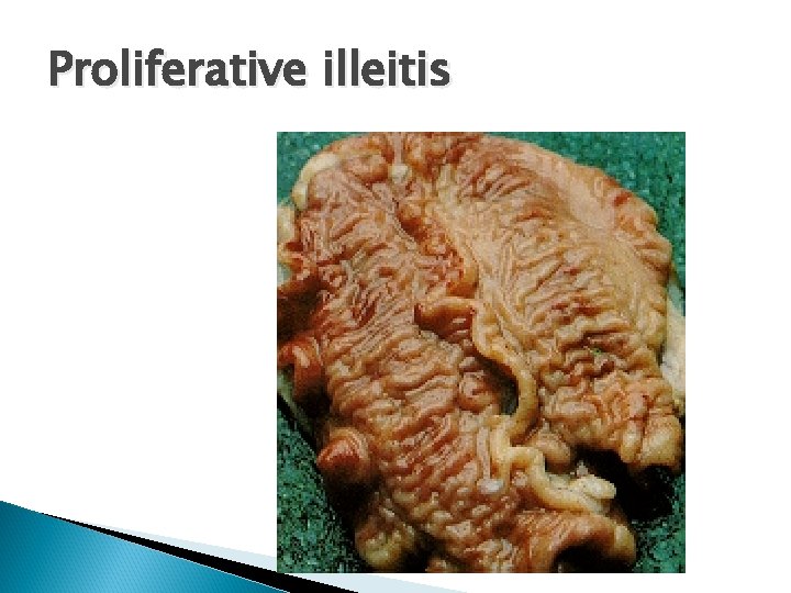
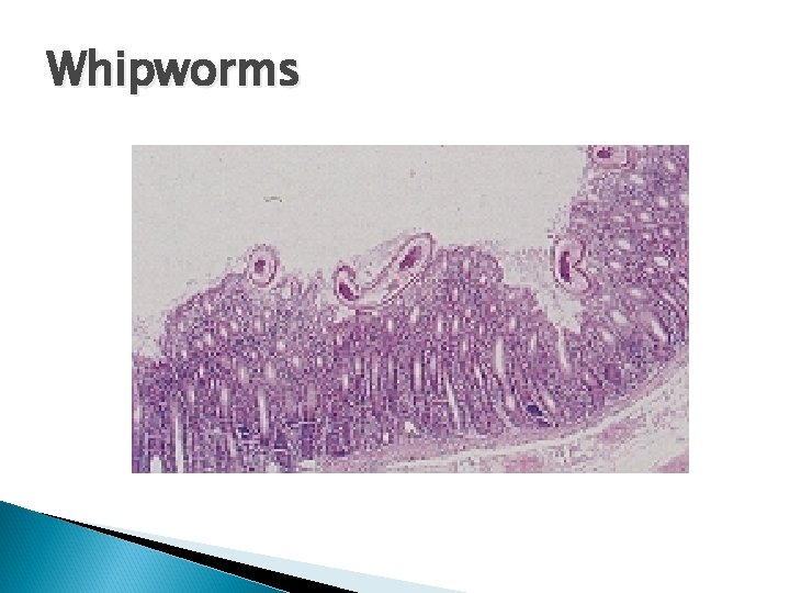
Proliferative enteropathy cont. . . Diagnosis Necropsy - “garden hose” ilium and colon can be hemorrhagic or fibrinonecrotic Histopathology - intracellular, silver positive DNA probes Treatment and control No specific treatment Reduce stress Medicate feed - tylosin, tetracyclines, lincomycin, tiamulin, and carbadox Live vaccine in water
Proliferative illeitis
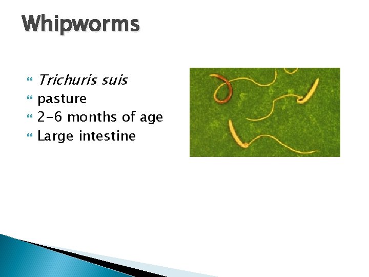
Whipworms Trichuris suis pasture 2 -6 months of age Large intestine
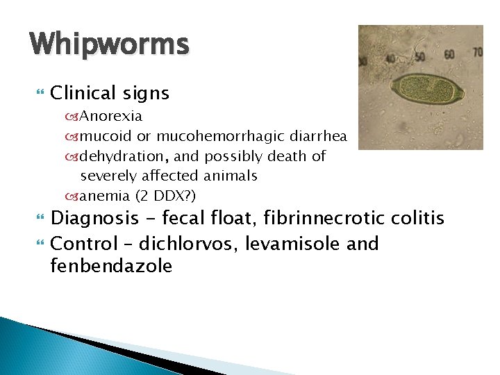
Whipworms Clinical signs Anorexia mucoid or mucohemorrhagic diarrhea dehydration, and possibly death of severely affected animals anemia (2 DDX? ) Diagnosis - fecal float, fibrinnecrotic colitis Control – dichlorvos, levamisole and fenbendazole
Whipworms
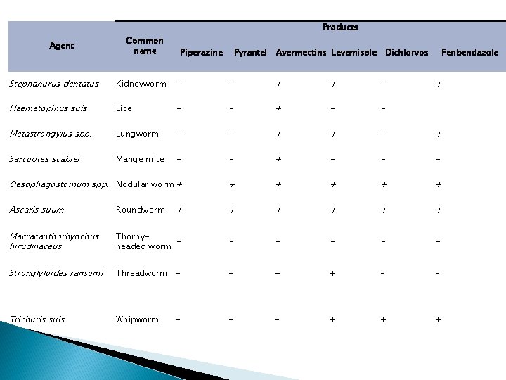
Products Agent Common name Piperazine Pyrantel Avermectins Levamisole Dichlorvos Fenbendazole Stephanurus dentatus Kidneyworm - - + + - Haematopinus suis Lice - - + - - Metastrongylus spp. Lungworm - - + + - + Sarcoptes scabiei Mange mite - - + - - - Oesophagostomum spp. Nodular worm + + + Ascaris suum Roundworm + + + Macracanthorhynchus hirudinaceus Thornyheaded worm - - - Stronglyloides ransomi Threadworm - - + + - - Trichuris suis Whipworm - - + + + - +
Don’t forget Salmonella! Salmonella typhimurium Salmonella cholersuis associated with rectal strictures? Can be large intestine Fibrinonecrotic colitis Rectal strictures Culture
Parasites of pigs Trichuris suis - colon Ascaris suum - small intestine, milk spots Stephanurus edentatus - kidney Macrocanthorynchus hirudinaceous -small intestine
Neurological diseases Hypoglycemia Streptococcus suis: Gram-positive Salt poisoning Edema disease
Hypoglycemia Newborn piglets Blood glucose <50 mg/d. L may develop signs Clinical signs convulsions shivering hypothermia gait abnormalities
Hypoglycemia cont. . . Diagnosis Blood glucose Empty stomach Treatment 20 ml/kg 5% dextrose, warm fluids Control make sure the milk is flowing
Salt poisoning Usually due to water deprivation rather than too much Na Causes hyperosmalarity of CNS resulting in swelling and edema Clinical signs thirst, constipation depression, blindness, convulsions
Salt poisoning cont. . . Diagnosis History Clinical pathology-eosinopenia, hypernatremia Histopathology - eosinophilic meningitis Treatment None Control provide free access to water reduce salt in diet
Musculoskeletal diseases Arthritis S. suis, Erysipelothrix, A. pyogenes Mycoplasma hyosynoviae Myodegenerative disease Malignant hyperthermia (PSE) White muscle disease
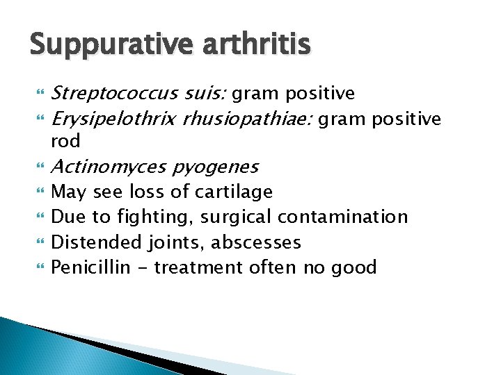
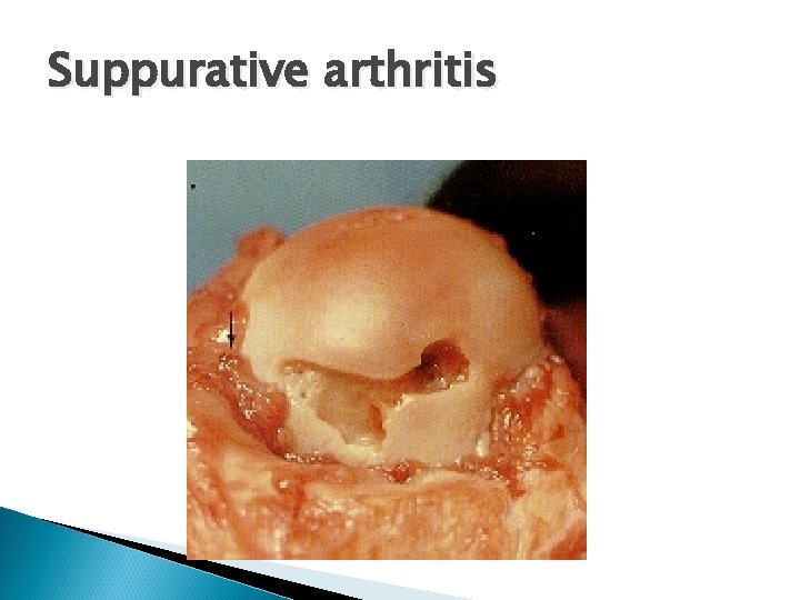
Suppurative arthritis Streptococcus suis: gram positive Erysipelothrix rhusiopathiae: gram positive Actinomyces pyogenes rod May see loss of cartilage Due to fighting, surgical contamination Distended joints, abscesses Penicillin - treatment often no good
Suppurative arthritis
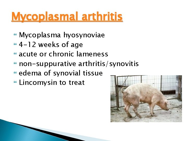
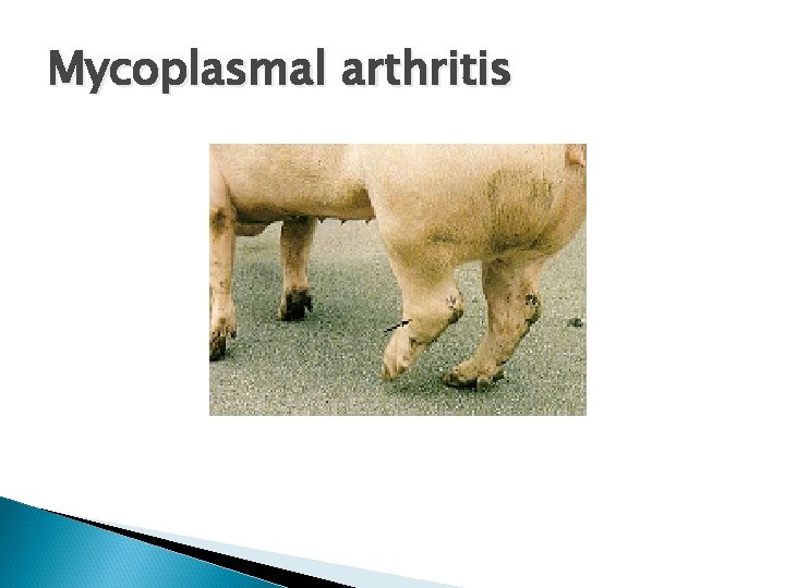
Mycoplasmal arthritis Mycoplasma hyosynoviae 4 -12 weeks of age acute or chronic lameness non-suppurative arthritis/synovitis edema of synovial tissue Lincomysin to treat
Mycoplasmal arthritis
Reproductive disease Parvovirus Leptospirosis: gram - spirochete PRRS: Arterivirus Brucellosis: gram negative
Porcine parvovirus 100% prevalence: endemic Important signs ◦ ◦ ◦ large numbers of mummified fetuses increase in the number of returns to estrus small litters failures to farrow, decreased farrowing rate, rarely abortion Transmission: secretions, oro-nasal, transplacental Poor conception rates, reabsorbed litters, mummies and small litters
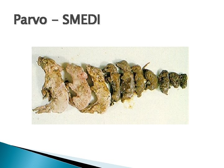
Porcine parvovirus Transient leukopenia Signs depend on time of infection <30 days - embryo resorbed 30 -70 days - mummy >70 days - dead or weak, survive normally no other signs of illness SMEDI - stillbirth, mummy, embryonic death, infertility Diagnosis - detection of virus in mummy by immunofluorescence or by rising titer
Parvo - SMEDI
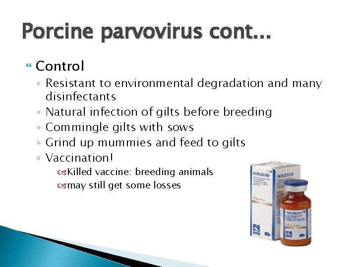
Porcine parvovirus cont. . . Control ◦ Resistant to environmental degradation and many disinfectants ◦ Natural infection of gilts before breeding ◦ Commingle gilts with sows ◦ Grind up mummies and feed to gilts ◦ Vaccination! Killed vaccine: breeding animals may still get some losses
Leptospirosis ◦ Leptospira interrogans (serovars pomona, icterohaemorrhagiae, canicola, and bratislava) ◦ Leptospira borgpetersenii (serovars sejroe and tarassovi) ◦ Leptospira kirschneri (serovar grippotyphosa) ◦ serovar L. hardjo : bovines and has been reported to infect pigs in close proximity Zoonosis
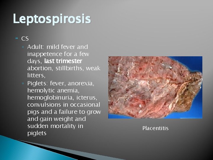
Leptospirosis CS ◦ Adult: mild fever and inappetence for a few days, last trimester abortion, stillbirths, weak litters, ◦ Piglets: fever, anorexia, hemolytic anemia, hemoglobinuria, icterus, convulsions in occasional pigs and a failure to grow and gain weight and sudden mortality in piglets Placentitis
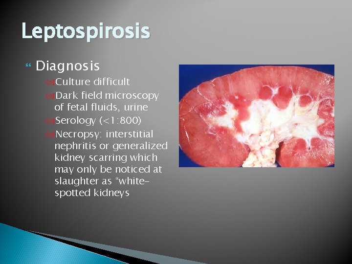
Leptospirosis Diagnosis Culture difficult Dark field microscopy of fetal fluids, urine Serology (<1: 800) Necropsy: interstitial nephritis or generalized kidney scarring which may only be noticed at slaughter as “whitespotted kidneys
Leptospirosis cont. . . Treatment Chlortetracycline in feed/ oxytetracycline, tylosin, and erythromycin Control Vaccination Gilts twice before first breeding Sows before every breeding Rodent control
PRRS Porcine reproductive/respiratory syndrome ◦ Arterivirus Premature farrowing Small weak piglets or stillborns increased numbers of mummies Delayed or abnormal estrus Serology to diagnose Vaccination for prevention Abortions, mummies and weak pigs
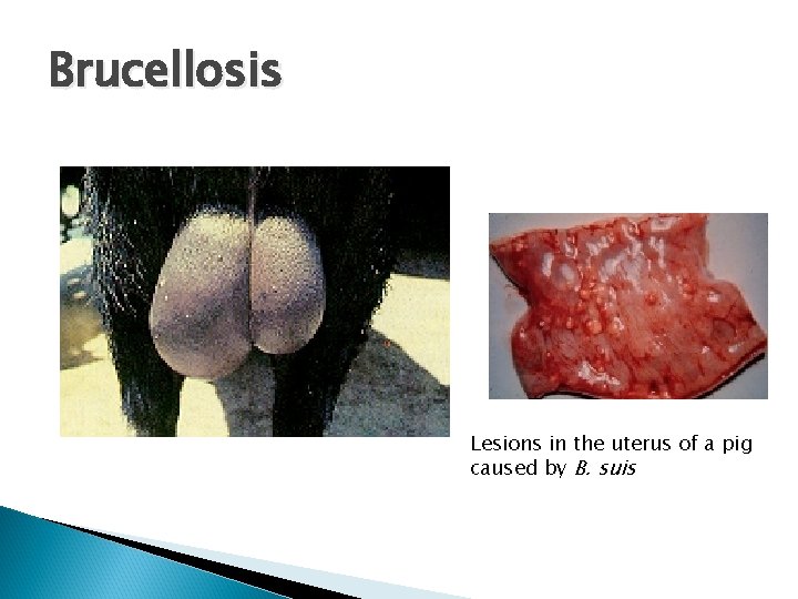
Brucellosis Brucella suis: gram negative Zoonotic Ist agent to be weaponized by US ~ 1950 Rare in US ◦ A cooperative, 3 -stage, State-Federal-Industry eradication program was initiated with a goal of eradication of brucellosis. ◦ Goal is nearly accomplished in domestic herds, but feral swine remain a reservoir in the US.
Brucellosis Transmission: direct contact ◦ ingesting aborted fetuses, fetal membranes or fluids discharged at the time of abortion Clinical signs abortion at any time in gestation infertility - many sows coming back into heat (abortions in first trimester) infected sows recover and deliver normally Lesions mild endometritis arthritis orchitis
Brucellosis Lesions in the uterus of a pig caused by B. suis
Brucellosis cont. . . Diagnosis The buffered, acidified plate antigen (BAPA) test and the standard card test (SCT) have been used extensively as presumptive test confirmation, either the standard tube test (STT) or the particle concentration fluorescence immunoassay (PCFIA) Culture: most accurate Treatment and control ◦ Test and slaughter (depopulate ~ 2 -3 months)
Abortions/stillbirths Parvo virus PRRS Pseudorabies Lepto
References http: //www. aphis. usda. gov/animal_health/an imal_dis_spec/swine/ http: //www. ncsu. edu/project/swine_extensio n/ncporkconf/2002/roberts. htm http: //www. vetmed. wisc. edu/pbs/zoonoses/ Erysipelas/erysipelasindex. html http: //vetmed. iastate. edu/vdpam/newvdpam-employees/food-supply-veterinarymedicine/swine-diseases/haemophilus -parasuishttp: //vetpath. wordpress. com/category/necr opsy-cases/
References http: //www. fmv. utl. pt/atlas/figado/pages_us /figad 015_ing. htm http: //www. cfsph. iastate. edu/Disease. Info/dis ease. php? name=influenza&lang=en http: //microgen. ouhsc. edu/a_pleuro/a_pleur o_home. htm http: //www. merckvetmanual. com/mvm/inde x. jsp? cfile=htm/bc/51205. htm&word=leptos pirosis%2 Cin%2 Cpigs