Stroke VolumeSV Frank Starling Principle End diastolic volume
 Frank – Starling Principle • End diastolic volume: The amount of blood")


 ml/min = heart rate (HR) beats/min ₓstroke volume")


- Slides: 12
Stroke Volume(SV) Frank – Starling Principle • End diastolic volume: The amount of blood that remains in the ventricle just before ventricular early systole is the EDV • End systolic volume: The amount of blood that remains in the ventricle at the end of ventricular systole is the ESV SV = EDV - ESV
Frank – Starling Principle • The Frank–Starling law of the heart (also known as Starling's law and the Frank–Starling mechanism) represents the relationship between stroke volume and end diastolic pressure • This principle illustrates the relationship between cardiac output and left ventricular end diastolic volume • The law states that the stroke volume of the heart increases in response to an increase in the volume of blood in the ventricles, before contraction (the end diastolic volume), when all other factors remain constant. • As a larger volume of blood flows into the ventricle, the blood stretches the cardiac muscle fibers, leading to an increase in the force of contraction. • The Frank-Starling mechanism allows the cardiac output to be synchronized with the venous return, arterial blood supply • The physiological importance of the mechanism lies mainly in maintaining left and right ventricular output equality • If this mechanism did not exist and the right and left cardiac outputs were not equivalent, blood would accumulate in the pulmonary circulation (were the right ventricle producing more output than the left) or the systemic circulation (were the left ventricle producing more output than the right).
Stroke volume and Cardiac output Dr. Arwa rawashdeh
Objectives v. Stroke volume v. Cardiac output and regulation
Factors affecting SV Exercise • Prolonged aerobic exercise training may also increase stroke volume • Reduced heart rate prolongs ventricles end-diastolic volume Preload • The degree to which the ventricles are stretched prior to contracting. • An increase in the volume or speed of venous return will increase preload Afterload • Commonly measured as the aortic pressure during systole • Not usually affecting stroke volume in healthy individuals • Increased afterload will hinder the ventricles in ejecting blood, causing reduced stroke volume. • Increased afterload may be found in aortic stenosis and arterial hypertension.
Ventricular volumes Stroke volume • In a healthy 70 -kg man, ESV is approximately 50 m. L and EDV is approximately 120 m. L, giving a difference of 70 m. L for the stroke volume. SV = EDV - ESV Ejection fraction (EF) • • Volumetric fraction of blood ejected from a heart with each contraction (heartbeat). EF is widely used as a measure of the pumping efficiency of the heart and is used to classify heart failure types. • . The EF of the right heart, or right ventricular ejection fraction (RVEF), is a measure of the efficiency of pumping into the pulmonary circulation • The EF of the left heart (LVEF) is an indicator of the effectiveness of pumping into the systemic circulation
Cardiac output q Cardiac output (CO) ml/min = heart rate (HR) beats/min ₓstroke volume (SV) ml/beat q Volume of blood pumped/min. by each ventricle. q Ohm’s Law q q q where F= Cardiac output R= Resistence = difference in vascular pressure
• Blood flows down a pressure gradient • The absolute value of the pressure is not important to flow, but the difference in pressure (DP or gradient) is important to determining flow.
Regulation of cardiac output Frank-Starling Mechanism
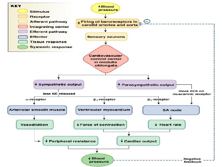
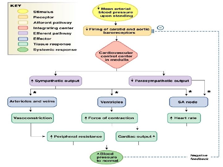
KEY Medullary cardiovascular control center Stimulus Sensor/receptor Integrating center Efferent pathway Change in blood pressure Effector Parasympathetic neurons Carotid and aortic baroreceptors Sympathetic neurons SA node Ventricles Veins Arterioles