Septic arthritis Septic arthritis Infected Joint Inflamation Infection


")


")
")
 : 1. non-weight-bearing on affect side. 2.")


 CRP ESR Imaging: : X-Ray : -widening of")
 In this child the left hip is subluxated and the soft tissues")


- Slides: 25
Septic arthritis
Septic arthritis Infected Joint Inflamation Infection in a joint
Causative agents : - Staphylococcus aureus - Group A streptococcus - Neisseriae Gonorrhoea - H. influenzae ( is an important pathogen unless they have been vaccinated against this organism. ) First most common cause
Route for bacterial entry: Injection into the joint space Skin infection (S. aureus ) Penetrating injuries STI surgery under septic technique Osteomyelitis URTI LRTI
Pathology: �There is an acute synovitis with a purulent joint effusion and Synovial membrane becomes edematous, swollen and hyperemic and an increase in synovial fluid. �As pus appears in the joint, articular cartilage is destroyed by bacterial and cellular enzymes. �If the infection is not arrested the cartilage may be completely destroyed. �Pus may burst out of the joint to form abscesses and sinuses. �With healing there may be : � (1) complete resolution and a return to normal �(2) partial loss of articular cartilage and fibrosis of the joint �(3) loss of articular cartilage and bony ankylosis �(4) bone destruction and permanent deformity of the joint. 5
With any Monoarthritis , it's important to role out septic arthritis. ? ! ? hy W A delay in diagnosis and treatment, particularly with septic arthritis, can have catastrophic results including sepsis, bacteremia, joint destruction, or death.
Septic arthritis is Medical Emergency , require rapid diagnosis &immediate management to avoid Irreversible joint damage. infection spreads through the joint there is an acute synovitis with a purulent joint Effusion. articular cartilage is eroded; the articular cartilage is attacked by bacterial and cellular enzymes. If the infection is not arrested, the cartilage may be completely destroyed Healing start , the raw articular surfaces may adhere. Healing then leads to fibrous or bony ankylosis.
Clinical features: 1. acute pain and swelling in a single large joint -any joint can be affected , Knee is the most commonly affected joint in adults, hip in children. All movements are grossly restricted and often completely abolished by pain and spasm (pseudoparesis). 2. Signs of Inflammation : Redness Warmth Tenderness 3. Fever 4. 'silent’ joint infection: - rheumatoid arthritis - corticosteroid treatment Suspicion may be aroused by an unexplained deterioration in the patient’s general condition
In adults • Often involve a superficial joint (knee, wrist, finger, ankle or toe) • Joint painful, swollen and inflamed • Unable to bear weight • Warm and tender • Evidence of gonococcal infection or drug abuse • Patients with RA and on corticosteroids may develop silent joint infection
In children • • Acute pain in a single large joint (commonly the hip) Reluctance to move the limb (pseudoparesis) Fever Rapid pulse Joint swelling and redness Joint tenderness All movements are restricted and often completely abolished by pain and spasm.
Kocher criteria: (for child with painful hip) : 1. non-weight-bearing on affect side. 2. ESR greater than 40 mm/hr. 3. fever. 4. WBC count of >12, 000 mm 3. - when 4/4 criteria are met, there is a 99% chance that the child has septic arthritis. • when 3/4 criteria are met, there is a 93% chance of septic arthritis. • - when 2/4 criteria are met, there is a 40% chance of septic arthritis. • - when 1/4 criteria are met, there is a 3% chance of septic arthritis.
� epiphyseal plate prevents infection from entering joint space in older children. � but apparently does not act as a barrier in infants. � synovial membrane inserting distally to epiphysis. � allowing bacteria to spread directly from themetaphysis to joint space. � metaphysis of shoulder, hip, radial head, and ankle remain intracapsular during early childhood. � the hip joint seems especially prone to sepsis from adjacent osteomyelitis. � synovial reflections over the metaphyseal bone decrease with age.
Examination �pain in groin area that occasionally radiates down the medial side of thigh. - progressive accompanied by spasm of the hip muscles. - hip in flexion and external rotation & decreased internal rotation compared to the normal hip. - patient resists all attempts to move hip. - palpate the SI joint for local tenderness.
Investigation: : CBC -----( elevated WBC) CRP ESR Imaging: : X-Ray : -widening of the joint space (due to the effusion) -Later, when the articular cartilage is attacked, the ‘joint space’ is narrowed confirmed by joint aspiration and immediate microbiological culture and sensitivity. Blood cultures - In advanced cases there are signs of bone destruction. Radio-isotope imaging and MRI are helpful for detecting signs in difficult sites such as the sternoclavicular and sacroiliac joints.
x-ray: (a) In this child the left hip is subluxated and the soft tissues are swollen. (b) If the infection persists untreated, the cartilaginous epiphysis may be entirely destroyed leaving a permanent pseudarthrosis (Tom Smith’s dislocation).
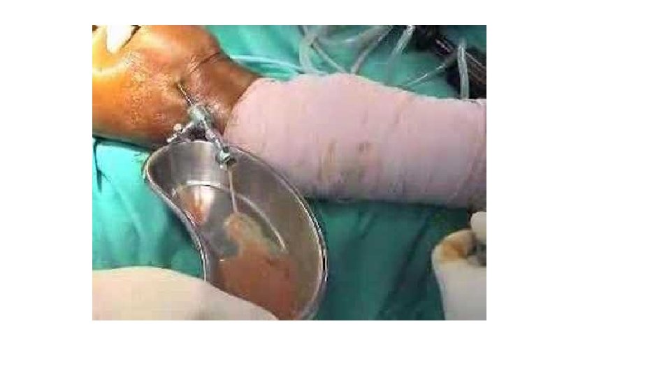
Septic arthritis in an adult knee joint. 16
Position of patient with septic arthritis flexion and external rotation max stretch to the joint capsule 17
Osteomyelitis near a joint may be indistinguishable from septic arthritis; the safest is to assume that both are present. An acute haemarthrosis, either post-traumatic or due to a haemophilic bleed, can closely resemble infection. The history is helpful and joint aspiration will resolve any doubt. Gout and pseudogout in adults can be indistinguishable from joint infection and cellulitis. Aspirated fluid may look turbid but the presence of urate or pyrophosphate crystals will confirm the diagnosis. Mono articulate joint pain
Complications: : ■ Dislocation: a tense effusion may cause dislocation. ■ Epiphyseal destruction: in neglected infants the largely cartilaginous epiphysis may be destroyed, leaving an unstable pseudoarthrosis. ■ Growth disturbance: physeal damage may result in shortening or deformity. ■ Ankylosis: if articular cartilage is eroded, healing may lead to ankylosis.
Treatment : : Treatment is then started without further delay Antibiotics Intravenous ------ a third-generation cephalosporin will cover both Gram-positive and Gram-negative organisms ------Once the bacterial sensitivity is known the appropriate drug is substituted. Intravenous administration is continued for several weeks and is followed by oral antibiotics for a further 2 or 3 weeks. Splintage The joint must be rested either on a splint or in a widely split plaster.
Drainage pus is drained and the joint washed out -------open operation (the best to done ) -------repeated needle aspiration and irrigation(superficial joint ) -------arthroscopy (knee joint) Once the patient’s general condition is good and the joint is no longer inflamed, gentle and gradually increasing movements are encouraged. But if articular cartilage has been destroyed, the aim is to keep the joint immobile in the optimum position while ankylosis is awaited.
Summary • Acute monoarthritis should be evaluated emergently to roll out septic arthritis. • Untreated septic arthritis can lead to a very serious complications. (joint destruction an sepsis) • Appropriate history and examination are crucial. • Aspiration is the key method for diagnosis and identifying the organism. • Early treatment with IV antibiotics and leg splinttage.