Premalignant disease of the cervix prof maysoon sharief


 v Definition of CIN: CIN is a pre-invasive changes of")

: - Between 1/3 -2/3 of the epithelium")

 • • • Demographic risk factors: Ethnicity (Latin American countries, U.")
 • • • Behavioural risk factors Infrequent or absent cancer screening")
 Tobacco smoking : - ↑ the risk of cervical cancer among")
: • Initiation of screening:")

 to the cervix:")


- Slides: 24
Premalignant disease of the cervix prof. maysoon sharief
Introduction • It is rising in young women. • There are 450, 000 cases of cervical cancer/year. • There are 300, 000 death/year. • It is the second most common cancer. • It is a preventable disease (screening programme-cervical smear).
Cervical Intraepithelial Neoplasia (CIN) v Definition of CIN: CIN is a pre-invasive changes of the cervical epithelium where the epithelium thickness is replaced by abnormal cells in varying degree without breaching the cell membrane. symptomless. v Abnormal cytological changes of CIN (immature and disorganized cells) are: Increased Nuclear/Cytoplasmic ratio. Prominence of nuclear chromatin. Multinucleation.
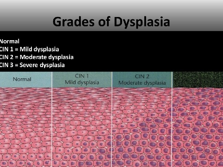
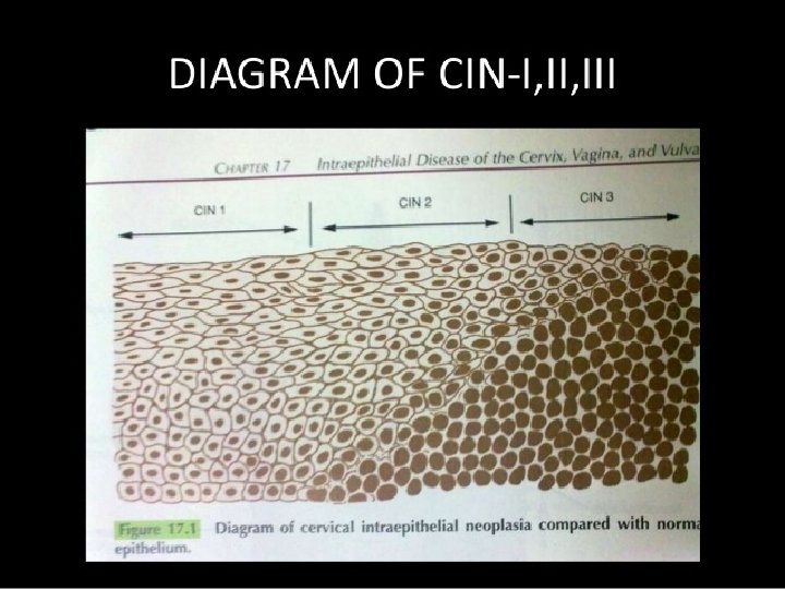
Grades of CIN is graded according the proportion of epithelium occupied by the abnormal cells. • CIN 1 (mild dysplasia): - One-third or less is occupied by the abnormal cells. - Progress to (CIS) in 6%. - Regressed or disappeared in 62%,
Grades of CIN • CIN 2(modrate dysplasia): - Between 1/3 -2/3 of the epithelium is occupied by the abnormal cells. - Become invasive in 13%. • CIN 3 (severe dysplasia): - The whole thickness of the squamous epithelium is occupied by the abnormal cells. - It is regarded as carcinoma-in-situ (CIS). - It could arise as CIN 3 or progress from CIN 1 or CIN 2. - Become invasive in 29%.
Incidence of CIN v The incidence of CIN: is 4 to 5% of Pap tests. v The incidence of CIN vary according to the: (1) population studied, as the peak incidence being between 25 and 29 years of age, (2)socioeconomic factors, and (3) risk-related behaviours. v The true incidence and prevalence can only be estimated, as screening cytology and colposcopy lack complete sensitivity.
Risk factors (Epidemiology) • • • Demographic risk factors: Ethnicity (Latin American countries, U. S. minorities). Low socioeconomic status. Older age. Medical risk factors: Cervical human papilloma virus infection. Parity. Immunosuppression.
Risk factors (Epidemiology) • • • Behavioural risk factors Infrequent or absent cancer screening Pap tests. Early coitus. Multiple sexual partners. Male partner who has had multiple sexual partners. Tobacco smoking. Dietary deficiencies.
Risk factors (Epidemiology) Tobacco smoking : - ↑ the risk of cervical cancer among HPV-positive women. - Nicotine and its major metabolite cotinine are found in the cervical mucus of women and in the semen of men who smoke → suppression of local cervical immunity and promotion of HPV-driven cellular transformation and neoplasia. Dietary deficiencies: - Vitamins such as A, C, E, beta carotene, and folic acid may alter cellular resistance to HPV infection → persistent viral infection and cervical neoplasia.
Identification of CIN v Cervical Cytology (The Pap test ): • Initiation of screening: 3 years after onset of vaginal intercourse; no later than age 21. • Screening intervals: (1) age < 30 years: annually, (2) age > 30 years: : every 2 to 3 years after 3 consecutive negative tests. (3) patients with HIV or other immunocompromised state: 2 tests during the first year, then annually. • Discontinuation of screening: Age 65 to 70 in women not at high risk (history of cervical cancer, DES, HPV, HIV, & immunocompromised state).
CIN Management A. Abnormal cervical smear: • Due to infection: treat then repeat the smear. • Atrophic smear: give oestrogen then repeat the smear. • Colposcopy. B. Colposcopy: • To view the cervix telescopically at magnification range of 6 to 40 times. • 4% of acetic acid is applied to the cervix which coagulates proteins of the epithelial cells and abnormal epithelium appears white; biopsies should be taken from that area. or
CIN Management B. Colposcopy: • Apply Lugol's iodine solution (Schiller test) to the cervix: - Iodine 2 gm, potassium iodide 4 gm and distilled water 300 ml - Malignant cells lack glycogen so they fail to take iodine (stainless). - Normal epithelium (rich in glycogen) stains darkbrown. - Biopsies taken from the non-staining areas. - Squamo-columnar junction should be seen entirely.
CIN Management C. Cone biopsies: § indications: - squamo-columnar junction not seen. - negative colposcopic examination with positive repeated cervical smear. - microinvasion or invasion is suspected. § Size: kept to a minimum with normal tissue, tailored & cut with a knife. § D/C is should be done. § Haemostasis: is achieved by using Dexon at 3 & 9 0’clock of the cervix. § Complications: primary & 2 nd bleeding & scarring → stenosis → impaired fertility, cryptomenorrhoea, abortion, preterm labour & cervical dystocia.
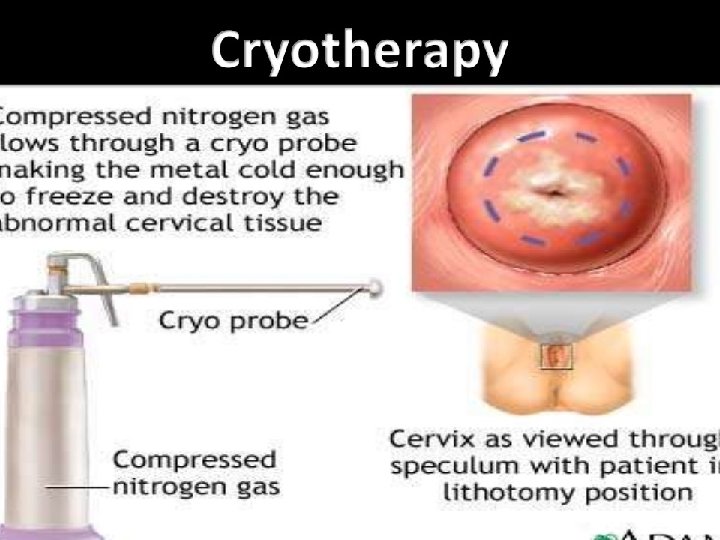
CIN treatment • • Knife Cone biopsy. Total hysterectomy: Persistent lesion after conization. Lesion extends to the upper vagina (colposcopy). Coexisting indication (menorrhagia or prolapse). Local destruction (ablation): (for young, unmarried or wanting children): Cryocautery: freezing the tissue. Electrocautery: burning the transformation zone. Large loop excision of the transformation zone: using electrodiathermy. Cold coagulation: destruction of the transformation zone with a probe heated to 100 to 120Ċ.