Practical Electrocardiography Myocardial Ischemia and Acute Myocardial Infarction


- Slides: 49

Practical Electrocardiography – Myocardial Ischemia and Acute Myocardial Infarction Scott Ewing, D. O. Cardiology Fellow August 30, 2006


Assess Initial 12 -Lead EKG Findings • ST elevation or new or presumably new LBBB: strongly suspicious for injury • ST-elevation AMI • ST depression or dynamic T-wave inversion: strongly suspicious for ischemia • High-risk unstable angina/ non–ST-elevation AMI • Nondiagnostic EKG: absence of changes in ST segment or T waves • Intermediate/low-risk unstable angina Classify patients with acute ischemic chest pain

Current-of-injury patterns with acute ischemia A. B. Resultant ST vector is directed toward the inner layer of the affected ventricle and the ventricular cavity. Overlying leads therefore record ST depression (Transmural or epicardial injury), ST vector is directed outward. Overlying leads record ST elevation. Reciprocal ST depression can appear in contralateral leads.

Acute Ischemia / Non-Q Wave MI / Non-ST Elevation MI Evolving ST-T changes over time without the formation of pathologic Q waves n Localization of non-Q wave MI by the particular leads showing ST-T changes is probably only valid with ST segment elevation pattern n Evolving ST-T changes may include any of the following patterns: n Convex downward ST segment depression n T wave flattening or inversion n Biphasic T wave changes n

Ischemic T Wave Changes

Ischemic T Wave Inversion

Ischemic ST Changes

Ischemic Biphasic T Wave Changes

Acute Ischemia - T Wave Changes

Acute Ischemia – ST Depression

Acute Ischemia – ST Depression

Acute Ischemia – ST Depression

60 -year-old Male

Anterior Ischemia n n EKG shows sinus rhythm with ventricular ectopy, left axis deviation, consistent with left anterior fascicular block (hemiblock), and T wave inversions in V 2 -V 5 with subtle upward bowing of the ST segments ST-T abnormalities in I and a. VL Symmetric T wave inversions, especially with upward bowing of the ST segments is highly suggestive of ischemia in the left anterior descending distribution (LAD) in this context Most expeditious test to order is a cardiac catheterization, which showed significant LAD (and obtuse marginal) disease

Elderly Male

Severe Ischemia n n n NSR at about 65 bpm with profound precordial ischemic ST segment depression, consistent with severe subendocardial ischemia and probable non-Q wave myocardial infarction Q waves in the infero-lateral leads are consistent with prior myocardial infarction(s) Profound ST depressions of this type usually indicate severe multivessel disease, and sometimes left main coronary disease Patient experienced severe chest pain and was transferred from an outside facility in cardiogenic shock En route to the cardiac catheterization laboratory, he developed refractory PEA and ventricular fibrillation

84 -year-old Female

NQWMI n n Left ventricular hypertrophy (LVH) plus left atrial abnormality (LAA) QRS axis is somewhat leftward (-7 degrees) Although LVH alone may be associated with ST-T abnormalities (sometimes referred to as a "strain pattern"), like those in lead a. VL, the prominent horizontal or downsloping ST depressions in other leads (I, II, a. VF, V 5, V 6) here are strongly suggestive of ischemia superimposed on LVH The patient had positive cardiac enzymes and underwent cardiac catheterization showing left main and three vessel coronary disease, followed by coronary artery bypass graft surgery.

Current-of-injury patterns with acute ischemia A. B. Resultant ST vector is directed toward the inner layer of the affected ventricle and the ventricular cavity. Overlying leads therefore record ST depression (Transmural or epicardial injury), ST vector is directed outward. Overlying leads record ST elevation. Reciprocal ST depression can appear in contralateral leads.

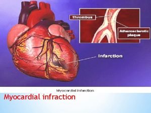
Acute Myocardial Infarction / ST Elevation MI / Q Wave MI n n n n Most acute MI's are located in the left ventricle In the setting of a proximal RCA occlusion, however, up to 50% may also have a component of RV infarction as well In general, the more leads of the 12 -lead EKG with MI changes (Q waves and ST elevation), the larger the infarct size and the worse the prognosis LAD and it's branches usually supply the anterior and anterolateral walls of the LV and the anterior two-thirds of the septum LCX and its branches usually supply the posterolateral wall of the LV RCA supplies the RV, the inferior (diaphragmatic) and true posterior walls of the LV, and the posterior third of the septum RCA also gives off the AV nodal coronary artery in 85 -90% of individuals; in the remaining 10 -15%, this artery is a branch of the LCX

Acute Myocardial Infarction / ST Elevation MI / Q Wave MI n n Normal EKG prior to MI Hyperacute T wave changes - increased T wave amplitude and width; may also see ST elevation Marked ST elevation with hyperacute T wave changes (transmural injury) Pathologic Q waves, less ST elevation, terminal T wave inversion (necrosis) n n n Pathologic Q waves are usually defined as duration >0. 04 s or >25% of R-wave amplitude Pathologic Q waves, T wave inversion (necrosis and fibrosis) Pathologic Q waves, upright T waves (fibrosis)

Evolution of EKG Changes

Evolution of EKG Changes

Infarct - ST Elevation

Inferior Infarct – ST Elevation

Inferior Infarct – ST Elevation

Posterior Infarct – ST Elevation!!!

Old Infarct - Anterior Q Waves

Old Infarct - Inferior Q Waves

Persistent ST Changes

Persistent T Wave Changes

34 Year Old Male With Chest Pain

34 -year-old male

Acute Anterior MI Classic findings of acute anterior wall Q wave myocardial infarction n Reciprocal inferior ST depressions n Hyperacute T waves n Distribution of changes is consistent with a proximal LAD occlusion n Confirmed at cardiac catheterization and treated with PTCA and stenting n

68 -year-old female

Acute Anterior MI n n n Note Q waves and loss of R waves V 1 - V 4 ST elevation in V 2 - V 5/V 6 Left anterior fascicular block is also present, but does not account for the loss of R wave progression The patient had a very recent anterior MI Cardiac catheterization revealed 3 -vessel disease with a 90% mid-LAD "culprit" lesion

53 -year-old female

Acute Lateral MI ST elevations in I and a. VL n Probable reciprocal ST depressions inferiorly consistent with acute lateral MI n Remember: ST elevations like this are never reciprocal but indicate the primary region of ischemia (diagonal or circumflex lesion) n Confirmed left circumflex occlusion at catheterization n

* 36 -year-old male

Acute Pericarditis n n Always consider myocardial infarction first when you see ST elevations But don't forget the differential diagnosis of ST elevations n n n Ischemic heart disease Pericarditis Left bundle branch block (LBBB) Normal ("early repolarization") variant Two features here point to pericarditis n n First, diffuseness of the ST elevations (I, III, a. VF, V 3 V 6) Second, PR depression in II, a. VF, V 4 -V 6 and PR elevation seen in a. VR (attributed to subepicardial atrial injury)

49 -year-old male

Acute Pericarditis n n n Diffuse ST segment elevations (I, II, a. VF, V 2 -V 6) Subtle PR segment deviations (elevated in a. VR and depressed in the inferolateral leads) ST elevations are due to a ventricular current of injury from the pericardial inflammation PR changes are due to an associated atrial current of injury Note that the PR and ST segment vectors point in opposite directions, i. e. , PR up and ST down in a. VR and PR down and ST up in inferolateral leads

Middle aged female

Acute Myocardial Infarction n n n Marked inferior and lateral ST segment elevation ST segment depression in anterior leads V 1 -V 4 ST elevations (“current of injury” pattern) indicate transmural ischemia of the infero-lateral wall ST depression most consistent with reciprocal change from the ST elevation generated by the acute posterior and lateral ischemia Remember, acute pericarditis causes diffuse ST segment elevation (e. g. , leads I, III, a. VL, a. VF, and the precordial leads) Reciprocal ST depressions of the type seen here (V 1 -V 4), are never a feature of pericarditis alone Cardiac catheterization revealed acute occlusion of a dominant left circumflex coronary artery (along with occlusion of a smaller RCA)

52 -year-old male

ST Elevation Myocardial Infarction Slight inferior ST elevation with T wave inversion n Minimal reciprocal ST depression in a. VL n Relatively low limb lead voltage makes these findings more subtle n

 Myocardial ischemia meaning
 Pico question myocardial infarction
 Myocardial injury
 Wall stemi
 Pancreas wiki
 Acute pericarditis
 Transmural ischemia
 Ischemia-guided strategy
 What is this
 Ischemia acuta degli arti
 Gross
Gross Unifocal pvc
 Latin and greek prefixes
 Cellular events of acute inflammation
 Classify each triangle as acute equiangular obtuse or right
 Vascular and cellular events of acute inflammation
 Common chronic and acute conditions chapter 18
 Morphology pattern of acute inflammation
 Difference between acute and subacute rehab
 Differential diagnosis leukocytosis
 Phoenix abscess
 Practical extraction and report language
 Ideal transformer and practical transformer
 Grade 4 term 3 natural science
 Strategic gender needs and practical gender needs
 Write the difference between ideal and practical op-amp.
 Practical research nature of inquiry and research
 Practical extraction and report language
 Differentiate between ideal and practical transformer
 Russian formalism slideshare
 Elseif perl
 Name 2 objects with acute angles
 Chronic blood loss
 Complementary supplementary vertical and adjacent angles
 Acute glomerulonephritis causes
 A quadrilateral with 4 acute angles
 Acute bronchospasm
 Right angled isosceles triangle
 Obtuse angle in real life
 What is hyl congruence theorem
 Acute spasmodic laryngitis
 Acute productive cough differential diagnosis
 Respiratory acidosis
 Acute tubular necrosis
 Acute glomerulonephritis causes
 Parallelogram rectangle rhombus square trapezoid properties
 Negative explanatory style
 Site:slidetodoc.com
 Acute stress disorder diagnosis
 Globulin types
