
INHIBITING AIM 2 IN ORDER TO RESTORE FUNCTION
INHIBITING AIM 2 IN ORDER TO RESTORE FUNCTION IN INJURED CARDIOMYOCYTES DUE TO ISCHEMIA & REPERFUSION Ankita Thakur BNFO 300
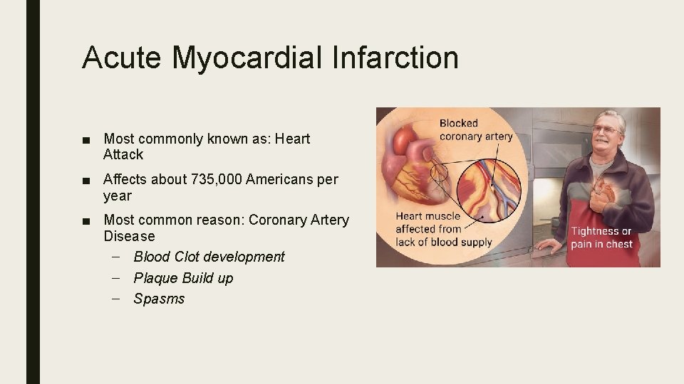
Acute Myocardial Infarction ■ Most commonly known as: Heart Attack ■ Affects about 735, 000 Americans per year ■ Most common reason: Coronary Artery Disease – Blood Clot development – Plaque Build up – Spasms
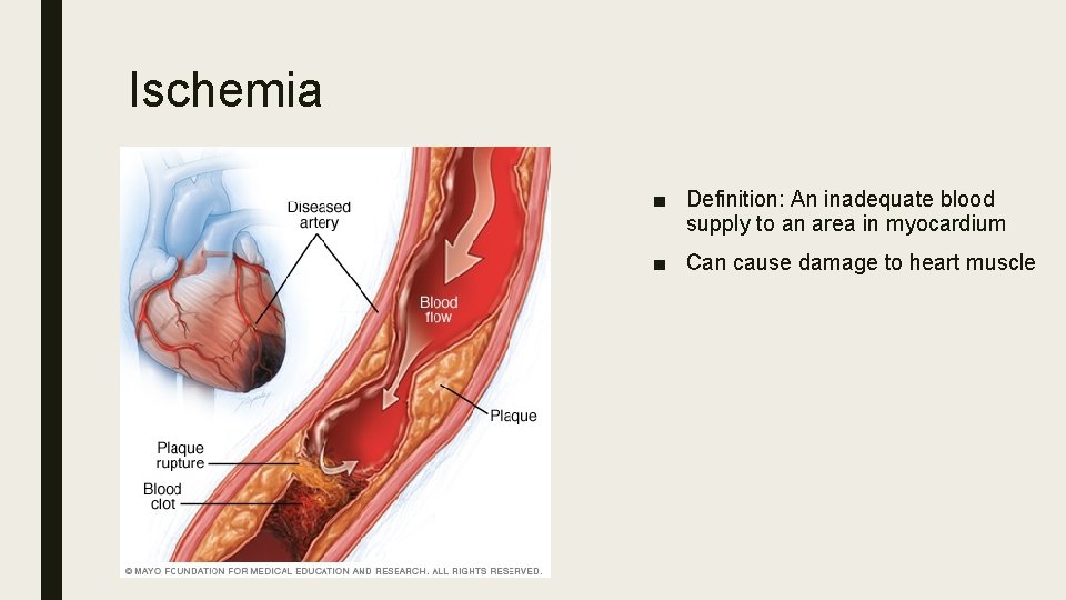
Ischemia ■ Definition: An inadequate blood supply to an area in myocardium ■ Can cause damage to heart muscle
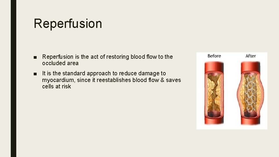
Reperfusion ■ Reperfusion is the act of restoring blood flow to the occluded area ■ It is the standard approach to reduce damage to myocardium, since it reestablishes blood flow & saves cells at risk
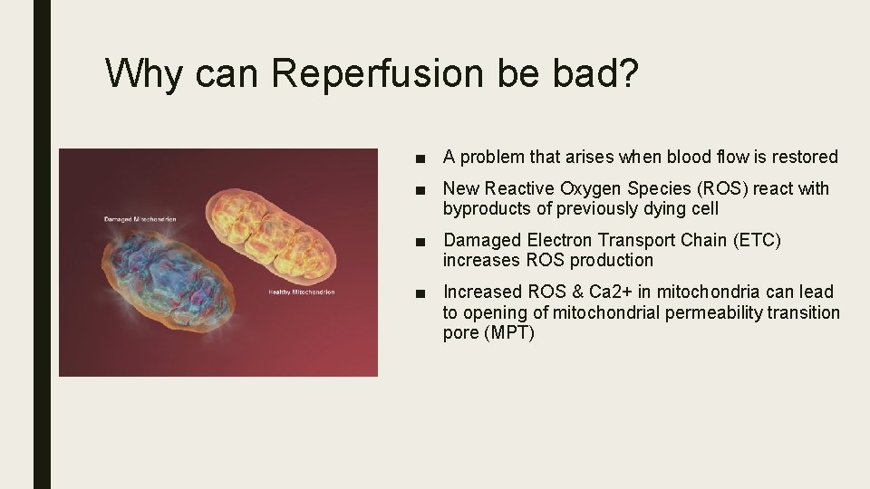
Why can Reperfusion be bad? ■ A problem that arises when blood flow is restored ■ New Reactive Oxygen Species (ROS) react with byproducts of previously dying cell ■ Damaged Electron Transport Chain (ETC) increases ROS production ■ Increased ROS & Ca 2+ in mitochondria can lead to opening of mitochondrial permeability transition pore (MPT)
Reperfusion Injury – continued ■ Damage to mitochondria changes ion homeostasis & causes ATP deficiency ■ Leads to cell death ■ ATP & other damage associated molecular patterns (DAMPs) are released from dying cell ■ Formation of inflammasome triggered ■ Pyroptosis initiated
Why is AIM 2 important? ■ AIM 2 – DNA sensor that forms AIM 2 inflammasome after detecting cytoplasmic ds. DNA ■ Normally recognizes ds. DNA of viral or bacterial origin in a sequenceindependent manner – Which is why it can recognize ds. DNA found in cytoplasm as a substrate
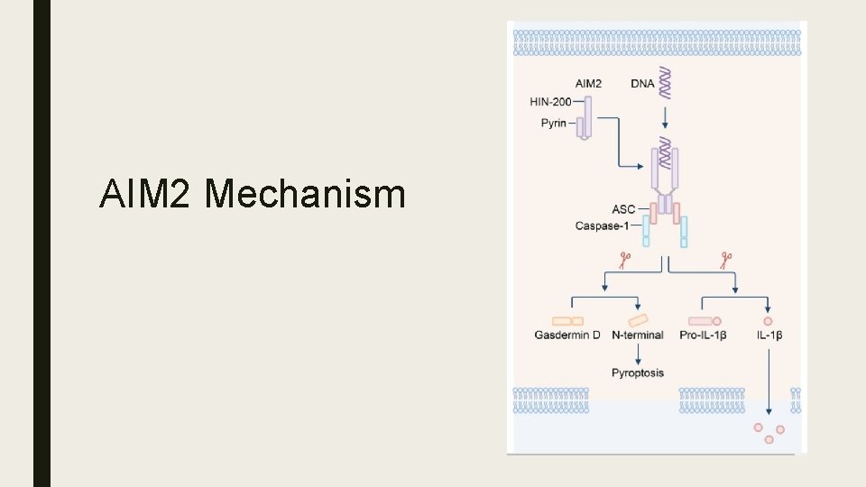
AIM 2 Mechanism
Mitochondrial DNA ■ mt. DNA can be released from damaged mitochondria ■ mt. DNA enters cytoplasm of cell that underwent myocardial ischemia reperfusion injury ■ AIM 2 can recognize mt. DNA, since similar to bacterial DNA
Hypothesis Mitochondrial DNA may be acting as a DAMP, which is what leads to the activation of AIM 2. ■ How can this be approached assuming the release of mt. DNA is inevitable due to damage?
Overview of Study Three Checkpoints 1. Reduce AIM 2 expression in the mouse & Confirm AIM 2 inhibition i. pay attention to caspase-1, indicator for inflammasome formation 2. Induce Ischemia/Reperfusion Injury & Measure Infarct size + Heart Function 3. Measure Mitochondrial DNA presence
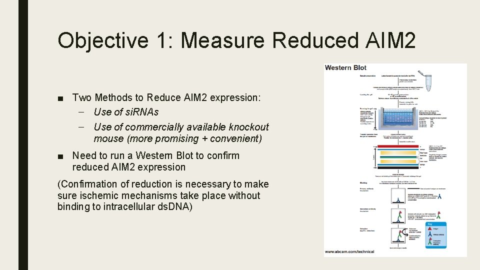
Objective 1: Measure Reduced AIM 2 ■ Two Methods to Reduce AIM 2 expression: – Use of si. RNAs – Use of commercially available knockout mouse (more promising + convenient) ■ Need to run a Western Blot to confirm reduced AIM 2 expression (Confirmation of reduction is necessary to make sure ischemic mechanisms take place without binding to intracellular ds. DNA)
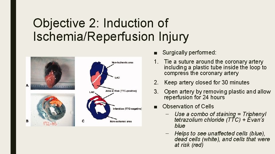
Objective 2: Induction of Ischemia/Reperfusion Injury ■ Surgically performed: 1. Tie a suture around the coronary artery including a plastic tube inside the loop to compress the coronary artery 2. Keep artery closed for 30 minutes 3. Open artery by removing plastic and allow reperfusion for 24 hours ■ Observation of Cells – Use a combo of staining = Triphenyl tetrazolium chloride (TTC) + Evan’s blue – Helps to see unaffected cells (blue), dead cells (white), and cells that were at risk (red)
Objective 3: Measure mt. DNA release ■ It needs to be measured to see if the mitochondrial DNA is released into the cytoplasm ■ Best measured by Real Time Polymerase Chain Reaction (q. PCR) ■ Sample for test will be from the affected mouse heart cells, from which there will be a mitochondrial and cytoplasmic extract prepared ■ DNA from extracts should be precipated ■ q. PCR should be run on cytoplasmic fraction to measure how much mt. DNA can be found ■ Quantitative analysis should be performed using a known amount of mt. DNA to prepare a standard curve to quantify the DNA
Discussion ■ AIM 2 Silencing/Knocking out – Hope to not spend too much confirming reduced AIM 2 expression – Need to confirm reduced caspase-1 activity: Noted through improved heart function & reduction of cell death after reperfusion due to lack of pyroptosis in the area affected during ischemia ■ Mitochondrial DNA presence – Expected q. PCR results: Inhibited AIM 2 sample would have intact mt. DNA, as opposed to sample with AIM 2 expressed ■ Hopes – If AIM 2 expression reduction shows to have significant impact on reduction of reperfusion injury, then there will be a need to develop specific AIM 2 inhibitors to reduce AIM 2 activity after AMI
References Buja, L. M. (2005) Myocardial ischemia and reperfusion injury. Cardiovascular Pathology, 14. 170 -175. doi: 10. 1016/j. carpath. 2005. 03. 006 Hausenloy, D. , Yellon, D. (2013) Myocardial ischemia-reperfusion injury: a neglected therapeutic target. The Journal of Clinical Investigation, 123(1) : 92– 100. doi: 10. 1172/JCI 62874 Jabir, M. , Hopkins, L. , Ritchie, N. , Ullah, I. , Bayes, H. , Li, D. , Tourlomousis, P. , Lupton, A. , Puleston, D. , Simon, A. , Bryant, C. , and Evans, T. (2015) Mitochondrial damage contributes to Pseudomonas aeruginosa activation of the inflammasome and is downregulated by autophagy. Autophagy, 11(1). pp. 166 -182. http: //dx. doi. org/10. 4161/15548627. 2014. 981915 Lian, Q. , Xu, J…. . Geng, M. (2017) Chemotherapy-induced intestinal inflammatory responses are mediated by exosome secretion of double-strand DNA via AIM 2 inflammasome activation. Springer Nature: Cell Research. 1 -17. doi: 10. 1038/cr. 2017. 54 Mangino, M. J, Tian, T. , Lindell, S. L. (2012). Ezring functionality and ischemia-reperfusion (I/R) injury in transplantation. The FASEB Journal, 26: 56. 7. Retrieved from: http: //www. fasebj. org/content/26/1_Supplement/56. 7. short Marchetti, C. , Chojnacki, J. , Toldo, S. , . . . Abbate, A. (2014) A novel pharmacologic inhibitor of the nlrp 3 inflammasome limits myocardial injury following ischemia-reperfusion in the mouse. J Cardiovasc Pharmacol. 63(4): 316– 322. doi: 10. 1097/FJC. 000000053. Mezzaroma, E. , Toldo, S. , . . Abbate, A. (2011) The inflammasome promotes adverse cardiac remodeling following acute myocardial infarction in the mouse. PNAS, 108(49). 19725 -19730. doi: 10. 1073/pnas. 1108586108. Ming Man, S. , Karki, R. , Kanneganti, T. (2016) AIM 2 inflammasome in infection, cancer, and autoimmunity: Role in DNA sensing, inflammation, and innate immunity. European Journal of Immunology, 46. 269 -280. DOI: 10. 1002/eji. 201545839
References Murphy, E. , Steenberg, C. (2008) Mechanisms underlying acute protection from cardiac ischemia-reperfusion injury. Physiology Review, 88. 581 -609. doi: 10. 1152/physrev. 00024. 2007. Toldo, S. , Arrosa, J. F. , . . . Quader, M. (2016) Determination of optimal coronary flow for the preservation of “donation after circulatory death” heart. Circulation, (134): A 18989. Retrieved from: http: //circ. ahajournals. org/content/134/Suppl_1/A 18989 Toldo, S. , Marchetti, C. , . . . , Abbate, A. (2016) Inhibition of NLRP 3 inflammasome limits the inflammatory injury following myocardial ischemia–reperfusion in the mouse. International Journal of Cardiology, 209: 215 -220. doi: http: //dx. doi. org/10. 1016/j. ijcard. 2016. 02. 043 Trankle, C. , Thurber, C. J. , Toldo, S. , Abbate, A. (2016) Mitochondrial membrane permeability inhibitors in acute myocardial infarction: still awaiting translation. JACC: Basic To Translational Science, 1(6): 524 -535. doi: http: //dx. doi. org/10. 1016/j. jacbts. 2016. 012 Protocols: http: //docs. abcam. com/pdf/protocols/general-western-blot-protocol. pdf https: //www. thermofisher. com/content/dam/Life. Tech/Documents/PDFs/PG 1503 -PJ 9169 -CO 019861 -Update-q. PCR-Handbook-branding-Americas. FLR. pdf Protocol Image: http: //www. abcam. com/ps/pdf/protocols/Western_blot_diagram. pdf Other Images: http: //www. mayoclinic. org/diseases-conditions/myocardial-ischemia/multimedia/causes-of-myocardial-ischemia/img-20008606 http: //www. mayoclinic. org/diseases-conditions/myocardial-ischemia/basics/definition/con-20035096 https: //www. researchgate. net/figure/5862083_fig 7_Figure-3 -Excised-heart-stained-with-Evans-blue-dye-and-TTC-The-pictures-demonstrate-the
- Slides: 17