Posterior view Superior articular process Inferior articular process


 1 - A")
 The transverse processes are thick, strong and are")


 surface - This is a concave and relatively smooth surface.")

 surface 1 - This is a convex rough surface. 2 - four")


- Slides: 28
Posterior view Superior articular process Inferior articular process Superior articular process
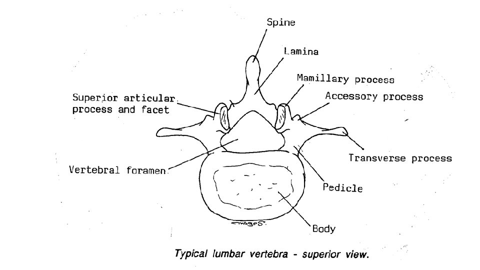
General features of the typical lumbar vertebrae (L 1 -L 4) 1 - A large body which increases in size gradually from the 1 st to the 4 th. 2 - A wide triangular vertebral foramen. 3 - A thin (long and tapering) transverse process. 4 An accessory process behind the root of the transverse process. 5 - The superior articular process is curved with the superior articular facet concave medially. 6 - The inferior articular process is curved with the inferior articular facet convex laterally. - The distance between two superior articular processes (facets) is wider than the inferior. 7 - mammillary process on posterior edge of superior articular process. 8 - Spine is broad and quadrilateral,
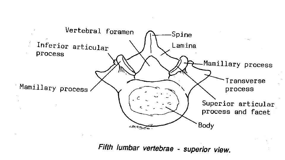
** 5 th Lumbar Vertebra 1) The transverse processes are thick, strong and are attached to the whole sides of the pedicles. 2) Smaller size of its spine. 3) The distance between two superior articular processes is nearly equal to the inferior articular processes
Lateral view
Ligaments 1 2 4 5 6 3
** Ligaments of the vertebrae 1 - Intervertebral discs between the bodies. 2 - Anterior longitudinal ligament in front of the bodies. 3 - Posterior longitudinal ligament on the back of the bodies inside the vertebral canal. 4 - Interspinous ligament between The spines 5 - Supraspinous Ligament on The tips of the spines 6 - Ligamentum flavum between the laminae
Base of sacrum Anterior surface of sacrum Lumbosacral angle Superior articular process Sacrum - This is a triangular bone formed by fusion of 5 sacral vertebrae. - It has an apex which is directed downwards. Base is directed upwards. Ala of sacrum Promontory Ant. Sacral foramina Ridges Apex of sacrum
s u l. I iac Piriformis Coccygeus
Base of the sacrum - It is formed by the first sacral piece. - The anterior border of its body forms the promontory of the sacrum. - The expanded sides of the base called ala of the sacrum gives origin to iliacus muscle. - The body articulates with the 5 th lumbar vertebrae, this articulation forms the lumbosacral angle. - The superior articular processes of the first piece articulate with the inferior articular processes of the fifth lumbar vertebra.
Anterior (Ventral or pelvic) surface - This is a concave and relatively smooth surface. - It presents ridges which mark the lines of fusion of the sacral vertebral bodies. - It presents four anterior (ventral) sacral foramina on each side that give Exit for the ventral rami of the upper four sacral nerves to form sacral plexus. - The middle three pieces give origin to the piriformis muscle. - The cococygeus muscle inserted into the lower part of anterior surface.
Posterior surface of sacrum Sacral canal Lateral sacral crest = T. processes Median sacral crest = spines Intermediate sacral crest = A. processes Sacral hiatus Post. Sacral foramina
Posterior (Dorsal) surface 1 - This is a convex rough surface. 2 - four posterior (dorsal) sacral foramina on each side. 3 - Median sacral crest are produced by fusion of spines. 4 - Intermediate sacral crest on the medial sides of the posterior sacral foramina are produced by fusion of the articular processes. 5 - Lateral sacral crest on the lateral sides of the posterior sacral foramina are produced by fusion of the transverse processes.
Sacral canal - The sacral canal is the lower part of the vertebral canal. - The canal ends by an opening called the sacral hiatus. **Contents of the sacral canal 1 - Cauda equine, Roots and trunks of the sacral and coccygeal nerves. 2 - Filum terminale (extension of the pia matter) to the tip of the coccyx. 3 - The dura and arachnoid mater extending to the 2 nd sacral vertebrae. 4 - Internal vertebral plexus of veins. 5 - Epidural fat surrounding all the structures
Origin: powerful Ms on the side of the spines between median and lateral crest Stabilizing of the vertebral joints Powerful Groups of muscles, , from the median and lateral crest, causes Low back pain
Differences between male and female sacrum Male sacrum Female sacrum 1 - long and narrow 1 - Short and wide. 2 - Curved 2 - Straight (upper half) but its lower half is curved. 3 - the auricular surface of ilium 3 - The auricular surface of ilium articulates with 2. 5 pieces of sacrum articulates with 2 pieces of sacrum. 4 - the articular surface on the base of 4 - The articular surface on the base of sacrum equal 1/2 the base sacrum Equal 1/3 the base.
Sagittal section in female pelvis Rectum - continuation of the sigmoid colon at S 3. ** End: one inch below and infront the tip of the coccyx • Anal canal about 4 cm long
Sagittal section in male pelvis
L. S. Sphincters of anal canal Ano-rectal ring Deep Superficial Surgical importance, division of the ring produces rectal incontinence. Subcutaneous
Sphincters of the anal canal Internal surrounds the upper 2/3 of the anal circular smooth muscle layer involuntary muscle External surrounds the whole length of the anal canal striated muscle fibers voluntary muscle supplied by inferior rectal nerve ** Parts of the external anal sphincter; 1 - Subcutaneous part (under the skin), 2 - Superficial part: above the subcutaneous part, 3 - Deep part: above the superficial part.
Position of urinary bladder Male between the symphysis pubis and rectum Female between the symphysis pubis and uterus & vagina
Surface and angles Apex Base
** Position: 1 - During childhood, it is an abdominal organ because the pelvis is narrow. 2 - At puberty, it lies in the pelvic cavity. - When the bladder is distended, it raises above the upper border of the symphysis pubis and becomes behind the anterior abdominal wall. Liable to rupture in accident when it is full ** Shape : - It has 4 surfaces, superior, posterior (base) and right and left inferior-lateral. - 4 angles, 1 - Apex - It is directed anteriorly and lies behind the upper border of the symphsis pubis. - It is continuous with the umbilicus by a median umbilical ligament (urachus). 2 - Neck (inferior angle): - It is pierced by internal urethral orifice. 3 - Postero-superior angles receive the ureters.
Male urethra begin 3 cm 2 cm 15 cm end
Difference between male and female urethra Male Urethra about 18 - 20 cm long about 4 cm long Three parts Prostatic, Membranous and Penile One part Female Urethra ** Begins, from internal urethral orifice in the urinary bladder. ** Ends: external urethral orifice at the tip of the ** Ends: external urethral orifice into the vestibule. glans penis. - It is surrounded by a- Internal sphincter - It is surrounded by an internal urethral sphincter only at the bladder neck (voluntary control). (involuntary) to prevent reflux of the semen into the urinary bladder. b- External sphincter surrounding the membranous part (voluntary control). passage for the urine and semen. passage for the urine only.
Th ank Qu you est ion s I/Azzam - 2004