PAIN definition of pain an unpleasant sensory or

 Mechanical nociceptors: activated by strong stimuli such as pinch, and")

")


 pain: Pain following peripheral nerve injury. Greater loss of small fibers than")


 of positron emission tomography data revealed statistically reliable relationships")

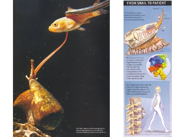
 May act at the site of injury and decrease the pain associated")

 weight drop or contusion; 2)")
- Slides: 29
PAIN: -definition of pain: an unpleasant sensory or emotional experience -perception of pain is a product of brain’s abstraction and elaboration of sensory input. -perception of pain varies with individuals and circumstances (soldier injured) -activation of nociceptors does not necessarily lead to experience of pain (asymbolia for pain; patient under morphine) -pain can be perceived without activation of nociceptors (phantom limb pain, thalamic pain syndrome) -important for survival, protect from damage: congenital and acquired insensitivity (diabetic neuropathy, neurosyphilis) to pain can lead to permanent damage -pain reflexes can be stopped if not appropriate (step on nail near precipice, burn hands while holding a baby. Pain can be suppressed if not needed for survival (soldier…). In general 2 clinical states of pain: Physiological (nociceptive) pain direct stimulation of nociceptors. Neuropathic (intractable) pain result from injury to the peripheral or central nervous system that causes permanent changes in circuit sensitivity and CNS connections.
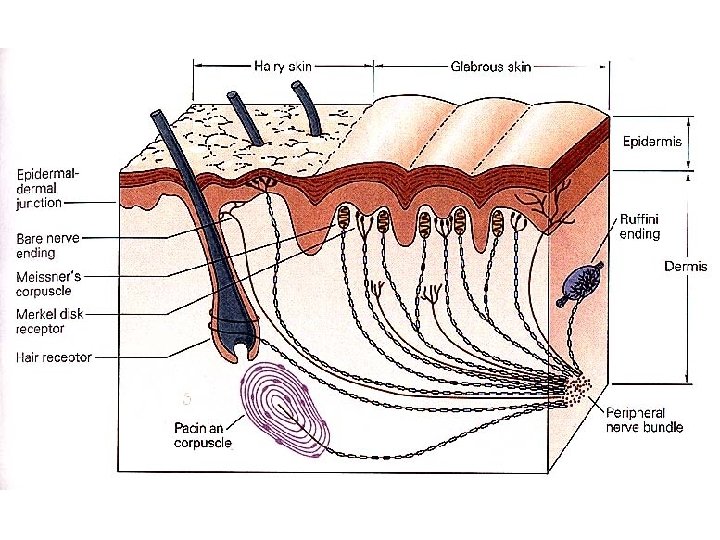
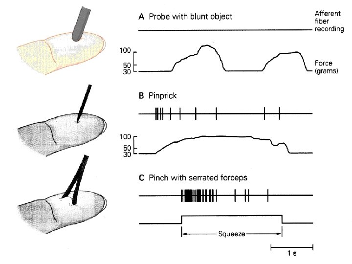
Nociceptors (Free nerve ending) Mechanical nociceptors: activated by strong stimuli such as pinch, and sharp objects that penetrate, squeeze, pinch the skin. sharp or pricking pain, via A-delta fibers. Thermal nociceptors: activated by noxious heat (temp. above 45°C), noxious cold (temp. below 5°C), and strong mechanical stimuli. via A-delta fibers. Polymodal nociceptors: activated by noxious mechanical stimuli, noxious heat, noxious cold, irritant chemicals. slow dull burning pain or aching pain, via nonmyelinated C fibers. Persists long after the stimulus is removed. Research for a transduction protein: capsaicin (from chili peppers) bind to capsaicin receptor on nociceptor endings transducer for noxious thermal and chemical stimuli burning sensation associated with spicy food. Knockout mouse lacking capsaicin receptor drinks solution of capsaicin, has reduced thermal hyperalgesia
Mechanisms associated with peripheral sensitization to pain
Agents that Activate or Sensitize Nociceptors: Cell injury arachidonic acid prostaglandins vasc. permeability (cyclo-oxygenase) sensitizes nociceptor Cell injury arachidonic acid leukotrienes vasc. permeability (lipoxygenase) sensitizes nociceptor Cell injury tissue acidity kallikrein bradykinin vasc. permeability activates nociceptors synthesis & release of prostaglandins Substance P (released by free nerve endings) sensitize nociceptors vasc. perm. , plasma extravasation (neurogenic inflammation) releases histamine (from mast cells) Calcitonin gene related peptide (free nerve endings) dilation of peripheral capillaries Serotonin (released from platelets & damaged endothelial cells) activates nociceptors Cell injury potassium activates nociceptors
Peripheral sensitization to pain: Some definitions: Hyperalgesia increased sensitivity to an already painful stimulus Allodynia normally non painful stimuli are felt as painful (i. e. light touch of a sun-burned skin)
Peripheral sensitization to pain: CGRP
To summarize peripheral sensitization to pain: -Sensitization results from the release of various chemicals by the damaged cells and tissues (bradykinin, prostaglandins, leukotrienes…). These chemicals alter the type and number of membrane receptors on free nerve endings, lowering the threshold for nociceptive stimuli. -The depolarized nociceptive sensory endings release substance P and CGRP along their branches (axon reflex), thus contributing to the spread of edema by producing vasodilation, increase in vascular permeability and plasma transvasation, and the spread of hyperalgesia by leading to the release of histamine from mast cells. -Aspirin and NSAID block the formation of prostaglandins by inhibiting the enzyme cyclooxygenase. -Local anesthetic preferentially blocks C fiber conduction, cold decreases firing of C fibers, ischemia blocks first the large myelinated fibers.
Pain input to the spinal cord: -Projecting neurons in lamina I receive A-delta and C fibers info. -Neurons in lamina II receive input from C fibers and relay it to other laminae. -Projecting neurons in lamina V (wide-dynamic range neurons) receive A-delta, C and A-beta (low threshold mechanoceptors) fibers information. How is pain info sent to the brain: hypotheses pain is signaled by lamina I and V neurons acting together. If lamina I cells are not active, the info about type and location of a stimulus provided by lamina V neurons is interpreted as innocuous. If lamina I cells are active then it is pain. Thus: lamina V cells details about the stimulus, and lamina I cells whether it is painful or not -A-delta and C fibers release glutamate and peptides on dorsal horn neurons. -Substance P (SP) is co-released with glutamate and enhances and prolongs the actions of glutamate. -Glutamate action is confined to nearby neurons but SP can diffuse and affect other populations of neurons because there is no specific reuptake.
Mechanisms of early-onset central sensitization: Windup homosynaptic activity-dependent plasticity characterized by a progressive increase in firing from dorsal horn neurons during a train of repeated low-frequency Cfiber or nociceptor stimulation. During stimulation, glutamate + substance P + CGRP elicit slow synaptic potentials lasting several-hundred milliseconds. Windup results from the summation of these slow synaptic potentials. This produces a cumulative depolarization that leads to removal of the voltage-dependent Mg 2+ channel blockade in NMDA receptors and entry of Ca 2+. Increasing glutamate action progressively increases the firing-response to each individual stimulus (behavioral correlate: repeated mechanical or noxious heat are perceived as more and more painful even if the stimulus intensity does not change.
Centrally mediated hyperalgesia: Under conditions of persistent injury, C fibers fire repetitively and the response of dorsal horn neurons increase progressively (“wind-up” phenomenon). This is due to activation of the N-methyl-D-aspartate (NMDA)-type glutamate receptor and diffusion of substance P that sensitizes adjacent neurons. Blocking NMDA receptors can block the wind-up. Noxious stimulation can produce these long-term changes in dorsal neurons excitability (central sensitization) which constitute a memory of the C fiber input. Can lead to spontaneous pain and decreases in the threshold for the production of pain. Carpal tunnel syndrome: median nerve frequently injured at the flexor retinaculum. Pain ends up affecting the entire arm. (rat model partial ligature of sciatic nerve or nerve wrapped with irritant solution)
Neuropathic (intractable) pain: Pain following peripheral nerve injury. Greater loss of small fibers than large diameter fibers. Axons of surviving Abeta fibers sprout new branches and make connection to neurons vacated by the lost C fibers. Nonpainful stimuli become painful. Change from innocuous to noxious sensation is called allodynia. Thalamic pain syndrome: usually following stroke in the ventral basal thalamus. Rearrangement of local circuit leads to excruciating pain. Phantom limb pain: A-beta Pain Signaling neurons C fibers N
Phantom limb pain: during amputation under general anesthesia the spinal cord can still “experience” the insult produced by the surgical procedure and central sensitization occurs. To try to prevent it, local infiltration of anesthetics in the site of surgery. But studies show also rearrangement of cortical circuits (cortical region of the missing limb receives afferents from other site of the skin) Phantom Pain intensity as a function of Cortical Reorganization.
Gate Control Theory of Pain:
Gate Control Hypothesis: Wall & Melzack 1965 Hypothesized interneurons activated by A-beta fibers act as a gate, controlling primarily the transmission of pain stimuli conveyed by C fibers to higher centers. i. e. rubbing the skin near the site of injury to feel better. i. e. Transcutaneous electrical nerve stimulation (TENS). i. e. dorsal column stim. i. e. Acupuncture
• Rheumatoid and Osteo-arthritis • Back pain • Menstrual Pain • Labour Pain • Peripheral Nerve Injuries • Shingles • Headache and Migraine • Cancer Pain • Trigeminal Neuralgia • Phantom Limb Pain • Sports Injuries • Sciatica • Aching Joints • Post Operative Pain • Muscular Pain • Whiplash and Neck Injury and many others
Referred Pain:
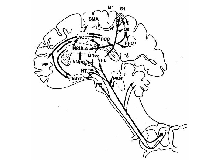
Ascending Pathways: ->localization, intensity, type of pain stimulus ->arousal, emotion; involves limbic system, amygdala, insula, cingulate cortex, hypothalamus. Mediate descending control of pain (feedback loop)
New pathway for visceral pain: selective lesion of fibers in the ventral part of the fasciculus gracilis reduces dramatically the perception of pain from the viscera. General problems with surgery: Rhizotomy (cutting dorsal root) Anterolateral cordotomy (cutting ALS) In both cases, pain come back, excruciating. Thalamus: lesion VPL, VPM thalamic syndrome. Intralaminar nuclei (arousal + limbic) Cortex: S 1 cortex localization, quality and intensity of pain stimuli. Lesion of cingulate gyrus and insular cortex asymbolia for pain
Multiple regression analysis (left panel) of positron emission tomography data revealed statistically reliable relationships between perceived pain intensity and activation of a functionally diverse group of brain regions, including those important in sensation, motor control, affect, and attention. Pain intensity-related activation occurred bilaterally in the cerebellum (CB), putamen, thalamus (Thal), insula, anterior cingulate cortex (ACC), and secondary somatosensory cortex (SII), contralaterally in the primary somatosensory cortex (SI) and supplementary motor area (SMA), and ipsilaterally in the frontal operculum (PMv).
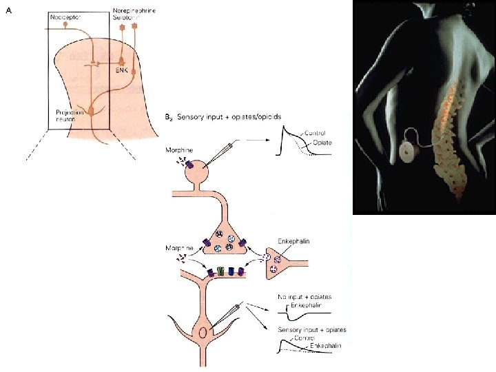
Descending pathways regulating the transmission of pain information: intensity of pain varies among individuals and depends on circumstances (i. e. soldier wounded, athlete injured, during stress). Stimulation of PAG causes analgesia so profound that surgery can be performed. PAG stimulation can ameliorate intractable pain. PAG receives pain information via the spinomesencephalic tract and inputs from cortex and hypothalamus related to behavioral states and to whether to activate the pain control system. PAG acts on raphe & locus ceruleus to inhibit dorsal horn neurons via interneurons and morphine receptors. Application: Intrathecal morphine pumps
Analgesics: 1) May act at the site of injury and decrease the pain associated with an inflammatory reaction (e. g. non-steroidal anti-inflammatory drugs (NSAID) such as: aspirin, ibuprofen, diclofenac). Believed to act through inhibition of cyclo-oxygenase (COX). COX-2 is induced at sites of inflammation. Inhibition of COX-1 causes the unwanted effects of NSAID, i. e. gastrointestinal bleeding and nephrotoxicity. Selective COX-2 inhibitor are now used. 2) May alter nerve conduction (e. g. local anesthetics): block action potentials by blocking Na channels. Used for surface anesthesia, infiltration, spinal or epidural anesthesia. Used in combination to steroid to reduce local swelling (injection near nerve root). Local anesthetic preferentially blocks C fiber conduction, cold decreases firing of C fibers, ischemia blocks first the large myelinated fibers. 3) May modify transmission in the dorsal horn (e. g. opioids: endorphin, enkephalin, dynorphin…). Opioids act on G-protein coupled receptors: Mu, Delta and Kappa. Opioid agonists reduce neuronal excitability (by increasing potassium conductance) and inhibit neurotransmitter release (by decreasing presynaptic calcium influx) 4) May affect the central component and the emotional aspects of pain (e. g. opioids, antidepressant). Problems of tolerance and dependence
Molecular tools in Pain research: “Toxin to kill targeted cells”: to use receptor-mediated endocytosis to selectively deliver cytotoxins to specific types of neurons. The effector toxin of choice is the ribosome inactivating protein (RIP), saporin (SAP). i. e. SAP combined with substance P to kill neurons expressing neurokinin-1 (NK-1) receptor. “Antisense oligonucleotide (ASO)-mediated knockdown”: an ASO, typically 15 -25 nucleotides in length, is designed to bind a complementary sequence on the target RNA. As a consequence, the protein product coded by that particular RNA is not synthesized. i. e. knock down of PSD-93/chapsin-110 “Knockout/transgenic mice”: create mice that either overexpress or do not express presumably pain-related proteins. i. e. mice lacking the capsaicin receptor; mice lacking PKCgamma; mice lacking neurokinin-1 (NK-1) receptor…(others NGF, Trk. A, p 75, interleukin-6, interferon-gamma, prostaglandin receptors, bradykinin receptor, substance P, PPT-A, neurokinin 1, adenosine-2 a, B-endorphin, enkephalin, u-opioid receptors, delta-opioid receptors, kappaopioid receptors, orphanin. FQ/nociceptin receptor, adrenergic receptors, serotonin receptors, PKA-RIB, PKC-gamma, nitrix oxyde, Go, NR 1 -NMDA… “Fusion molecule”: i. e. use recombinant techniques to couple the extracellular domain of trk. A receptor to the Fc portion of human immunoblobin G, to produce a fusion protein that binds and neutralize the effects of NGF.
Animal models of chronic pain: Central pain models: 1) weight drop or contusion; 2) photochemical SCI; 3) excitotoxic SCI Peripheral nerve injury models: 1) nerve transection; 2) chronic constriction injury (Bennett); 3) partial sciatic nerve ligation (Seltzer); 4) L 5/L 6 spinal nerve ligation; 5) L 5 spinal nerve ligation; 6) sciatic cryoneurolysis; 7) inferior caudal trunk resection; 8) sciatic inflammatory neuritis. Peripheral neuropathy induced by diseases: 1) postherpetic neuralgia; 2) diabetic neuropathic Cancer pain models: 1) chemotherapy-induced peripheral neuropathy; 2) vincristine-induced peripheral neuropathy; 3) taxol-induced peripheral neuropathy; 4) cisplatin-induced peripheral neuropathy; 5) cancer invasion; 6) bone cancer; 7) mouse femur bone cancer; 8) mouse calcaneus bone cancer; 9) rat tibia bone cancer.