OUTCOMES AND MORPHOLOGICAL PATTERNS OF ACUTE INFLAMMATION OUTLINE


. This")

- Slides: 25
OUTCOMES AND MORPHOLOGICAL PATTERNS OF ACUTE INFLAMMATION
OUTLINE • Inflammation • Acute inflammation • Recognition of microbes and damaged cells • Reactions of blood vessels in acute inflammation • Leukocyte recruitment to sites of inflammation • Morphological patterns • Outcomes
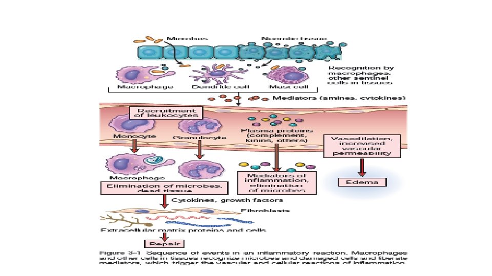
Inflammation • Inflammation is a response of vascularized tissues to infections and damaged tissues that brings cells and molecules of host defense from the circulation to the sites where they are needed, in order to eliminate the offending agents. • It serves to rid the host of both the initial cause of cell injury (e. g. , microbes, toxins) and the consequences of such injury (e. g. , necrotic cells and tissues). The mediators of defense include phagocytic leukocytes, antibodies and complement proteins • In addition to inflammatory cells, components of innate immunity include other cells, such as natural killer cells, dendritic cells, and epithelial cells, as well as soluble factors such as the proteins of the complement system • The major participants in the inflammatory reaction in tissues are blood vessels and leukocytes • Causes of inflammation include; infections, tissue necrosis, foreign bodies, immune reactions, etc
Acute Inflammation • The initial, rapid response to infections and tissue damage is called acute inflammation • It typically develops within minutes or hours and is of short duration, lasting for several hours or a few days • Its main characteristics are the exudation of fluid and plasma proteins (edema) and the emigration of leukocytes, predominantly neutrophils (also called polymorphonuclear leukocytes). • It has 3 components; • Dilation of small blood vessels • Increased permeability of microvascuature • Emigration of leukocytes from microvasculature, accumulation in injury focus.
Recognition of Microbes and Damaged cells • Recognition of offending agents is the first step in all inflammatory reactions. • The cells and receptors that perform this function of recognizing invaders are very adapted and the responses they trigger are critical for the survival of the organisms. • Several cellular receptors and circulating proteins are capable of recognizing microbes and products of cell damage and triggering inflammation. • Cellular receptors for microbes • Sensors of cell damage • Other cellular receptors involved in inflammation
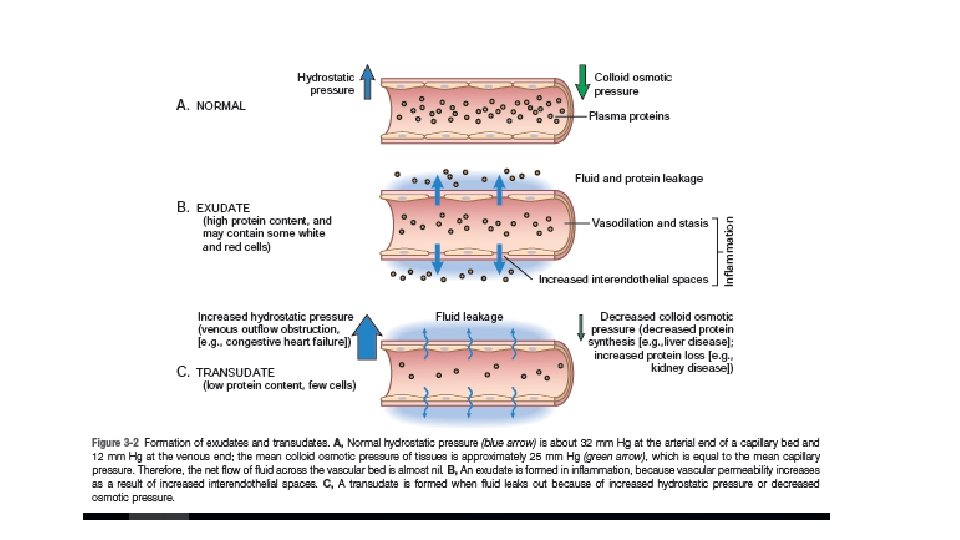
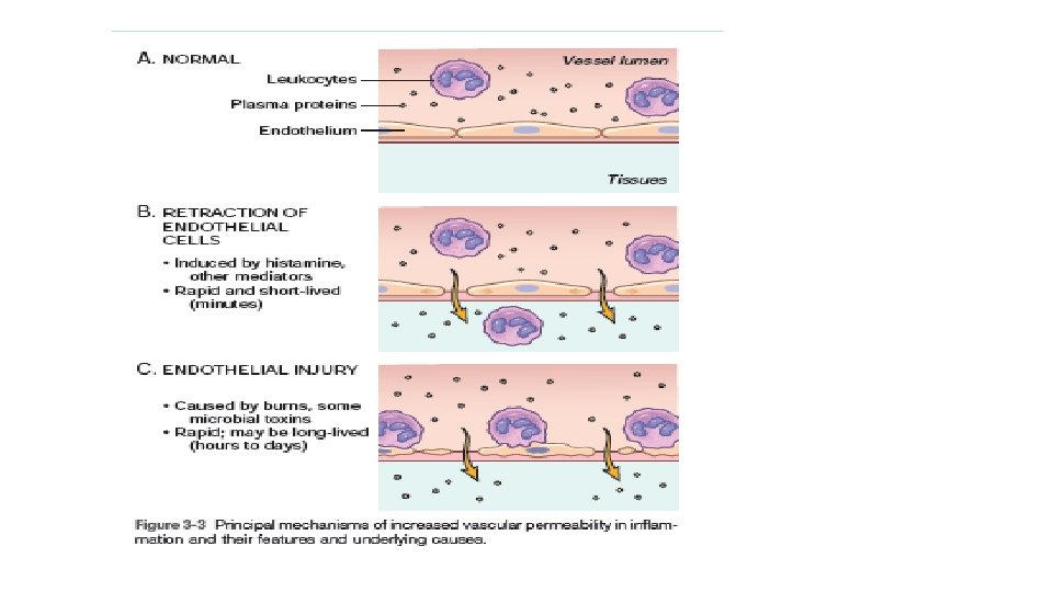
Reactions of Blood Vessels in Acute Inflammation • The vascular reactions of acute inflammation consist of changes in the flow of blood and the permeability of vessels, both designed to maximize the movement of plasma proteins and leukocytes out of the circulation and into the site of infection or injury. • This includes : • Changes in vascular flow and caliber begin early after injury and consist of Vasodilation which is induced by the action of several mediators, notably histamine, on vascular smooth muscle, stasis and accumulation of blood leukocytes along the vascular endothelium • Increased Vascular Permeability (Vascular Leakage) Several mechanisms are responsible for the increased permeability of postcapillary venules, a hallmark of acute inflammation • Responses of Lymphatic Vessels and Lymph Nodes In addition to blood vessels, lymphatic vessels also participate in acute inflammation
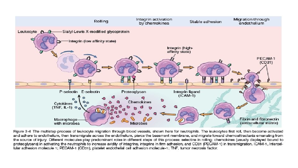
Leukocyte recruitment to sites of Inflammation • The changes in blood flow and vascular permeability are quickly followed by an influx of leukocytes into the tissue. • The most important leukocytes in typical inflammatory reactions are the ones capable of phagocytosis, namely neutrophils and macrophages. They also produce growth factors that aid in repair. • A price that is paid for the defensive potency of leukocytes is that, when strongly activated, they may induce tissue damage and prolong inflammation, because the leukocyte products that destroy microbes and help “clean up” necrotic tissues can also injure normal bystander host tissues.
• The journey of leukocytes from the vessel lumen to the tissue is a multistep process that is mediated and controlled by adhesion molecules and cytokines called chemokines. It is divided into phases • In the lumen: margination, rolling, and adhesion to endothelium. • Migration across the endothelium and vessel wall • Migration in the tissues toward a chemotactic stimulus
Morphological patterns of Acute Inflammation • The morphologic hallmarks of acute inflammatory reactions are dilation of small blood vessels and accumulation of leukocytes and fluid in the extravascular tissue. • However, special morphologic patterns are often superimposed on these general features, depending on the severity of the reaction, its specific cause, and the particular tissue and site involved. • They include : • • Serous inflammation Fibrinous inflammation Purulent inflammation Ulcers
Serous Inflammation • Serous inflammation is marked by the exudation of cellpoor fluid into spaces created by cell injury or into body cavities lined by the peritoneum, pleura, or pericardium. • Typically, the fluid in serous inflammation is not infected by destructive organisms and does not contain large numbers of leukocytes (which tend to produce purulent inflammation, described later). • In body cavities the fluid may be derived from the plasma (as a result of increased vascular permeability) or from the secretions of mesothelial cells (as a result of local irritation); accumulation of fluid in these cavities is called an effusion. (Effusions also occur in noninflammatory conditions, such as reduced blood outflow in heart failure, or reduced plasma protein levels in some kidney and liver diseases. ) • The skin blister resulting from a burn or viral infection represents accumulation of serous fluid within or immediately beneath the damaged epidermis of the skin
SEROUS INFLAMMATION; Skin blister showing the epidermis separated from the dermis by a focal collection of serous effusion.
Fibrinous Inflammation • With greater increase in vascular permeability, large molecules such as fibrinogen pass out of the blood, and fibrin is formed and deposited in the extracellular space. • A fibrinous exudate develops when the vascular leaks are large or there is a local procoagulant stimulus (e. g. , cancer cells). A fibrinous exudate is characteristic of inflammation in the lining of body cavities, such as the meninges, pericardium, and pleura. Histologically, fibrin appears as an eosinophilic meshwork of threads or sometimes as an amorphous coagulum. • Fibrinous exudates may be dissolved by fibrinolysis and cleared by macrophages. If the fibrin is not removed, over time it may stimulate the ingrowth of fibroblasts and blood vessels and thus lead to scarring. • Conversion of the fibrinous exudate to scar tissue (organization) within the pericardial sac leads to opaque fibrous thickening of the pericardium and epicardium in the area of exudation and, if the fibrosis is extensive, obliteration of the pericardial space
FIBROUS PERICARDITIS; Deposits of fibrin on the pericardium. A pink meshwork of fibrin exudate (F) overlies the pericardial surface (P)
Purulent Inflammation • Purulent inflammation is characterized by the production of pus, an exudate consisting of neutrophils, the liquefied debris of necrotic cells, and edema fluid. • The most frequent cause of purulent (also called suppurative) inflammation is infection with bacteria that cause liquefactive tissue necrosis, such as staphylococci; these pathogens are referred to as pyogenic (pus producing) bacteria. A common example of an acute suppurative inflammation is acute appendicitis. • Abscesses are localized collections of purulent inflammatory tissue caused by suppuration buried in a tissue, an organ, or a confined space. They are produced by seeding of pyogenic bacteria into a tissue. • Abscesses have a central region that appears as a mass of necrotic leukocytes and tissue cells. There is usually a zone of preserved neutrophils around this necrotic focus, and outside this region there may be vascular dilation and parenchymal and fibroblastic proliferation, indicating chronic inflammation and repair. In time the abscess may become walled off and ultimately replaced by connective tissue.
PURULENT INFLAMMATION; Multiple bacterial abscesses in the lung in a case of bronchopneumonia The abscess contains neutrophils and cellular debris and is surrounded by congested blood vessels
Ulcers • An ulcer is a local defect, or excavation, of the surface of an organ or tissue that is produced by the sloughing (shedding) of inflamed necrotic tissue. • Ulceration can occur only when tissue necrosis and resultant inflammation exist on or near a surface. It is most commonly encountered in (1) the mucosa of the mouth, stomach, intestines, or genitourinary tract, and (2) the skin and subcutaneous tissue of the lower extremities in older persons who have circulatory disturbances that predispose to extensive ischemic necrosis. • Ulcerations are best exemplified by peptic ulcer of the stomach or duodenum, in which acute and chronic inflammation coexist. During the acute stage there is intense polymorphonuclear infiltration and vascular dilation in the margins of the defect. • With chronicity, the margins and base of the ulcer develop fibroblastic proliferation, scarring, and the accumulation of lymphocytes, macrophages, and plasma cells.
Morphology of an ulcer; Chronic duodenal ulcer Cross section view of a duodenal ulcer crater with an acute inflammatory exudate in the base
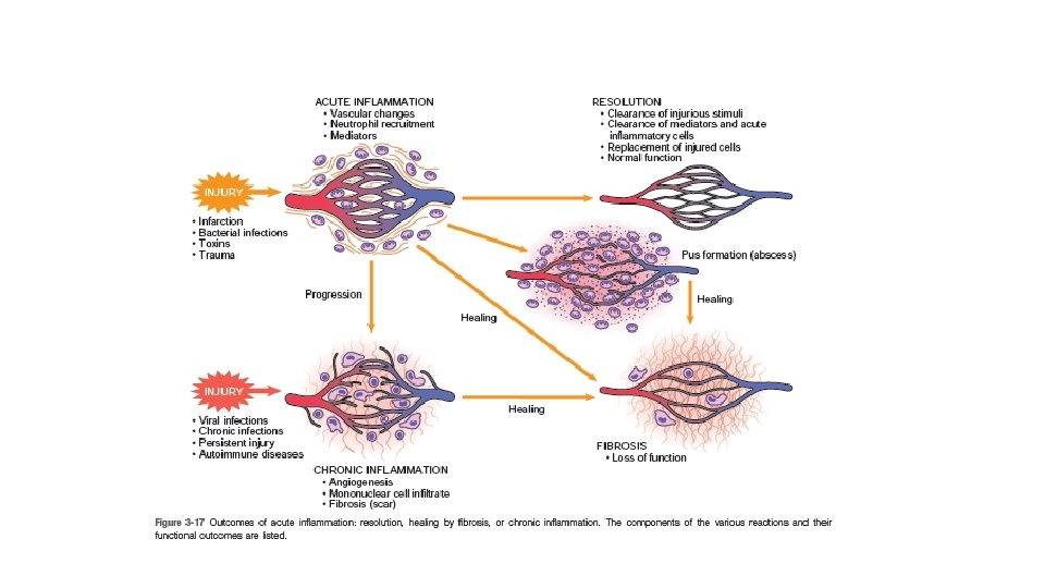
OUTCOMES OF ACUTE INFLAMMATION • Although, as might be expected, many variables may modify the basic process of inflammation, including the nature and intensity of the injury, the site and tissue affected, and the responsiveness of the host, all acute inflammatory reactions typically have one of three outcomes: • Complete resolution. In a perfect world, all inflammatory reactions, once they have succeeded in eliminating the offending agent, should end with restoration of the site of acute inflammation to normal. This is called resolution and is the usual outcome when the injury is limited or short lived or when there has been little tissue destruction and the damaged parenchymal cells can regenerate. Resolution involves removal of cellular debris and microbes by macrophages, and resorption of edema fluid by lymphatics
OUTCOMES OF ACUTE INFLAMMATION • Healing by connective tissue replacement (scarring, or fibrosis). This occurs after substantial tissue destruction, when the inflammatory injury involves tissues that are incapable of regeneration, or when there is abundant fibrin exudation in tissue or in serous cavities (pleura, peritoneum) that cannot be adequately cleared. In all these situations, connective tissue grows into the area of damage or exudate, converting it into a mass of tissue, a process also called Organization. • Progression of the response to chronic inflammation (discussed later). Acute to chronic transition occurs when the acute inflammatory response cannot be resolved, as a result of either the persistence of the injurious agent or some interference with the normal process of healing.
CONCLUSION • Microscopic patterns are important to recognize because they often provide valuable clues about the underlying cause. • The outcomes of acute inflammation includes; Resolution, Healing by connective tissue replacement or Progression to chronic inflammation.