NERVE SUPPLY OF THE FACE 5 TH 7


. Ø Fibers: 1. General somatic afferent:")
 & 4 other muscles 3 sensory + 1 Motor")
. Ø General somatic afferent: 1. 2.")


 Ø 1. 2. 3. Divides into 3 branches: Frontal, (in the")
 Ø Supplies: 1. Upper teeth, gums & maxillary air sinus: (posterior")
 Ø SENSORY BRANCHES: 1. Lingual: receives General sensations from anterior 2/3 the")


: receives taste")
!!! Ø by")


- Slides: 26
NERVE SUPPLY OF THE FACE 5 TH & 7 TH CRANIAL NERVES Prof. Saeed Abuel Makarem
OBJECTIVES By the end of the lecture, you should be able to: Ø List the nuclei of deep origin of the trigeminal and facial nerves in the brain stem. Ø Describe the site and type of each nucleus. Ø Describe the superficial attachment of these 2 nerves to the brain stem. Ø Describe the main points in the course and distribution of these 2 nerves to the face. Ø Describe the main motor & sensory manifestation in case of lesion of these 2 nerves.
TRIGEMINAL NERVE Ø Type: Mixed: (sensory & motor). Ø Fibers: 1. General somatic afferent: Carrying general sensations from the face and anterior part of the scalp. 2. Special visceral efferent: Supplying muscles developed from the 1 st pharyngeal arch, (8 muscles).
TRIGEMINAL NERVE NUCLEI (Deep origin) & 4 other muscles 3 sensory + 1 Motor
Ø Four nuclei: (3 sensory + 1 Motor). Ø General somatic afferent: 1. 2. 3. Principal (main) sensory nucleus, (pons): receives touch fibers from face and anterior part of the scalp. Mesencephalic nucleus (Pons & midbrain): receives proprioceptive fibers from muscles of mastication. Spinal nucleus, (Pons, medulla & upper 2 or 3 cervical segments of the spinal cord): receives pain & temperature sensations from face & scalp. Ø Special visceral efferent: 4. 1. 2. Motor nucleus (Pons), supplies: 8 Ms. : Four Muscles of mastication: (temporalis, masseter, medial pterygoid & lateral pterygoid). Other 4 muscles: (Anterior belly of digastric, mylohyoid, tensor palati & tensor tympani). TRIGEMINAL NERVE NUCLEI
TRIGEMINAL GANGLION Ø Site: Ø Occupies a depression in the middle cranial fossa called trigeminal impression, (apex of petrous temporal bone). Ø Importance: Contains cell bodies: 1. Whose dendrites carry sensations from the face & scalp. 2. Whose axons form the sensory root of trigeminal nerve.
TRIGEMINAL NERVE Ø Emerges from the middle of the ventrolateral surface of the pons by 2 roots (Large Lateral sensory root & small medial motor root). Ø Divides into 3 divisions (dendrites of trigeminal ganglion): 1. Ophthalmic. 2. Maxillary. 3. Mandibular. Ø Axons of cells of motor nucleus join only the mandibular division.
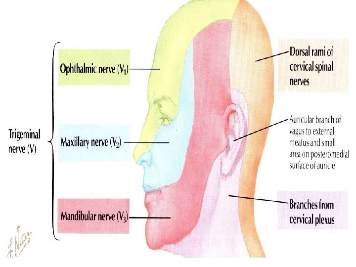
OPHTHALMIC (PURE SENSORY) Ø 1. 2. 3. Divides into 3 branches: Frontal, (in the middle). Lacrimal, (most lateral). Nasociliary, (most medial). All pass through superior orbital fissure to reach the orbit. Frontal: supplies skin of face & scalp. Lacrimal: supplies skin of face & lacrimal gland (sensory)!!!!!!. Nasociliary: supplies skin of face, nasal cavity & eyeball. 1 2 3
MAXILLARY (PURE SENSORY) Ø Supplies: 1. Upper teeth, gums & maxillary air sinus: (posterior superior middle superior, anterior superior alveolar nerves). 2. Face: (zygomaticofacial & infraorbital nerves).
MANDIBULAR (MIXED) Ø SENSORY BRANCHES: 1. Lingual: receives General sensations from anterior 2/3 the of tongue. 2. Inferior alveolar: supplies Lower teeth, gums & face. 3. Buccal: supplies the skin and mucous membrane of a small area of the cheek of upper jaw. 4. Auriculotemporal: supplies auricle, temple, parotid gland & Temporomandibular joint (TMJ). Ø MOTOR BRANCHES: to 8 muscles (4 muscles of mastication & other 4 muscles).
Trigeminal Neuralgia • Compression, degeneration or inflammation of the 5 th cranial nerve may result in a condition called trigeminal neuralgia or tic douloureux (spasmodic contraction of the muscles in the face) • This condition is characterized by recurring episodes of intense severe stabbing excruciating pain radiating from the angle of the jaw along a branches of the trigeminal nerve. • Usually involves maxillary & mandibular branches, rarely in the ophthalmic division.
• Type: Mixed: 1. Motor, 2. Special sensory, 3. Parasympathetic. Ø Fibers: 1. Special visceral afferent: carrying taste sensation from the anterior 2/3 of the tongue. 2. Special visceral efferent: To muscles developed from the 2 nd pharyngeal arch. 3. General visceral efferent: supplying parasympathetic secretomotor fibers to the: 1. Submandibular, 2. Sublingual, 3. Lacrimal, 4. Nasal & 5. Palatine glands. FACIAL NERVE Parasymp. taste sensation motor
FACIAL NERVE NUCLEI Ø 3 Nuclei: 1. Special visceral afferent: (nucleus solitarius): receives taste from the anterior 2/3 of the tongue. 2. Special visceral efferent: (motor nucleus of facial nerve): supplies muscles of 2 nd pharyngeal arch: muscles of face, posterior belly of digastric, stylohyoid, platysma, stapedius, and occipitofrontalis. 3. General visceral efferent: superior salivatory nucleus: sends preganglionic parasympathetic secretory fibers to: Ø Pterygopalatine ganglion and Ø Submandibular ganglion. Then the Postganglionic fibers pass to the lacrimal, nasal, palatine sublingual, submandibular glands. 2 1 3
COURSE OF FACIAL NERVE Ø Emerges from the cerebellopontine angle, (very important)!!! Ø by 2 roots: 1. Medial motor root: contains motor fibers. 2. Lateral root (nervous intermedius): contains parasympathetic & taste fibers.
COURSE OF FACIAL NERVE Ø Ø Passes through internal acoustic meatus to the inner ear where it runs in the facial canal. Emerges from the stylomastoid foramen & enters the parotid gland where it ends.
Ø In facial canal: 1. Greater petrosal nerve: carries preganglionic parasympathetic fibers to pterygopalatine ganglion then to the lacrimal, nasal & palatine glands. 2. Chorda tympani: carries: a) preganglionic parasympathetic fibers to submandibular ganglion then to submandibular & sublingual salivary glands. b) taste fibers from anterior 2/3 of the tongue. 3. Nerve to stapedius. control the amplitude of sound waves from external environment to inner ear. N. B: Geniculate ganglion: Lies in internal acoustic meatus. contains cell bodies of neurones ; its Fibers carrying taste sensations from anterior 2/3 of tongue; ending in the solitary nucleus in medulla oblongata. BRANCHES 3 1 2
BRANCHES OF FACIAL NERVE Ø Just as it emerges from the stylomastoid foramen it gives 2 branches: 1. Posterior auricular: to occipitofrontalis muscle. 2. Muscular branches to posterior belly of digastric & stylohyoid. Ø Inside parotid gland: it gives 5 terminal motor branches: Ø Temporal, Ø Zygomatic, Ø Buccal, Ø Mandibular & Ø Cervical…. To the muscles of the face.
Bell’s Palsy • Damage of the facial nerve results in paralysis of muscles of facial expressions: Facial (Bell’s) palsy; lower motor neuron lesion (whole face affected) • NB. In upper motor neuron lesion (upper face is intact). The face is distorted: Drooping of lower eyelid, Sagging of mouth angle, Dribbling of saliva, Loss of facial expressions, Loss of chewing, !!!!!! Loss of blowing, Loss of suckling, Unable to show teeth or close the eye on that side. § Hyperacusis. § § § § §
THANK YOU & BEST LUCK
SUMMARY Ø Both trigeminal & facial nerves are mixed. Ø Nuclei of trigeminal nerve are found in midbrain, pons & medulla. They are of the general somatic afferent & special visceral efferent types. Ø The trigeminal nerve emerges from the pons and divides into: ophthalmic, maxillary & mandibular divisions that receive sensory supply from the face (with an exception of a small area over ramus of mandible by great auricular nerve C 2, 3). Ø All motor fibers are only included in the mandibular division of the trigeminal nerve supply muscles of the 1 st pharyngeal arch.
SUMMARY Ø Nuclei of facial nerve are found in pons. They are of the special visceral afferent & efferent types, as well as general visceral efferent type. Ø The facial nerve emerges from the cerebellopontine angle, gives motor fibers to muscles of 2 nd pharyngeal arch, and secretory fibers to submandibular, sublingual, lacrimal, nasal & palatine glands & receives taste fibers from anterior 2/3 of tongue.
Lower Motor Neuron Lesion §Results from injury of the facial nerve fibers: § In Internal acoustic meatus; § In the middle ear; § In the facial canal or § In parotid gland. §Manifested by complete paralysis of facial muscles on the same side of lesion, (Whole face affection. Upper Motor Neuron Lesion §This occurs after injury to the pyramidal tract (corticonuclear) above facial nucleus. §Leads to paralysis of facial muscles of the lower ½ of face in the opposite side but the upper ½ of the face intact because: §Ms. of lower ½ of face receive pyramidal fibers from opposite cerebral cortex only, §While Ms. of upper ½ of face receive pyramidal fibers from both cerebral hemispheres (Bilateral representation).
TEST YOUR SELF ! Ø Stimulation of which of the following nerves could lead to salivation and lacrimation? a) Glossopharyngeal. b) Trigeminal. c) Facial. d) Vagus. Ø Lesion of the mandibular nerve may result in: a) Loss of sensation of skin over the nose. b) Loss of lacrimation. c) Loss of sensory supply of upper teeth. d) Loss of general sensations of anterior 2/3 of tongue.
TEST YOUR SELF ! • In bell palsy Hyperacusis is due to paralysis of which one of the following muscles? • Tensor tympani. • Tensor palati • Stapedius. • Auricularis superior.
THANK YOU AND GOOD LUCK