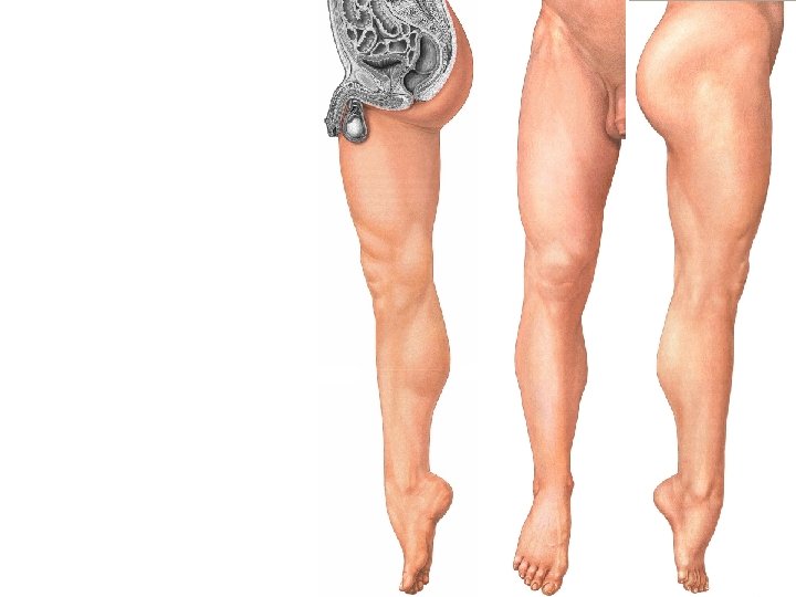
Origin ASIS Nerve Supply Femoral nerve Action Tailors
 Insertion: Upper")


 Nerve Supply: Obturator N. Action")
")


 Hamstring part: lower")


 Insertion Back of tibia above")

 Distal phalanges")
 Distal phalanx")
- Slides: 37
Origin: ASIS Nerve Supply Femoral nerve Action Tailor’s position (Cross leg position) Insertion: Upper part of the medial surface of tibia (SGST)
• • Rectus Femoris Vastus medialis Vastus Lateralis Vastus intermedius All are supplied by the Femoral N.
Origin: Nerve Supply: Femoral N. Straight H. : Anterior inferior iliac spine Reflected H. : groove above acetabulum Action Flexion of the hip & extension of the knee Insertion: Quadriceps tendon into upper, lat & medial borders of the patella.
Origin: Medial lip of the Linea aspra Origin: Lateral lip of the Linea aspra Action Insertion: Quadriceps tendon into upper, lat & medial borders of the patella Extension of the knee Insertion: Quadriceps tendon into upper, lat & medial borders of the patella
Origin: Upper ⅔ of the anterior & lateral surface of the femur Action Extension of knee Insertion: Quadriceps tendon into upper, lat & medial borders of the patella.
• • • Gracilis Adductor Longus Adductor Brevis Pectineus Adductor Magnus
Origin: Conjoint ramus (inferior pubic ramus & ischial ramus) Nerve Supply: Obturator N. Action Adduction Insertion: Upper part of the medial surface of the tibia (behind sartorius)
Origin: Front of the body of the pubis just below pubic tubercle (rounded tendon) Nerve Supply: Obturator N. Action Insertion: Linea aspra Anterior Adduction
Origin: Body & inferior ramus of the pubis below adductor longus Nerve Supply: Obturator N. Action Insertion: Linea aspra Adduction
Origin: Pectineal surface of pubis Nerve Supply: Femoral N. Insertion: Below lesser trochanter Action Adduction
Origin: Adductor part: conjoint ramus (inferior pubic ramus & ischial ramus) Hamstring part: lower lateral part of the ischial tuberosity Nerve Supply: Obturator N. & Sciatic N. Insertion: Linea aspra. Adductor tubercle Action Anterior Adduction Extends the hip joint Posterior
• Semitendinosus • Semimembranosus • Biceps Femoris
Origin: Lower medial part of the upper area of the ischial tuberosity (with long head of the biceps) Nerve Supply: Sciatic N. Action Extension of the hip Flexion of the knee Insertion: Surface of the tibia (behind gracilis)
Origin: Upper lateral part of the upper area of ischial tuberosity Nerve Supply: Sciatic N. Action Insertion: SGST tibia Extension of the hip & flexion of the knee
Origin: Long head: with semitendinosus Short head: linea aspra Nerve Supply: Sciatic N. Action Extension of the hip Flexion of the knee Locking of the knee Insertion: Head of fibula
• • Gluteus Maximus Gluteus Medius Gluteus Minimus Piriformis Obturator Internus 4 Sup & Inf gemelli Obturator Externus
Origin Hip bone behind the posterior gluteal line & back of the sacrum Insertion Gluteal tuberosity & iliotibial tract Nerve Supply Inferior gluteal N. Action Extension of the hip joint
Origin Insertion Nerve Supply Action Hip bone between the posterior gluteal lines anterior & Back of the greater trochanter Superior gluteal N. Abduction, medial rotaion of the hip & standing on one limb
Origin Insertion Hip bone between superior gluteal nerve & inferior gluteal lines Front of the greater trochanter Nerve Supply Superior gluteal N. Action Same as gluteus medius
Nerve Supply Action Origin Insertion S 1, 2 : Lateral rotation of the thigh Anterior of the middle 3 pieces of sacrum Upper border of the greater trochanter
Origin Pelvic wall & obturator membrane Insertion Medial surface of the greater trochanter Nerve Supply Nerve to obturator internus Action Lateral rotation of the thigh
Origin Upper & lower margins of the lesser sciatic notch Insertion With obturator internus Nerve Supply Action Superiorly: N. to obturator internus Inferiorly: N. to quadratus femoris Lateral rotation of the thigh
Posterior Origin Outer surface of the obturator foramen & membrane Insertion Trochanteric fossa of the femur Nerve Supply Obturator N. Action Lateral rotation of the thigh Anterior
Nerve Supply Action Origin Insertion Ant tibial N. Dorsiflexion & inversion of foot Upper 2/3 of lat surface of tibia Medial cuneiform
Origin Insertion Nerve Supply Action Upper ¾ of ant surf of fibula Extensor exp into middle & distal phalanges of lat 4 toes Ant tibial N. Dorsiflexion of foot & extension of lat 4 toes
Origin Insertion Nerve Supply Action Middle 2/4 of ant surface of fibula Base of distal phalanx of big toe Ant tibial N. Dorsiflexion of foot & extension of big toe
Origin Insertion Nerve Supply Action Lower ¼ of ant surface of fibula 5 th metatarsal bone Ant tibial N. Dorsiflexin & eversion of foot
Origin Insertion Nerve Supply Action Upper 2/3 of lat surface of fibula Med cuneiform & base of 1 st metatarsal Musculo-cutaneous N. Eversion & supports the arches of foot
Origin Insertion Nerve Supply Action Lower 2/3 of lat surface of fibula Base of 5 th metatarsal bone Musculo-cutaneous N. Eversion of foot
Origin Insertion Nerve Supply Action Med head: popliteal surface of femur above med condyle Lat head: lat epicondyle of femur Tendoachilis attached to mid back of calcaneous Med popliteal N. Flexion of knee & plantar flexion of ankle
Origin Insertion Nerve Supply Action Soleal line of tibia & upper 1/3 of back of fibula tendoachilis Med popliteal & tibial Ns Plantar flexion of ankle
Origin Insertion Nerve Supply Action Popliteal surface of femur above the lat condyle Post surface of calcaneous Med popliteal N. Flexion of knee & plantar flexion of foot
Origin Popliteal groove of femur (intracapsular inside knee joint) Insertion Back of tibia above soleal line Nerve Supply Action Med popliteal N Flexion & med rotation which accompanies flexion (unlocking of knee)
Origin Insertion Nerve Supply Action Back of tibia & fibula & interosseous membrane Tuberosity of navicular B. & all tarsal bones except talus Post tibial N Plantar flexion & inversion & keeps arches of foot
Origin Insertion Nerve Supply Action Back of tibia (med to vertical line) Distal phalanges of lat 4 toes (plantar surface) Post tibial N. Plantar flexion of foot & flexion of lat 4 fingers
Origin Insertion Nerve Supply Action Back of fibula (lat to the crest) Distal phalanx of big toe (plantar surface) Post tibial N. Plantar flexion of foot & flexion of big toe