MUSCLES OF MASTICATION 1272020 1 The muscles of


- Slides: 48

MUSCLES OF MASTICATION 12/7/2020 1

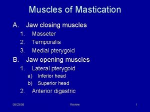
The muscles of mastication move the mandible during mastication and speech They are: 1. Masseter 2. Temporalis 3. The lateral pterygoid and 4. Medial pterygoid 12/7/2020 2

1. MASSETER: It is a quadrilateral muscle and situated between the face and parotid region and consists of three layers of fibers: a. Superficial b. Middle and c. Deep fibers 12/7/2020 3

a. Origin of Superficial fibers: It takes origin from the anterior two third of the lower border of the zygomatic arch and the adjoining zygomatic process of the maxilla Superficial fibers 12/7/2020 4

a. Insertion of Superficial fibers: Inserted into the lower and posterior part of the outer ramus of mandible Superficial fibers 12/7/2020 5

b. Origin and Insertion of Middle fibers: It arises from the deep surface of the anterior two thirds and lower border of the posterior one-third of the zygomatic arch and inserted into the middle of the mandibular ramus Middle fibers 12/7/2020 6

c. Origin and Insertion of deep fibers: It arises from the deep surface of the zygomatic arch and is inserted into the upper and anterior part of the lateral surface of the mandible Deep fibers 12/7/2020 7

Nerve supply: The muscle is supplied from the deep surface by the masseteric branch of the anterior division of the mandibular nerve 12/7/2020 8

Actions: 1. It is a strong elevator of the mandible 2. Superficial fibers help in protraction and deep fibers in the retraction of the mandible 12/7/2020 9

2. TEMPORALIS: It is a fan shaped muscle Origin: It arises from the floor of the temporal fossa below the inferior temporal line and from the overlying temporal fascia 12/7/2020 10

Insertion: It is inserted into the coronoid process of the mandible 12/7/2020 11

Nerve supply: It is supplied by deep temporal branches of the mandibular nerve 12/7/2020 12

Actions: 1. It elevates the mandible 2. It posterior fibers help retraction of the mandible 3. Temporalis muscles of both sides are involved in side-to-side movements 12/7/2020 13

Temporal Fascia: It is a strong sheet of fascia which covers the temporalis muscle above the zygomatic arch Temporal fascia 12/7/2020 14

3. Lateral pterygoid : It arises by two heads: i. Upper head: Arises from the infratemporalsuface of the greater wing of the sphenoid ii. Lower head: Arises from the lateral surface of the lateral pterygoid plate 12/7/2020 15

Upper head Lower head 12/7/2020 16

Insertion: Both heads converge to form a tendon which inserted mainly to a depression in front of the neck of the mandible 12/7/2020 17

Nerve supply: It is supplied by a branch from the anterior division of the mandibular nerve 12/7/2020 18

Actions: 1. It helps in opening of the mouth (Depression of the mandible) 2. Combined actions of the medial and lateral pterygoid muscles of both sides protrude the mandible 3. When the lateral and medial pterygoid muscles of one side act alternately with the other side produce side-to-side movements 12/7/2020 19

4. Medial pterygoid : It is a quadrilateral muscle and consists of : i. Deep head Arises from medial surface of the lateral pterygoid plate ii. Superficial head: From the tuberosity of the maxilla and the pyramidal process of platine bone 12/7/2020 20

Insertion: Into the medial surface of the ramus of the mandible 12/7/2020 21

Nerve supply: It is supplied by a branch derived from the trunk of mandibular nerve 12/7/2020 22

Actions: 1. It assists in elevation of the mandible 2. In combination with the lateral pterygoid – bilateral and simultaneous contraction help protrusion, and alternate contraction produce side -to-side chewing movements of the mandible 12/7/2020 23

All the three muscles: Masseter, temporalis and medial pterygoid elevate the mandible to close the mouth 12/7/2020 24

TEMPORO-MANDIBULAR JOINT 12/7/2020 25

Temporomandibular joint: This is a synovial joint of the condylar variety. The joints of both sides move as one unit and form a bicondylar articulation 12/7/2020 26

Bones forming the joint: i. Above: Articular tubercle and anterior articular part of the mandibular fossa of the temporal bone 12/7/2020 27

ii. Below: Head/condyle of the mandible 12/7/2020 28

Articular surfaces of the both bones are covered with fibro-cartilage. The joint cavity is divided completely by an articular disc into an upper menisco-temporal compartment and a lower menisco-mandibular compartment Meniscotemporal compartment Meniscomandibular compartment 12/7/2020 29

Ligaments: The joint presents the following ligaments 1. Capsular ligament with synovial membrane 2. Articular disc 3. Lateral or temporomandibular ligament 4. Accessory ligaments –sphenomandibular and stylomandibular ligaments 12/7/2020 30

1. Capsular ligament & synovial membrane: It envelops the joint and presents the attachments: i. Above: In front: Articular tubercle Behind: Squamo-tympanic fissure; and periphery of the articular fossa between them 12/7/2020 31

ii. Below: Attached around the neck of the mandible 12/7/2020 32

The synovial membrane lines the inner aspect of the capsule of each compartment of the joint, but fails to cover the articular cartilages and the articular disc 12/7/2020 33

2. Articular disc: It is an oval plate of fibro-cartilage which caps the head of the mandible and divides the joint completely into two compartments 12/7/2020 34

3. Lateral or temporo-mandibular ligament: It blends with the lateral part of fibrous capsule, and extends from the tubercle of the root of the zygoma to the lateral surface and posterior margin of the neck of the mandible Lateral ligament 12/7/2020 35

Lateral ligament 12/7/2020 36

4. Accessory ligaments: a. Sphenomandibular ligament: It is derived from the fibrous envelope of the Meckle’s cartilage of the first branchial arch Sphenomandibular ligament 12/7/2020 37

b. Stylomandibular ligament: It is formed by the thickening of deep cervical fascia Stylomandibular ligament 12/7/2020 38

Arterial supply: : Branches of superficial temporal and maxillary arteries 12/7/2020 39

Nerve supply: : It is supplied by: i) Auriculo-temporal from posterior division of mandibular nerve ii) Masseteric nerve from anterior division of mandibular 12/7/2020 40

Movements : Movements permitted at the temporomandibul ar joints are: Protrusion & Retraction Depression & Elevation 12/7/2020 41

Protrusion 12/7/2020 42

In slight opening of mouth: 12/7/2020 43

In wide opening of the mouth: 12/7/2020 44

Chewing movements: 12/7/2020 45

Muscles producing the movements : ü Depression: Is brought about mainly by the lateral pterygoids. The digastrics, geniohyoids, and mylohyoids help when the mouth is opened wide or against resistance ü Elevation: Is brought about mainly by the masseter. The temporalis, and the medial pterygoid muscles of both sides 12/7/2020 46

Muscles producing the movements : ü Protursion: Is done by both lateral pterygoid and medial pterygoid muscles ü Retraction: Is produced by the posterior fibers of the temporalis. It may be resisted by the middle and deep fibers of the masseter, the digastric and geniohyoid muscles ü Lateral or side-to-side movements: Are produced by the medial and lateral pterygoids of each side acting alternately 12/7/2020 47

Thank you 12/7/2020 48
 Masticatory myositis
 Origin and insertion of temporalis muscle
 Caput mandibulae
 Anisognathie
 Muscle of mastication
 Degultation
 Introduction definition
 Tư thế ngồi viết
 Gấu đi như thế nào
 Thẻ vin
 Thơ thất ngôn tứ tuyệt đường luật
 Các châu lục và đại dương trên thế giới
 Từ ngữ thể hiện lòng nhân hậu
 Khi nào hổ con có thể sống độc lập
 Diễn thế sinh thái là
 Thứ tự các dấu thăng giáng ở hóa biểu
 Vẽ hình chiếu vuông góc của vật thể sau
 Slidetodoc
 101012 bằng
 Lời thề hippocrates
 Chụp phim tư thế worms-breton
 đại từ thay thế
 Quá trình desamine hóa có thể tạo ra
 Cong thức tính động năng
 Thế nào là mạng điện lắp đặt kiểu nổi
 Các loại đột biến cấu trúc nhiễm sắc thể
 Bổ thể
 Vẽ hình chiếu đứng bằng cạnh của vật thể
 Biện pháp chống mỏi cơ
 Phản ứng thế ankan
 Khi nào hổ con có thể sống độc lập
 Thiếu nhi thế giới liên hoan
 Chúa yêu trần thế
 điện thế nghỉ
 Một số thể thơ truyền thống
 Trời xanh đây là của chúng ta thể thơ
 Số nguyên tố là
 Tỉ lệ cơ thể trẻ em
 Tia chieu sa te
 đặc điểm cơ thể của người tối cổ
 Các châu lục và đại dương trên thế giới
 Thế nào là hệ số cao nhất
 Hệ hô hấp
 ưu thế lai là gì
 Các môn thể thao bắt đầu bằng tiếng nhảy
 Tư thế ngồi viết
 Cái miệng nó xinh thế
 Hình ảnh bộ gõ cơ thể búng tay
 Cách giải mật thư tọa độ
