Muscle Specialized for Types Single contraction characteristics of


; multinucleate • Voluntary (activated by a")

 >")

 filaments • Each filament contains about 400 individual myosin molecules • Bundled")

 • Monomers of globular or g actin combine to form")


![Frequency of stimulus determines the overall cellular [Ca 2+]. The more Ca 2+, the](https://slidetodoc.com/presentation_image_h2/6000a39328e0e0d26b2c526bbffe7b0c/image-25.jpg "Frequency of stimulus determines the overall cellular [Ca 2+]. The more Ca 2+, the")
 • Slow twitch, fatigue resistant activated")


- Slides: 31
Muscle Specialized for: Types:
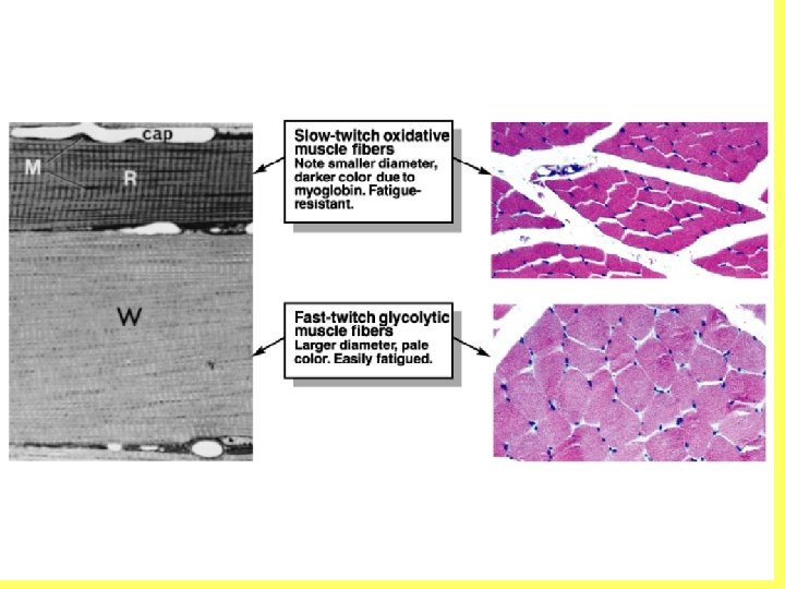
Single contraction characteristics of the three muscle types
Skeletal Muscle • Striated (sarcomere is functional unit); multinucleate • Voluntary (activated by a motor neurons) • Each fiber is a cell; many fibers to a muscle • Different types of fibers • Each fiber has a nerve connection
Typing based on succinate dehydrogenase activity Type I = low activity Type IIa = moderate activity Type IIb = high activity Typing based on speed of myosin (myosin ATPase activity Type I = low activity Type II = high activity
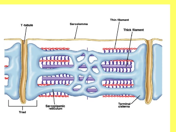
Basic Structure of a Skeletal Muscle Fiber Sarcolemma = cell membrane Fiber (cell) > myofibril (bundles of actin and myosin) > myofilamints (actin and myosin)
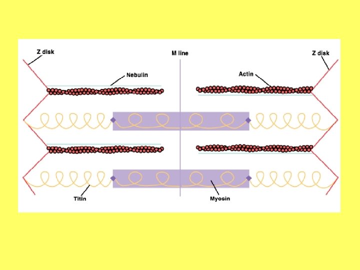
sarcomere • Functional unit of a myofibril is the sarcomere • Major filamentous proteins are actin and myosin, z proteins (in the z disk) • Many structural proteins; alpha actinin, myomesin, C protein, titin, nebulin • Cytoskeletal proteins; desmin, vimentin, filamin
Myosin (thick) filaments • Each filament contains about 400 individual myosin molecules • Bundled so that half of the molecules have their head facing one direction, the other half, the opposite direction Myosin heads are in a staggered arrangement along the filament
• Myosin is composed of 6 polypeptide chains, 2 H or heavy chains, and two L or light chains • Myosin head has ATPase activity • Myosin has a hinge region where the molecule is flexible • The myosin head has a high affinity for g actin • In smooth muscle, light chains regulate myosin action; in cardiac and skeletal muscle, light chains partially determine the speed of the myosin ATPase activity
Actin Filaments (thin filaments) • Monomers of globular or g actin combine to form long fibers of f actin. Two f actin molecules twist around one another to form a single thin filament • All actin myofilaments are anchored to the proteins of the z disk • Each g actin molecule has a binding site for the myosin head
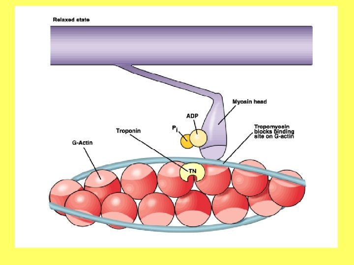
• Actin • Tropomyosin – covers active sites on g actin molecules • Troponin – regulates tropomyosin; three subunits – troponin c, troponin I, and troponin m • Troponin c has a binding site for calcium and is bound to the other two subunits • Troponin I keeps the tropomyosin over the myosin binding sites on G actin (inhibits actin/myosin binding) • Troponin m anchors the three subunits to the tropomyosin molecule
Ryanodine receptor
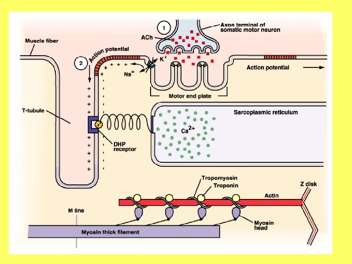
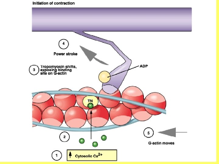
Excitation-contraction coupling 1. Motor neuron releases Ach onto the surface of the skeletal muscle fiber. The fiber’s nicotinic receptors are activated, opening K+ and Na+ channels. The cell membrane is depolarized. 2. The action potential moves away from the motor end plate in all directions, including down the t-tubule system. 3. Receptors in the t-tubules called dihydropyridine receptors (DHP) are activated by the change in voltage. They are connected to the ryanodine receptors in the lateral sacks of the sarcoplasmic reticulum (SR). 4. The ryanodine receptors are opened by the change in conformation of the DHP receptors and Ca 2+ is released from the SR. 5. Ca 2+ diffuses across the myofilaments. 6. The Ca 2+ binds to troponin C, causing it to change conformation, pulling on troponin I, which in turn pulls on tropomyosin. With the altered conformation in tropomyosin, the myosin binding sites on g actin molecules are exposed. 7. Myosin heads can now bind to g actin molecules and cross-bridge cycling begins, shortening the sarcomere by pulling on the actin filaments and drawing the z disks closer together
Cross-Bridge Cycling
Cross-bridge cycling • Continues as long as nerve depolarizes muscle membrane; also dependent on fuel supply as myosin power stroke is dependent on ATP • Pulls z lines closer together; shortens sarcomere • Myosin heads do not pull in a synchronized manner; random rowing Why?
Relaxation What must happen for a muscle to relax: 1. _ 2. _ 3. _ 4. _
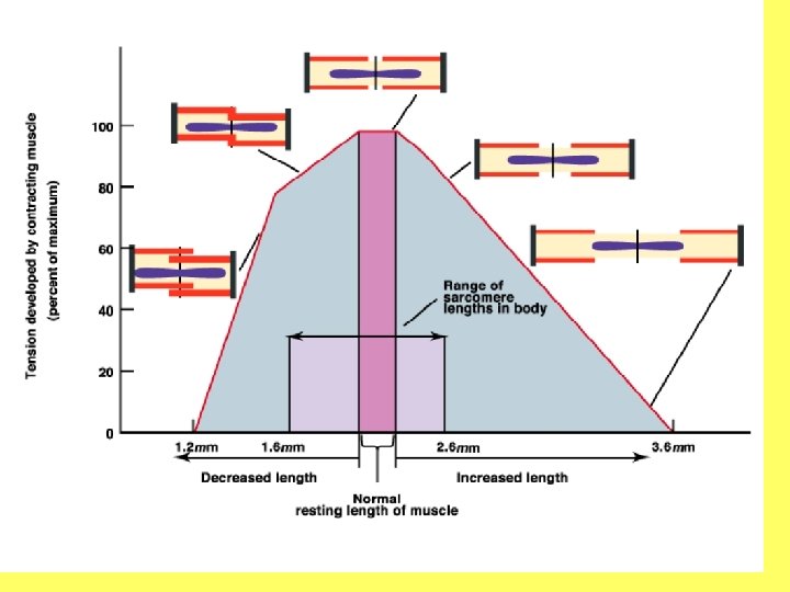
Factors Affecting Strength of Muscle Contraction • Energy supply • Anaerobic glycolysis; FA metabolism; phosphocreatine metabolism ATP + creatine • Muscle length phosphocreatine + ADP ATP + creatine
• Frequency of stimulation • Summation; tetanus Why does the force of contraction increase? What factors are changing at the cellular level?
Frequency of stimulus determines the overall cellular [Ca 2+]. The more Ca 2+, the greater the binding to troponin. The greater the binding, the more movement of tropomyosin. The more active sites exposed, the greater the number of myosin heads that can bind. The more myosin heads performing a powerstroke, the greater the strength of contraction.
• Number of motor units activated (recruitment) • Slow twitch, fatigue resistant activated first; weak stimulus activates them • Fast twitch, fatigueresistant (intermediate) activated as stimulus intensity increases • Fast twitch, glycolytic activated at stimuli of highest intensity What will happen if you maximally contract a muscle for an extended period of time? Why?
Types of Contractions 1. 2. Isotonic = same stretch; moves a load; as muscle contracts, it shortens • Concentric = muscle shortens as it moves a load • Eccentric = muscle lengthens as it moves a load Isometric = same length; load is not moved; muscle contracts but doesn’t shorten
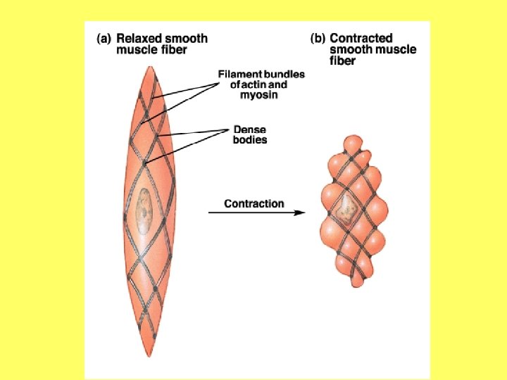
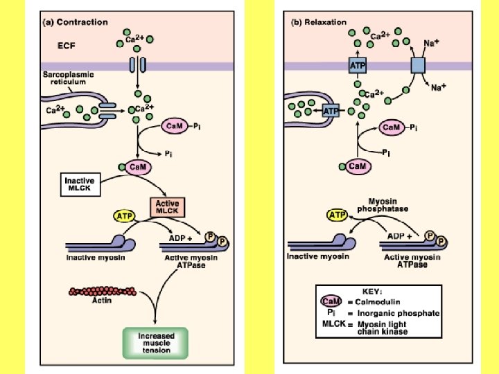
Smooth muscle • Small, not striated • Involuntary • Found in hollow organs; other organs important to homeostasis • Can be activated by stretch, neurons, hormones • Little SR; caveolae • Myosin has heads down entire length • Slow ATPase activity • Regulated by myosin light chains • Dense bodies anchor actin • Actin has no troponin or tropomyosin components
ACTIN REGULATED CONTRACTION VS. MYOSIN REGULATED CONTRACTION