Muscles Muscles Muscle cells specialized for contraction Allow


- Slides: 17

Muscles

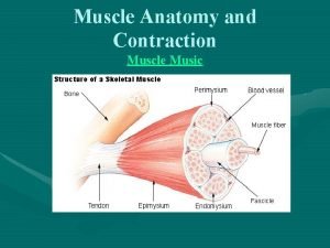
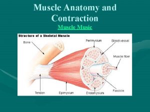
Muscles • Muscle cells specialized for contraction • Allow movement & bodily functions such as respiration & digestion

3 Types of Muscle Skeletal • Attached to bones • Control locomotion & movement • Voluntary • Striated Smooth • Walls of organs • Control digestion, respiratory • Involuntary • Non-striated Cardiac • Only in heart • Contractions pump blood • Involuntary • Striated

3 Types of Muscle Skeletal • Attached to bones • Control locomotion & movement • Voluntary • Striated Smooth • Walls of organs • Control digestion, respiratory • Involuntary • Non-striated Cardiac • Only in heart • Contractions pump blood • Involuntary • Striated

3 Types of Muscle Skeletal • Attached to bones • Control locomotion & movement • Voluntary • Striated Smooth • Walls of organs • Control digestion, respiratory • Involuntary • Non-striated Cardiac • Only in heart • Contractions pump blood • Involuntary • Striated

Skeletal Muscle Fiber Structure • Each muscle fiber has 1000’s of myofibrils • Sarcomere: Repeating structural unit of a myofibril btw two Z lines

Skeletal Muscle Fiber Structure Myosin (thick filaments) & Actin (thin filaments) give striated appearance

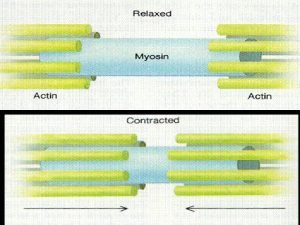
Sliding Filament Theory of Muscle Contraction Contracting muscle shortens but the filaments stay the same length; Instead, they slide past each other

ATP needed for muscle contraction • Each myosin has a “tail” region and a “head” region • Head region of myosin binds to ATP (lowenergy configuration)

Hydrolysis of ATP converts myosin to high-energy form

Myosin head can now bind to actin by a cross-bridge

Myosin head returns to low-energy form as it pulls the actin towards sarcomere center

New ATP binds to myosin head, breaking cross-bridge, releasing actin

Role of Action Potentials & Calcium in Contraction • Synaptic terminal of motor neuron releases acetylcholine (neurotransmitter), depolarizing muscle fiber’s membrane • Depolarization causes action potential to move across fiber and into it along transverse (T) tubules • Action potential triggers release of Ca 2+ from sarcoplasmic reticulum into cytosol

Regulatory Proteins • Muscle fiber at rest: tropomyosin & troponin complex binds to actin, blocking myosin-binding sites so no cross-bridge can form

Regulatory Proteins • Muscle fiber at rest: tropomyosin & troponin complex binds to actin, blocking myosin-binding sites so no cross-bridge can form • Motor neurons enable contraction by triggering release of Ca 2+ into cytosol • Ca 2+ binds to troponin complex, exposing myosinbinding sites, allowing cross-bridges

Summary of Contraction in Skeletal Muscle Fiber
 Smooth muscle contraction
 Myofibril
 Muscle contraction
 Muscle contraction
 Spread of cardiac impulse
 Muscle contraction animation mcgraw hill
 Muscle tissue ppt
 Latent phase muscle contraction
 3 phases of muscle contraction
 Drug acting on eye
 Sliding filament theory worksheet
 Phases of muscle contraction
 Sarcoplasmic reticulum
 Direct phosphorylation
 Muscle spasm
 Muscle contraction
 Whole muscle contraction
 Muscle contraction
