Muscle Contraction Put the following structures in order


- Slides: 40
Muscle Contraction Put the following structures in order of size, starting with the biggest: • • • Sarcomere Bundle of muscle fibres Myofibril Muscle fibre I band
Muscle Contraction Put the following structures in order of size, starting with the biggest: • • • Muscle Bundle of muscle fibres Muscle fibre Myofibril Sarcomere I band
The Sarcomere These filaments are arranged in an interlocking pattern within the sarcomere, producing the characteristic banding pattern of the myofibrils. thin filament (actin) thick filament (myosin) Z-line
The Sliding Filament Theory • • • When the muscle contracts, sarcomeres shorten. The filaments do not change in length. They slide past each other (overlap) Actin filaments slide between myosin filaments The zone of overlap increases during contraction. What will change in a sarcomere when a muscle contracts? What will stay the same?
Evidence for the sliding filament theory • Sarcomeres shorten during contraction – Z lines get closer together • The I band becomes narrower • The A band (length of myosin filament) does not change • The H zone becomes narrower
Proteins in muscle contraction Myosin • fibrous protein made up of several hundred molecules • globular protein formed into two bulbous structures at one end
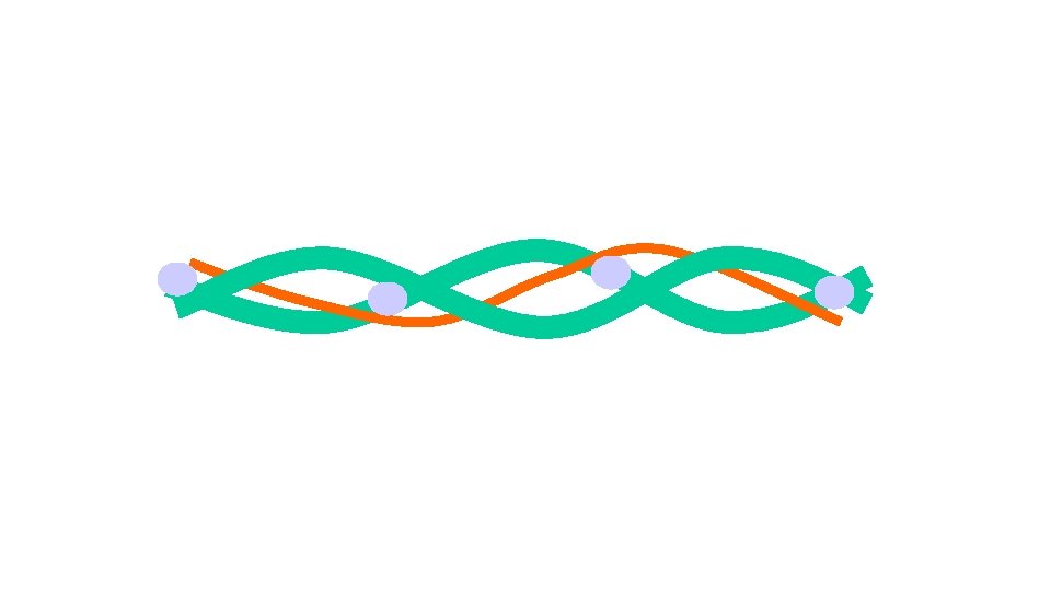
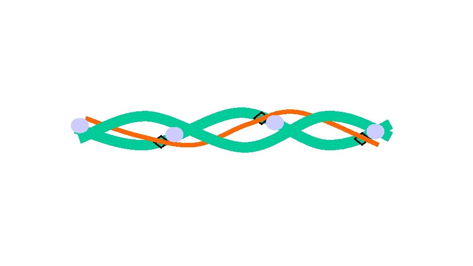
Proteins in muscle contraction Actin • Globular protein arranged into two long chains to form a helical strand
Proteins in muscle contraction Tropomyosin • Long thing threads that wind around the actin filaments
Muscle contraction • The action potential reaches the muscle through a system of tubules (T tubules) that branch throughout the sarcoplasm (cytoplasm of the muscle) • The tubules are in contact with the endoplasmic reticulum of the muscle (sarcoplasmic reticulum) which has actively transported Ca 2+ from the sarcoplasm. • The action potential opens the Ca 2+ channels and the calcium ions rush into the sarcoplasm down a conc gradient…
actin
actin troponin
tropomyosin actin troponin
tropomyosin actin troponin
myosin binding site actin tropomyosin troponin
Ca 2+
Ca 2+ Calcium ions are released from the sarcolemma after stimulation from the T system
Ca 2+
Ca 2+ the calcium ions cause the tropomyosin to change its shape
Ca 2+
Ca 2+
Ca 2+
Ca 2+ This exposes the myosin binding sites on the actin molecule
Myosin Head Ca 2+ Pi Pi A Ca 2+
Pi Pi A Ca 2+
Pi Pi A Ca 2+ the bulbous heads of the myosin attach to the binding sites on the actin filaments Ca 2+
Pi Pi A Ca 2+
Pi Pi A Ca 2+ the myosin heads change their angle to achieve a lower energy state and pull the actin filaments past the stationary myosin ADP molecule is released
Pi Pi A Ca 2+ the myosin heads change their angle to achieve a lower energy state and pull the actin filaments past the stationary myosin ADP molecule is released
Ca 2+ Pi Pi Pi A
Pi Pi Pi A Pi Pi Pi Ca A 2+ Ca 2+ Pi Pi Pi A Ca 2+
Pi Pi Pi A Pi Pi Pi Ca A 2+ Ca 2+ Pi Pi Pi A Ca 2+ ATP binds to the bulbous heads and causes it to become detached
Pi Pi Pi A Ca 2+ ATP binds to the bulbous heads and causes it to become detached
Pi Pi Pi A Ca 2+ The calcium ions activate the enzyme ATPase which hydrolyses ATP. This provides the energy to return the heads to their original position
Pi Pi Pi A Pi Ca 2+ Pi Pi Pi A Ca 2+ The myosin head returns to its original position and then can attach itself further along the actin filament as long as calcium conc remains high
Pi Pi Pi A Pi Ca 2+ Pi Pi A Ca 2+
Muscle Relaxation • When nervous stimulation stops, Ca 2+ are actively transported back into endoplasmic reticulum using energy from hydrolysis of ATP • This means that tropomyosin blocks binding sites on actin so myosin heads can no longer bind
1. 2. 3 4. 5. An action potential arrives at the neuromuscular junction. This causes the release of the neurotransmitter acetylcholine. This initiates an action potential in the sarcolemma. Action potential is carried into the large muscle cell via T-tubules. Causes the sarcoplasmic reticulum to release its store of Ca 2+ into myofibrils. 6. Causes tropomyosin to be displaced. Uncovers myosin binding sites on actin. 7. Actinmyosin cross bridges can now attach, the cross bridge cycle takes place. 8. When muscle relaxes calcium ions actively transported back into sarcoplasmic reticulum (using energy from hydrolysis of ATP ) 9. Tropomyosin blocks the actin filaments again. 10. Myosin heads unable to bind to actin filaments so contraction ceases.