Microscopes History Parts Functions Early Microscope History of


, glass")


- Slides: 19
Microscopes History, Parts, & Functions
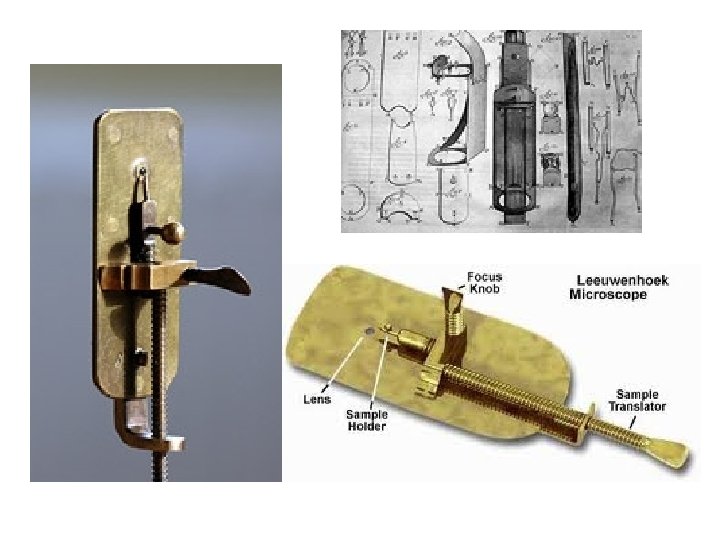
Early Microscope
History of the Microscope • During the 1 st century AD (year 100), glass had been invented and the Romans were looking through the glass and testing it. – They experimented with different shapes of clear glass and one of their samples was thick in the middle and thin on the edges. – They discovered that if you held one of these “lenses” over an object, the object would look larger.
History of the Microscope • These early lenses were called magnifiers or burning glasses. • The word lens by the way, is derived from the Latin word lentil, as they were named because they resembled the shape of a lentil bean
History of the Microscope • These magnifiers became popular in the 13 th century when spectacle makers were producing lenses to be worn as glasses. • In 1590, two Dutch spectacle makers started experimenting with these lenses. – Zaccharias Janssen and his father Hans • Experiments led by these men and other scientists led to the discovery of the compound microscope
History of the Microscope • Many great people experimented with lenses, such as Galileo. But one man became the “father of microscopy” – his name is Anthony Leeuwenhoek (1632 -1723) – He saw bacteria, yeast, blood cells and many tiny animals swimming about in a drop of water. – Compound microscopes had been invented in the 1590 s, nearly forty years before Leeuwenhoek was born, however there were technical difficulties in building them.
The Modern Compound Microscope • A compound light microscope is a microscope with more than one lens and its own light source. – is an instrument commonly used inside the laboratory to view specimens mounted on a glass slide – The compound microscope magnifies the object in the slide in such a way that the smallest structures become visible and clear. – allowing structures in the slide to be magnified for up to 2, 000 times their actual size.
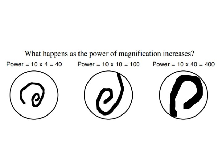
Magnification • If the magnification of a lens is 2 X then it roughly doubles the size of the image of the object.
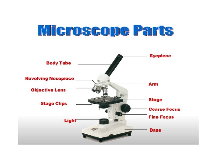
Parts of the Microscope • Eyepiece: the lens at the top that you look through. • Body Tube: Connects the eyepiece to the objective lenses
Parts of the Microscope • Stage Clips: clips that hold your slide in place • Objectives: Usually you will find 3 or 4 objective lenses on a microscope. They almost always consist of 4 X, 10 X, 40 X and 100 X powers.
Parts of the Microscope • Coarse Adjustment Knob: Moves the stage up and down for FOCUSING • Fine Adjustment Knob: Moves the stage slightly to SHARPEN the image
Parts of the Microscope • Light: A steady light source to help illuminate the slides • Stage: The flat platform where you place your slides.
Parts of the Microscope • Nosepiece: This is the part that holds two or more objective lenses and can be rotated to easily change power.
Parts of the Microscope Arm • Arm: used to support the microscope when carried • Base: bottom of the microscope. Supports it. Base
• Let’s see the world of microscopes!